Filters
Clonality
Type
Reactivity
Gene Name
Isotype
Host
Application
Clone
10000 results for "Goat IgG Fc " - showing 1400-1450


 Western Blot (WB)
(Western blot analysis of lysates from WiDr,Jurkat,mouse NIH/3T3,rat PC-12,Hela cell line (from left to right), using ATP5B Antibody (Center). It was diluted at 1:1000 at each lane. A goat anti-rabbit IgG H&L(HRP) at 1:10000 dilution was used as the secondary antibody.)
Western Blot (WB)
(Western blot analysis of lysates from WiDr,Jurkat,mouse NIH/3T3,rat PC-12,Hela cell line (from left to right), using ATP5B Antibody (Center). It was diluted at 1:1000 at each lane. A goat anti-rabbit IgG H&L(HRP) at 1:10000 dilution was used as the secondary antibody.)
ATP5B, Polyclonal Antibody (Cat# AAA9231807)
Full Name
ATP5B Antibody (Center)
Gene Names
ATP5F1B; ATP5B; ATPMB; ATPSB; HEL-S-271
Reactivity
Human, Mouse, Rat
Predicted: Bovine
Predicted: Bovine
Applications
WB, IHC, IF, FC/FACS, EIA
Pricing
$425/0.1 mL | $1,615/5x0.1 mL


 Immunocytochemistry/Immunofluorescence (ICC/IF)
(Immunocytochemistry/Immunofluorescence analysis using Mouse Anti-Dityrosine Monoclonal Antibody, Clone 10A6. Tissue: Embryonic kidney epithelial cell line (HEK293). Species: Human. Fixation: 5% Formaldehyde for 5 min. Primary Antibody: Mouse Anti-Dityrosine Monoclonal Antibody at 1:50 for 30-60 min at RT. Secondary Antibody: Goat Anti-Mouse Alexa Fluor 488 at 1:1500 for 30-60 min at RT. Counterstain: Phalloidin Alexa Fluor 633 F-Actin stain; DAPI (blue) nuclear stain at 1:250, 1:50000 for 30-60 min at RT. Localization: Cytoplasmic. Magnification: 20X (2X Zoom). (A,C,E,G) - Untreated. (B,D,F,H) - Cells cultured overnight with 50 uM H2O2. (A,B) DAPI (blue) nuclear stain. (C,D) Phalloidin Alex Fluor 633 F-Actin stain. (E,F) Dityrosine Antibody. (G,H) Composite. Courtesy of: Dr. Robert Burke, University of Victoria.)
Immunocytochemistry/Immunofluorescence (ICC/IF)
(Immunocytochemistry/Immunofluorescence analysis using Mouse Anti-Dityrosine Monoclonal Antibody, Clone 10A6. Tissue: Embryonic kidney epithelial cell line (HEK293). Species: Human. Fixation: 5% Formaldehyde for 5 min. Primary Antibody: Mouse Anti-Dityrosine Monoclonal Antibody at 1:50 for 30-60 min at RT. Secondary Antibody: Goat Anti-Mouse Alexa Fluor 488 at 1:1500 for 30-60 min at RT. Counterstain: Phalloidin Alexa Fluor 633 F-Actin stain; DAPI (blue) nuclear stain at 1:250, 1:50000 for 30-60 min at RT. Localization: Cytoplasmic. Magnification: 20X (2X Zoom). (A,C,E,G) - Untreated. (B,D,F,H) - Cells cultured overnight with 50 uM H2O2. (A,B) DAPI (blue) nuclear stain. (C,D) Phalloidin Alex Fluor 633 F-Actin stain. (E,F) Dityrosine Antibody. (G,H) Composite. Courtesy of: Dr. Robert Burke, University of Victoria.)
Dityrosine, Monoclonal Antibody (Cat# AAA808880)
Full Name
Dityrosine Antibody, Clone 10A6
Reactivity
Species Independent
Applications
WB, ICC, IF, FC/FACS, EIA
Purity
Protein G Purified
Pricing
$505/0.1 mg | $2,050/5x0.1 mg


 Immunocytochemistry/Immunofluorescence (ICC/IF)
(Immunocytochemistry/Immunofluorescence analysis using Mouse Anti-Dityrosine Monoclonal Antibody, Clone 10A6. Tissue: Embryonic kidney epithelial cell line (HEK293). Species: Human. Fixation: 5% Formaldehyde for 5 min. Primary Antibody: Mouse Anti-Dityrosine Monoclonal Antibody at 1:50 for 30-60 min at RT. Secondary Antibody: Goat Anti-Mouse Alexa Fluor 488 at 1:1500 for 30-60 min at RT. Counterstain: Phalloidin Alexa Fluor 633 F-Actin stain; DAPI (blue) nuclear stain at 1:250, 1:50000 for 30-60 min at RT. Localization: Cytoplasmic. Magnification: 20X (2X Zoom). (A,C,E,G) - Untreated. (B,D,F,H) - Cells cultured overnight with 50 uM H2O2. (A,B) DAPI (blue) nuclear stain. (C,D) Phalloidin Alex Fluor 633 F-Actin stain. (E,F) Dityrosine Antibody. (G,H) Composite. Courtesy of: Dr. Robert Burke, University of Victoria.)
Immunocytochemistry/Immunofluorescence (ICC/IF)
(Immunocytochemistry/Immunofluorescence analysis using Mouse Anti-Dityrosine Monoclonal Antibody, Clone 10A6. Tissue: Embryonic kidney epithelial cell line (HEK293). Species: Human. Fixation: 5% Formaldehyde for 5 min. Primary Antibody: Mouse Anti-Dityrosine Monoclonal Antibody at 1:50 for 30-60 min at RT. Secondary Antibody: Goat Anti-Mouse Alexa Fluor 488 at 1:1500 for 30-60 min at RT. Counterstain: Phalloidin Alexa Fluor 633 F-Actin stain; DAPI (blue) nuclear stain at 1:250, 1:50000 for 30-60 min at RT. Localization: Cytoplasmic. Magnification: 20X (2X Zoom). (A,C,E,G) - Untreated. (B,D,F,H) - Cells cultured overnight with 50 uM H2O2. (A,B) DAPI (blue) nuclear stain. (C,D) Phalloidin Alex Fluor 633 F-Actin stain. (E,F) Dityrosine Antibody. (G,H) Composite. Courtesy of: Dr. Robert Burke, University of Victoria.)
Dityrosine, Monoclonal Antibody (Cat# AAA808882)
Full Name
Dityrosine Antibody, Clone 10A6: ATTO 488
Reactivity
Species Independent
Applications
WB, ICC, IF, FC/FACS, EIA
Purity
Protein G Purified
Pricing
$560/0.1 mg | $2,275/5x0.1 mg


 Immunofluorescence (IF)
(Immunofluorescent analysis of 4% paraformaldehyde-fixed, 0. 1% Triton X-100 permeabilized U-2 OS (human bone osteosarcoma cell line) cells labeling Pdx1 at 1/25 dilution, followed by Dylight 488-conjugated goat anti-mouse IgG (NA166821) secondary antibody at 1/200 dilution (green). Immunofluorescence image showing cytoplasm staining on U-2 OS cell line. Cytoplasmic actin is detected with Dylight 554 Phalloidin (PD18466410) at 1/100 dilution (red). The nuclear counter stain is DAPI (blue).)
Immunofluorescence (IF)
(Immunofluorescent analysis of 4% paraformaldehyde-fixed, 0. 1% Triton X-100 permeabilized U-2 OS (human bone osteosarcoma cell line) cells labeling Pdx1 at 1/25 dilution, followed by Dylight 488-conjugated goat anti-mouse IgG (NA166821) secondary antibody at 1/200 dilution (green). Immunofluorescence image showing cytoplasm staining on U-2 OS cell line. Cytoplasmic actin is detected with Dylight 554 Phalloidin (PD18466410) at 1/100 dilution (red). The nuclear counter stain is DAPI (blue).)
PPP2R1B, Monoclonal Antibody (Cat# AAA9232165)
Full Name
PPP2R1B Antibody
Gene Names
PPP2R1B; PR65B; PP2A-Abeta
Reactivity
Human, Mouse, Rat
Applications
WB, EIA
Pricing
$435/0.1 mL | $1,655/5x0.1 mL


 Testing Data
(Staining of mouse spleen with Rat anti Mouse CD4)
Testing Data
(Staining of mouse spleen with Rat anti Mouse CD4)
CD4, Monoclonal Antibody (Cat# AAA219566)
Full Name
RAT ANTI MOUSE CD4
Gene Names
Cd4; L3T4; Ly-4
Applications
EIA, FC/FACS, IF, IP, WB
Pricing
$475/0.25 mg | $1,995/5x0.25 mg


 Immunofluorescence (IF)
(Immunofluorescent analysis of 4% paraformaldehyde-fixed, 0. 1% Triton X-100 permeabilized HepG2 (human liver hepatocellular carcinoma cell line) cells labeling Pdx1 with AP22330a at 1/25 dilution, followed by Dylight 488-conjugated goat anti-rabbit IgG (NK179883) secondary antibody at 1/200 dilution (green). Immunofluorescence image showing cytoplasm staining on HepG2 cell line. Cytoplasmic actin is detected with Dylight 554 Phalloidin (PD18466410) at 1/100 dilution (red). The nuclear counter stain is DAPI (blue).)
Immunofluorescence (IF)
(Immunofluorescent analysis of 4% paraformaldehyde-fixed, 0. 1% Triton X-100 permeabilized HepG2 (human liver hepatocellular carcinoma cell line) cells labeling Pdx1 with AP22330a at 1/25 dilution, followed by Dylight 488-conjugated goat anti-rabbit IgG (NK179883) secondary antibody at 1/200 dilution (green). Immunofluorescence image showing cytoplasm staining on HepG2 cell line. Cytoplasmic actin is detected with Dylight 554 Phalloidin (PD18466410) at 1/100 dilution (red). The nuclear counter stain is DAPI (blue).)
MKS1, Polyclonal Antibody (Cat# AAA9231277)
Full Name
MKS1 Antibody (N-Term)
Gene Names
MKS1; MES; MKS; BBS13; POC12; JBTS28
Reactivity
Human
Applications
IF, FC/FACS, WB, EIA
Purity
Purified through a protein A column, followed by peptide affinity purification.
Pricing
$185/0.05 mL | $405/0.2 mL | $1,585/5x0.2 mL


 Western Blot (WB)
(Figure 1. Western blot analysis of CCT3 using anti- CCT3 antibody (MBS178426). Electrophoresis was performed on a 5-20% SDS-PAGE gel at 70V (Stacking gel) / 90V (Resolving gel) for 2-3 hours. The sample well of each lane was loaded with 50ug of sample under reducing conditions. Lane 1: rat kidney tissue lysates, Lane 2: HELA whole cell lysates. After Electrophoresis, proteins were transferred to a Nitrocellulose membrane at 150mA for 50-90 minutes. Blocked the membrane with 5% Non-fat Milk/ TBS for 1.5 hour at RT. The membrane was incubated with rabbit anti- CCT3 antigen affinity purified polyclonal antibody at 0.5ug/mL overnight at 4 degree C, then washed with TBS-0.1%Tween 3 times with 5 minutes each and probed with a goat anti-rabbit IgG-HRP secondary antibody at a dilution of 1:10000 for 1.5 hour at RT. The signal is developed using an Enhanced Chemiluminescent detection (ECL) kit with Tanon 5200 system. A specific band was detected for CCT3 at approximately 61KD. The expected band size for CCT3 is at 61KD. )
Western Blot (WB)
(Figure 1. Western blot analysis of CCT3 using anti- CCT3 antibody (MBS178426). Electrophoresis was performed on a 5-20% SDS-PAGE gel at 70V (Stacking gel) / 90V (Resolving gel) for 2-3 hours. The sample well of each lane was loaded with 50ug of sample under reducing conditions. Lane 1: rat kidney tissue lysates, Lane 2: HELA whole cell lysates. After Electrophoresis, proteins were transferred to a Nitrocellulose membrane at 150mA for 50-90 minutes. Blocked the membrane with 5% Non-fat Milk/ TBS for 1.5 hour at RT. The membrane was incubated with rabbit anti- CCT3 antigen affinity purified polyclonal antibody at 0.5ug/mL overnight at 4 degree C, then washed with TBS-0.1%Tween 3 times with 5 minutes each and probed with a goat anti-rabbit IgG-HRP secondary antibody at a dilution of 1:10000 for 1.5 hour at RT. The signal is developed using an Enhanced Chemiluminescent detection (ECL) kit with Tanon 5200 system. A specific band was detected for CCT3 at approximately 61KD. The expected band size for CCT3 is at 61KD. )
CCT3, Polyclonal Antibody (Cat# AAA178426)
Full Name
Anti-CCT3 Antibody
Gene Names
CCT3; CCTG; PIG48; TRIC5; CCT-gamma; TCP-1-gamma
Reactivity
Human, Rat
No cross reactivity with other proteins.
No cross reactivity with other proteins.
Applications
WB
Purity
Immunogen affinity purified.
Pricing
$345/0.1 mg | $1,485/5x0.1 mg


 Immunohistochemistry (IHC)
(Formalin-fixed, paraffin-embedded human Tonsil stained with CD20 Mouse Monoclonal Antibody (L26).)
Immunohistochemistry (IHC)
(Formalin-fixed, paraffin-embedded human Tonsil stained with CD20 Mouse Monoclonal Antibody (L26).)
CD20/MS4A1, Monoclonal Antibody (Cat# AAA4381412)
Full Name
CD20/MS4A1 (B-Cell Marker)
Gene Names
MS4A1; B1; S7; Bp35; CD20; CVID5; MS4A2; LEU-16
Reactivity
Human
Applications
FC/FACS, IF, WB, IHC
Purity
Purified Ab with BSA and Azide at 200ug/ml OR Purified Ab WITHOUT BSA and Azide at 1.0mg/ml
Pricing
$390/0.1 mg (With BSA & Azide at 0.2mg/mL) | $390/0.1 mg (Without BSA & Azide at 1mg/mL) | $215/0.02 mg (With BSA & Azide at 0.2mg/mL) | $1,710/5x0.1 mg (With BSA & Azide at 0.2mg/mL) | $1,710/5x0.1 mg (Without BSA & Azide at 1mg/mL)


 Western Blot (WB)
(Western Blot analysis of 293 (lane 1), Jurkat cell line (lane 2) and mouse liver tissue (lane 3) lysates (35ug/lane) using MBS6003748. This demonstrates that MBS6003748 detected the FOXP3 protein (arrow).)
Western Blot (WB)
(Western Blot analysis of 293 (lane 1), Jurkat cell line (lane 2) and mouse liver tissue (lane 3) lysates (35ug/lane) using MBS6003748. This demonstrates that MBS6003748 detected the FOXP3 protein (arrow).)
FOXP3, Polyclonal Antibody (Cat# AAA6003748)
Full Name
FOXP3, CT (Forkhead Box Protein P3, AIID, IPEX, JM2, Scurfin)
Gene Names
FOXP3; JM2; AIID; IPEX; PIDX; XPID; DIETER
Reactivity
Mouse
Applications
EL/EIA, WB, IHC, FC
Purity
Purified by Protein A and affinity chromatography.
Pricing
$675/0.2 mL | $3,020/5x0.2 mL
IgG (H+L), Secondary Antibody (Cat# AAA128205)
Full Name
FITC Donkey Anti-Goat IgG (H+L) Antibody
Applications
ICC, FC/FACS
Purity
Affinity Purification
Pricing
$150/0.1 mL | $525/5x0.1 mL

 Flow Cytometry (FC/FACS)
(Flow cytometric analysis of Raji cells using CD74(green) compared to an isotype control of mouse IgG2b(blue). MBS9200181 was diluted at 1:25 dilution. An Alexa Fluor 488 goat anti-mouse lgG at 1:400 dilution was used as the secondary antibody.)
Flow Cytometry (FC/FACS)
(Flow cytometric analysis of Raji cells using CD74(green) compared to an isotype control of mouse IgG2b(blue). MBS9200181 was diluted at 1:25 dilution. An Alexa Fluor 488 goat anti-mouse lgG at 1:400 dilution was used as the secondary antibody.)
CD74, Monoclonal Antibody (Cat# AAA9200181)
Full Name
CD74 Antibody
Gene Names
CD74; II; DHLAG; HLADG; Ia-GAMMA
Reactivity
Human
Applications
WB, EIA, FC/FACS
Purity
Purified Mouse Monoclonal Antibody (Mab)
Pricing
$425/0.4 mL | $190/0.08 mL | $1,665/5x0.4 mL
 Western Blot (WB)
(Western blot analysis of extracts of Raji cells, using FCRL2 antibody at 1:1000 dilution.Secondary antibody: HRP Goat Anti-Rabbit IgG (H+L) (MBS128200) at 1:10000 dilution.Lysates/proteins: 25ug per lane.Blocking buffer: 3% nonfat dry milk in TBST.Detection: ECL Basic Kit.Exposure time: 1s.)
Western Blot (WB)
(Western blot analysis of extracts of Raji cells, using FCRL2 antibody at 1:1000 dilution.Secondary antibody: HRP Goat Anti-Rabbit IgG (H+L) (MBS128200) at 1:10000 dilution.Lysates/proteins: 25ug per lane.Blocking buffer: 3% nonfat dry milk in TBST.Detection: ECL Basic Kit.Exposure time: 1s.)
FCRL2, Polyclonal Antibody (Cat# AAA9130835)
Full Name
FCRL2 Polyclonal Antibody
Gene Names
FCRL2; FCRH2; IFGP4; IRTA4; SPAP1; CD307b; SPAP1A; SPAP1B; SPAP1C
Reactivity
Human
Applications
WB
Purity
Affinity Purification
Pricing
$180/0.02 mL | $210/0.05 mL | $285/0.1 mL | $430/0.2 mL | $1,790/5x0.2 mL

 Western Blot (WB)
(Western blot analysis of lysates from Ramos,Raji,Daudi cell line (from left to right), using TCL1A Antibody (Center). It was diluted at 1:1000 at each lane. A goat anti-rabbit IgG H&L(HRP) at 1:10000 dilution was used as the secondary antibody.Lysates at 20ug per lane.)
Western Blot (WB)
(Western blot analysis of lysates from Ramos,Raji,Daudi cell line (from left to right), using TCL1A Antibody (Center). It was diluted at 1:1000 at each lane. A goat anti-rabbit IgG H&L(HRP) at 1:10000 dilution was used as the secondary antibody.Lysates at 20ug per lane.)
TCL1A, Polyclonal Antibody (Cat# AAA9231916)
Full Name
TCL1A Antibody (Center)
Gene Names
TCL1A; TCL1
Reactivity
Human
Applications
WB, FC/FACS, EIA
Pricing
$425/0.1 mL | $1,615/5x0.1 mL


 Testing Data
(Staining of human erythrocytes with Mouse anti Human CD147:APC)
Testing Data
(Staining of human erythrocytes with Mouse anti Human CD147:APC)
CD147, Monoclonal Antibody (Cat# AAA215305)
Full Name
MOUSE ANTI HUMAN CD147:APC
Gene Names
BSG; M6; OK; 5F7; TCSF; CD147; EMMPRIN
Applications
FC/FACS
Pricing
$500/100 Tests | $2,090/5x100 Tests


 Western Blot (WB)
((2ug/ml) staining of K562 nuclear cell lysate (A) + peptide (B) and negative control Human Hippocampus (C) lysate (35ug protein in RIPA buffer). Detected by chemiluminescence.)
Western Blot (WB)
((2ug/ml) staining of K562 nuclear cell lysate (A) + peptide (B) and negative control Human Hippocampus (C) lysate (35ug protein in RIPA buffer). Detected by chemiluminescence.)
GATA1, Antibody (Cat# AAA423829)
Full Name
Goat Anti-GATA1 Antibody
Gene Names
GATA1; GF1; GF-1; NFE1; XLTT; ERYF1; NF-E1; XLANP; XLTDA; GATA-1
Reactivity
Human
Expected from sequence similarity: Human
Expected from sequence similarity: Human
Applications
EIA, IF, FC/FACS
Purity
Purified from goat serum by ammonium sulphate precipitation followed by antigen affinity chromatography using the immunizing peptide.
Pricing
$335/0.1 mg


 Western Blot (WB)
(Western blot analysis of extracts of NIH/3T3 cells, using MAP1LC3B antibody at 1:1000 dilution. NIH/3T3 cells were treated by Chloroquine (50 uM) for 20 hours.Secondary antibody: HRP Goat Anti-Rabbit IgG (H+L) (MBS128200) at 1:10000 dilution.Lysates/proteins: 25ug per lane.Blocking buffer: 3% nonfat dry milk in TBST.Detection: ECL Basic Kit.Exposure time: 3min.)
Western Blot (WB)
(Western blot analysis of extracts of NIH/3T3 cells, using MAP1LC3B antibody at 1:1000 dilution. NIH/3T3 cells were treated by Chloroquine (50 uM) for 20 hours.Secondary antibody: HRP Goat Anti-Rabbit IgG (H+L) (MBS128200) at 1:10000 dilution.Lysates/proteins: 25ug per lane.Blocking buffer: 3% nonfat dry milk in TBST.Detection: ECL Basic Kit.Exposure time: 3min.)
MAP1LC3B, Polyclonal Antibody (Cat# AAA9125044)
Full Name
MAP1LC3B Polyclonal Antibody
Gene Names
MAP1LC3B; LC3B; ATG8F; MAP1LC3B-a; MAP1A/1BLC3
Reactivity
Human, Mouse, Rat, Hamster
Applications
WB, IHC, ICC, IP, FC/FACS
Purity
Affinity Purification
Pricing
$180/0.02 mL | $210/0.05 mL | $285/0.1 mL | $430/0.2 mL | $1,790/5x0.2 mL


 Western Blot (WB)
(Western blot analysis of lysates from Hela cell line and human ovary tissue lysate (from left to right), using DIRAS3 Antibody. MBS9207832 was diluted at 1:1000 at each lane. A goat anti-rabbit IgG H&L(HRP) at 1:10000 dilution was used as the secondary antibody. Lysates at 35ug per lane.)
Western Blot (WB)
(Western blot analysis of lysates from Hela cell line and human ovary tissue lysate (from left to right), using DIRAS3 Antibody. MBS9207832 was diluted at 1:1000 at each lane. A goat anti-rabbit IgG H&L(HRP) at 1:10000 dilution was used as the secondary antibody. Lysates at 35ug per lane.)
DIRAS3, Polyclonal Antibody (Cat# AAA9207832)
Full Name
DIRAS3 Antibody
Gene Names
DIRAS3; ARHI; NOEY2
Reactivity
Human
Applications
WB, EIA, IHC, FC/FACS
Purity
Peptide Affinity Purified Rabbit Polyclonal Antibody (Pab)
Pricing
$405/0.4 mL | $185/0.08 mL | $1,585/5x0.4 mL


 Western Blot (WB)
(All lanes : CD5 Mouse mAb at 1/2000 dilution Lane 1 : Jurkat cells membrane fraction Lane 2 : THP1 cells membrane fraction Lysates/proteins at 20 ug per lane. Secondary Goat Anti-Mouse IgG-HRP, 5% milk conjugated at 1/10000 dilution Predicted band size : 55 KDa Observed band size : 55 KDa Blocking/Dilution buffer : 1x TBST.)
Western Blot (WB)
(All lanes : CD5 Mouse mAb at 1/2000 dilution Lane 1 : Jurkat cells membrane fraction Lane 2 : THP1 cells membrane fraction Lysates/proteins at 20 ug per lane. Secondary Goat Anti-Mouse IgG-HRP, 5% milk conjugated at 1/10000 dilution Predicted band size : 55 KDa Observed band size : 55 KDa Blocking/Dilution buffer : 1x TBST.)
CD5, Monoclonal Antibody (Cat# AAA435110)
Full Name
CD5 Mouse mAb
Gene Names
CD5; T1; LEU1
Reactivity
Human
Applications
WB, IP, FC/FACS
Pricing
$260/0.02 mL | $405/0.05 mL | $650/0.1 mL | $2,645/5x0.1 mL


 Western Blot (WB)
TIM50, Monoclonal Antibody (Cat# AAA435157)
Full Name
TIM50 (680) Mouse mAb
Gene Names
TIMM50; MGCA9; TIM50; TIM50L
Reactivity
Human
Applications
WB, IP, FC/FACS
Pricing
$260/0.02 mL | $405/0.05 mL | $650/0.1 mL | $2,645/5x0.1 mL


 Quality Control
(Western blot analysis of immunized recombinant protein, using anti-POLR2G monoclonal antibody. )
Quality Control
(Western blot analysis of immunized recombinant protein, using anti-POLR2G monoclonal antibody. )
POLR2G, Monoclonal Antibody (Cat# AAA120241)
Full Name
Mouse monoclonal antibody Anti-Human POLR2G
Gene Names
POLR2G; RPB7; RPB19; hRPB19; hsRPB7
Reactivity
Human
Applications
DB, WB, FC/FACS
Pricing
$460/0.1 mg | $2,070/5x0.1 mg

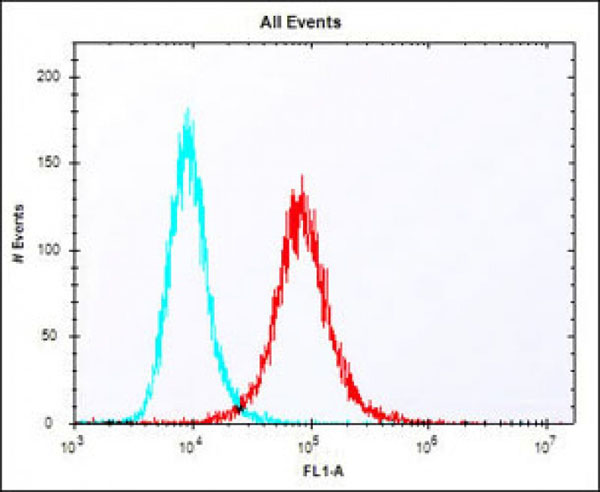 Flow Cytometry (FC/FACS)
(Overlay histogram showing NIH/3T3 cells stained at 37 degree C. The secondary antibody used was Alexa Fluor 488 goat anti-rabbit lgG (H+L) (1583138) at 1/400 dilution for 40 min at 37 degree C. Isotype control antibody (blue line) was rabbit IgG1 (1mug/1x10^6 cells) used under the same conditions. Acquisition of >10, 000 events was performed.)
Flow Cytometry (FC/FACS)
(Overlay histogram showing NIH/3T3 cells stained at 37 degree C. The secondary antibody used was Alexa Fluor 488 goat anti-rabbit lgG (H+L) (1583138) at 1/400 dilution for 40 min at 37 degree C. Isotype control antibody (blue line) was rabbit IgG1 (1mug/1x10^6 cells) used under the same conditions. Acquisition of >10, 000 events was performed.)
Klf4, Polyclonal Antibody (Cat# AAA9232000)
Full Name
Mouse Klf4 Antibody (Center)
Gene Names
Klf4; EZF; Zie; Gklf
Reactivity
Mouse
Applications
WB, FC/FACS, EIA
Pricing
$425/0.1 mL | $1,615/5x0.1 mL


 Western Blot (WB)
(Western blot analysis of lysates from MCF-7, HepG2 cell line (from left to right), using PSME2 Antibody (Center). MBS9214742 was diluted at 1:1000 at each lane. A goat anti-rabbit IgG H&L(HRP) at 1:5000 dilution was used as the secondary antibody. Lysates at 35ug per lane.)
Western Blot (WB)
(Western blot analysis of lysates from MCF-7, HepG2 cell line (from left to right), using PSME2 Antibody (Center). MBS9214742 was diluted at 1:1000 at each lane. A goat anti-rabbit IgG H&L(HRP) at 1:5000 dilution was used as the secondary antibody. Lysates at 35ug per lane.)
PSME2, Polyclonal Antibody (Cat# AAA9214742)
Full Name
PSME2 Antibody (Center)
Gene Names
PSME2; PA28B; REGbeta; PA28beta
Reactivity
Human (Predicted Reactivity: Bovine, Mouse, Pig)
Applications
EIA, IHC, FC/FACS, WB
Purity
Peptide Affinity Purified Rabbit Polyclonal Antibody (Pab)
Pricing
$405/0.4 mL | $185/0.08 mL | $1,585/5x0.4 mL


 Western Blot (WB)
(Western blot analysis of lysates from mouse liver,rat liver,human liver tissue (from left to right), using HMGCS1 Antibody (Center). It was diluted at 1:1000 at each lane. A goat anti-rabbit IgG H&L(HRP) at 1:10000 dilution was used as the secondary antibody.)
Western Blot (WB)
(Western blot analysis of lysates from mouse liver,rat liver,human liver tissue (from left to right), using HMGCS1 Antibody (Center). It was diluted at 1:1000 at each lane. A goat anti-rabbit IgG H&L(HRP) at 1:10000 dilution was used as the secondary antibody.)
HMGCS1, Polyclonal Antibody (Cat# AAA9231841)
Full Name
HMGCS1 Antibody (Center)
Gene Names
HMGCS1; HMGCS
Reactivity
Human, Mouse, Rat
Predicted: Chicken, Hamster
Predicted: Chicken, Hamster
Applications
WB, IHC, FC/FACS, EIA
Pricing
$425/0.1 mL | $1,615/5x0.1 mL

 SDS-Page
(SDS-PAGE Analysis Purified CD71 Mouse Monoclonal Antibody (TFRC/1396). Confirmation of Integrity and Purity of Antibody.)
SDS-Page
(SDS-PAGE Analysis Purified CD71 Mouse Monoclonal Antibody (TFRC/1396). Confirmation of Integrity and Purity of Antibody.)
CD71/Transferrin Receptor (TFRC), Monoclonal Antibody (Cat# AAA4381286)
Full Name
CD71/Transferrin Receptor (TFRC)
Gene Names
TFRC; T9; TR; TFR; p90; CD71; TFR1; TRFR; IMD46
Reactivity
Human
Applications
FC/FACS, IF, IHC
Purity
Purified Ab with BSA and Azide at 200ug/ml OR Purified Ab WITHOUT BSA and Azide at 1.0mg/ml
Pricing
$390/0.1 mg (With BSA & Azide at 0.2mg/mL) | $390/0.1 mg (Without BSA & Azide at 1mg/mL) | $215/0.02 mg (With BSA & Azide at 0.2mg/mL) | $1,710/5x0.1 mg (With BSA & Azide at 0.2mg/mL) | $1,710/5x0.1 mg (Without BSA & Azide at 1mg/mL)


 Western Blot (WB)
(Western blot analysis of lysates from human blood plasma tissue (from left to right),using LRRC45 Antibody (C-term). MBS9201779 was diluted at 1:1000 at each lane. A goat anti-rabbit IgG H&L(HRP) at 1:5000 dilution was used as the secondary antibody.Lysates at 35ug per lane.)
Western Blot (WB)
(Western blot analysis of lysates from human blood plasma tissue (from left to right),using LRRC45 Antibody (C-term). MBS9201779 was diluted at 1:1000 at each lane. A goat anti-rabbit IgG H&L(HRP) at 1:5000 dilution was used as the secondary antibody.Lysates at 35ug per lane.)
LRRC45, Polyclonal Antibody (Cat# AAA9201779)
Full Name
LRRC45 Antibody (C-term)
Reactivity
Human
Applications
EIA, IHC, FC/FACS, IF, WB
Purity
Peptide Affinity Purified Rabbit Polyclonal Antibody (Pab)
Pricing
$415/0.4 mL | $185/0.08 mL | $1,625/5x0.4 mL


 Western Blot (WB)
(Western blot analysis of lysates from HL-60,THP-1 cell line (from left to right), using MDH1 Antibody (C-term). It was diluted at 1:1000 at each lane. A goat anti-rabbit IgG H&L(HRP) at 1:10000 dilution was used as the secondary antibody.)
Western Blot (WB)
(Western blot analysis of lysates from HL-60,THP-1 cell line (from left to right), using MDH1 Antibody (C-term). It was diluted at 1:1000 at each lane. A goat anti-rabbit IgG H&L(HRP) at 1:10000 dilution was used as the secondary antibody.)
MDH1, Polyclonal Antibody (Cat# AAA9231817)
Full Name
MDH1 Antibody (C-term)
Gene Names
MDH1; MDHA; MOR2; MDH-s; HEL-S-32; MGC:1375
Reactivity
Human
Predicted: Bovine, Chicken, Mouse, Pig, Rat
Predicted: Bovine, Chicken, Mouse, Pig, Rat
Applications
WB, IHC, FC/FACS, EIA
Pricing
$425/0.1 mL | $1,615/5x0.1 mL


 Immunohistochemistry (IHC)
(IHC image of MBS7110797 diluted at 1:100 and staining in paraffin-embedded human lung tissue performed on a Leica BondTM system. After dewaxing and hydration, antigen retrieval was mediated by high pressure in a citrate buffer (pH 6.0). Section was blocked with 10% normal goat serum 30min at RT. Then primary antibody (1% BSA) was incubated at 4 degree C overnight. The primary is detected by a biotinylated secondary antibody and visualized using an HRP conjugated SP system.)
Immunohistochemistry (IHC)
(IHC image of MBS7110797 diluted at 1:100 and staining in paraffin-embedded human lung tissue performed on a Leica BondTM system. After dewaxing and hydration, antigen retrieval was mediated by high pressure in a citrate buffer (pH 6.0). Section was blocked with 10% normal goat serum 30min at RT. Then primary antibody (1% BSA) was incubated at 4 degree C overnight. The primary is detected by a biotinylated secondary antibody and visualized using an HRP conjugated SP system.)
CD163, Monoclonal Recombinant Antibody (Cat# AAA7110797)
Full Name
CD163 Antibody
Gene Names
CD163; M130; MM130; SCARI1
Reactivity
Human
Applications
EIA, IHC, FC/FACS
Purity
Affinity-chromatography
Pricing
$205/0.05 mL | $290/0.1 mL | $1,305/5x0.1 mL


 Western Blot (WB)
(Western blot analysis of lysates from Hela,A431,Jurkat,mouse NIH/3T3,HepG2,Raji,rat PC-12 cell line (from left to right), using PCNA Antibody (Center). It was diluted at 1:1000 at each lane. A goat anti-rabbit IgG H&L(HRP) at 1:10000 dilution was used as the secondary antibody.)
Western Blot (WB)
(Western blot analysis of lysates from Hela,A431,Jurkat,mouse NIH/3T3,HepG2,Raji,rat PC-12 cell line (from left to right), using PCNA Antibody (Center). It was diluted at 1:1000 at each lane. A goat anti-rabbit IgG H&L(HRP) at 1:10000 dilution was used as the secondary antibody.)
PCNA, Polyclonal Antibody (Cat# AAA9231806)
Full Name
PCNA Antibody (Center)
Gene Names
PCNA; ATLD2
Reactivity
Human, Mouse, Rat
Predicted: Bovine, Chicken, Hamster, Monkey
Predicted: Bovine, Chicken, Hamster, Monkey
Applications
WB, IHC, IF, FC/FACS, EIA
Pricing
$435/0.1 mL | $1,655/5x0.1 mL


 Western Blot (WB)
(All lanes : Anti-CBFB Antibody (Center) at 1:1000 dilutionLane 1: K562 whole cell lysatesLane 2: Jurkat whole cell lysatesLane 3: Raji whole cell lysatesLysates/proteins at 20 ug per lane.SecondaryGoat Anti-Rabbit IgG, (H+L),Peroxidase conjugated at 1/10000 dilutionPredicted band size : 22 kDaBlocking/Dilution buffer: 5% NFDM/TBST.)
Western Blot (WB)
(All lanes : Anti-CBFB Antibody (Center) at 1:1000 dilutionLane 1: K562 whole cell lysatesLane 2: Jurkat whole cell lysatesLane 3: Raji whole cell lysatesLysates/proteins at 20 ug per lane.SecondaryGoat Anti-Rabbit IgG, (H+L),Peroxidase conjugated at 1/10000 dilutionPredicted band size : 22 kDaBlocking/Dilution buffer: 5% NFDM/TBST.)
CBFB, Polyclonal Antibody (Cat# AAA9232041)
Full Name
CBFB Antibody (Center)
Gene Names
CBFB; PEBP2B
Reactivity
Human
Predicted: Mouse
Predicted: Mouse
Applications
WB, IHC, IF, FC/FACS, EIA
Pricing
$425/0.1 mL | $1,615/5x0.1 mL


 Western Blot (WB)
(All lanes : Anti-EEF1E1 Antibody at1:2000 dilutionLane 1: HepG2 whole cell lysateLane 2: A431 whole cell lysateLane 3: A549 whole cell lysateLane 4: Jurkat whole cell lysateLysates/proteins at 20 ug per lane. SecondaryGoat Anti-mouse IgG, (H+L), Peroxidase conjugated at 1/10000 dilution. Predicted band size : 20 kDaBlocking/Dilution buffer: 5% NFDM/TBST.)
Western Blot (WB)
(All lanes : Anti-EEF1E1 Antibody at1:2000 dilutionLane 1: HepG2 whole cell lysateLane 2: A431 whole cell lysateLane 3: A549 whole cell lysateLane 4: Jurkat whole cell lysateLysates/proteins at 20 ug per lane. SecondaryGoat Anti-mouse IgG, (H+L), Peroxidase conjugated at 1/10000 dilution. Predicted band size : 20 kDaBlocking/Dilution buffer: 5% NFDM/TBST.)
EEF1E1, Monoclonal Antibody (Cat# AAA9232191)
Full Name
EEF1E1 Antibody
Gene Names
EEF1E1; P18; AIMP3
Reactivity
Human
Applications
WB, IF, FC/FACS, EIA
Pricing
$435/0.1 mL | $1,655/5x0.1 mL


 Testing Data
(Immunoperoxidase staining of mouse lymph node cryosection using Rat anti Mouse CD11b antibody followed by horseradish peroxidase conjugated Goat anti Rat IgG (MBS235195). Medium power)
Testing Data
(Immunoperoxidase staining of mouse lymph node cryosection using Rat anti Mouse CD11b antibody followed by horseradish peroxidase conjugated Goat anti Rat IgG (MBS235195). Medium power)
CD11b, Monoclonal Antibody (Cat# AAA213144)
Full Name
RAT ANTI MOUSE CD11b
Gene Names
Itgam; CR3; CR3A; MAC1; Cd11b; Ly-40; Mac-1; Mac-1a; CD11b/CD18; F730045J24Rik
Applications
FC/FACS, IF, IP
Pricing
$340/0.1 mg | $1,375/5x0.1 mg


 Testing Data
(Immunoperoxidase staining of mouse lymph node cryosection using Rat anti Mouse CD11b antibody followed by horseradish peroxidase conjugated Goat anti Rat IgG (MBS235195). Medium power)
Testing Data
(Immunoperoxidase staining of mouse lymph node cryosection using Rat anti Mouse CD11b antibody followed by horseradish peroxidase conjugated Goat anti Rat IgG (MBS235195). Medium power)
CD11b, Monoclonal Antibody (Cat# AAA213143)
Full Name
RAT ANTI MOUSE CD11b
Gene Names
Itgam; CR3; CR3A; MAC1; Cd11b; Ly-40; Mac-1; Mac-1a; CD11b/CD18; F730045J24Rik
Applications
FC/FACS, IF, IP
Pricing
$475/0.25 mg | $1,995/5x0.25 mg


 Immunohistochemistry (IHC)-Paraffin
(Staining Epcam in Mouse colon tissue sections by Immunohistochemistry (IHC-P - paraformaldehyde-fixed, paraffin-embedded sections). Tissue was fixed with formaldehyde and blocked with 3% BSA for 0. 5 hour at room temperature; antigen retrieval was by heat mediation with a citrate buffer (pH6). Samples were incubated with primary antibody (1/25) for 1 hours at 37 degree C. A undiluted biotinylated goat polyvalent antibody was used as the secondary antibody.)
Immunohistochemistry (IHC)-Paraffin
(Staining Epcam in Mouse colon tissue sections by Immunohistochemistry (IHC-P - paraformaldehyde-fixed, paraffin-embedded sections). Tissue was fixed with formaldehyde and blocked with 3% BSA for 0. 5 hour at room temperature; antigen retrieval was by heat mediation with a citrate buffer (pH6). Samples were incubated with primary antibody (1/25) for 1 hours at 37 degree C. A undiluted biotinylated goat polyvalent antibody was used as the secondary antibody.)
Epcam, Polyclonal Antibody (Cat# AAA9232060)
Full Name
(Mouse) Epcam Antibody (C-term)
Gene Names
Epcam; EGP; Ly74; gp40; CD326; EGP-2; TROP1; Egp314; Ep-CAM; EpCAM1; Tacsd1; GA733-2; Tacstd1
Reactivity
Mouse
Applications
WB, IHC, FC/FACS, EIA
Pricing
$425/0.1 mL | $1,615/5x0.1 mL


 Western Blot (WB)
(Western blot analysis of CD27 expression in HELA whole cell lysates (lane 1). CD27 at 51KD was detected using rabbit anti- CD27 Antigen Affinity purified polyclonal antibody at 0.5 ug/mL. The blot was developed using chemiluminescence (ECL) method. )
Western Blot (WB)
(Western blot analysis of CD27 expression in HELA whole cell lysates (lane 1). CD27 at 51KD was detected using rabbit anti- CD27 Antigen Affinity purified polyclonal antibody at 0.5 ug/mL. The blot was developed using chemiluminescence (ECL) method. )
CD27, Polyclonal Antibody (Cat# AAA178431)
Full Name
Anti-CD27 Antibody
Gene Names
CD27; T14; S152; Tp55; TNFRSF7; S152. LPFS2
Reactivity
Human
No cross reactivity with other proteins.
No cross reactivity with other proteins.
Applications
WB, IHC
Purity
Immunogen affinity purified.
Pricing
$345/0.1 mg | $1,485/5x0.1 mg


 Immunocytochemistry/Immunofluorescence (ICC/IF)
(Immunocytochemistry/Immunofluorescence analysis using Mouse Anti-Methylglyoxal Monoclonal Antibody, Clone 9F11. Tissue: Embryonic kidney epithelial cell line (HEK293). Species: Human. Fixation: 5% Formaldehyde for 5 min. Primary Antibody: Mouse Anti-Methylglyoxal Monoclonal Antibody at 1:50 for 30-60 min at RT. Secondary Antibody: Goat Anti-Mouse Alexa Fluor 488 at 1:1500 for 30-60 min at RT. Counterstain: Phalloidin Alexa Fluor 633 F-Actin stain; DAPI (blue) nuclear stain at 1:250, 1:50000 for 30-60 min at RT. Magnification: 20X (2X Zoom). (A,C,E,G) - Untreated. (B,D,F,H) - Cells cultured overnight with 50 uM H2O2. (A,B) DAPI (blue) nuclear stain. (C,D) Phalloidin Alex Fluor 633 F-Actin stain. (E,F) Methylglyoxal Antibody. (G,H) Composite. Courtesy of: Dr. Robert Burke, University of Victoria.)
Immunocytochemistry/Immunofluorescence (ICC/IF)
(Immunocytochemistry/Immunofluorescence analysis using Mouse Anti-Methylglyoxal Monoclonal Antibody, Clone 9F11. Tissue: Embryonic kidney epithelial cell line (HEK293). Species: Human. Fixation: 5% Formaldehyde for 5 min. Primary Antibody: Mouse Anti-Methylglyoxal Monoclonal Antibody at 1:50 for 30-60 min at RT. Secondary Antibody: Goat Anti-Mouse Alexa Fluor 488 at 1:1500 for 30-60 min at RT. Counterstain: Phalloidin Alexa Fluor 633 F-Actin stain; DAPI (blue) nuclear stain at 1:250, 1:50000 for 30-60 min at RT. Magnification: 20X (2X Zoom). (A,C,E,G) - Untreated. (B,D,F,H) - Cells cultured overnight with 50 uM H2O2. (A,B) DAPI (blue) nuclear stain. (C,D) Phalloidin Alex Fluor 633 F-Actin stain. (E,F) Methylglyoxal Antibody. (G,H) Composite. Courtesy of: Dr. Robert Burke, University of Victoria.)
Methylglyoxal, Monoclonal Antibody (Cat# AAA808810)
Full Name
Methylglyoxal Antibody, Clone 9F11: ATTO 488
Reactivity
Species Independent
Applications
WB, ICC, IF, FC/FACS, EIA
Purity
Protein G Purified
Pricing
$560/0.1 mg | $2,275/5x0.1 mg


 Immunocytochemistry/Immunofluorescence (ICC/IF)
(Immunocytochemistry/Immunofluorescence analysis using Mouse Anti-Hexanoyl-Lysine adduct Monoclonal Antibody, Clone 5D9. Tissue: Embryonic kidney epithelial cell line (HEK293). Species: Human. Fixation: 5% Formaldehyde for 5 min. Primary Antibody: Mouse Anti-Hexanoyl-Lysine adduct Monoclonal Antibody at 1:50 for 30-60 min at RT. Secondary Antibody: Goat Anti-Mouse Alexa Fluor 488 at 1:1500 for 30-60 min at RT. Counterstain: Phalloidin Alexa Fluor 633 F-Actin stain; DAPI (blue) nuclear stain at 1:250, 1:50000 for 30-60 min at RT. Magnification: 20X (2X Zoom). (A,C,E,G) - Untreated. (B,D,F,H) - Cells cultured overnight with 50 uM H2O2. (A,B) DAPI (blue) nuclear stain. (C,D) Phalloidin Alex Fluor 633 F-Actin stain. (E,F) Hexanoyl-Lysine adduct Antibody. (G,H) Composite. Courtesy of: Dr. Robert Burke, University of Victoria.)
Immunocytochemistry/Immunofluorescence (ICC/IF)
(Immunocytochemistry/Immunofluorescence analysis using Mouse Anti-Hexanoyl-Lysine adduct Monoclonal Antibody, Clone 5D9. Tissue: Embryonic kidney epithelial cell line (HEK293). Species: Human. Fixation: 5% Formaldehyde for 5 min. Primary Antibody: Mouse Anti-Hexanoyl-Lysine adduct Monoclonal Antibody at 1:50 for 30-60 min at RT. Secondary Antibody: Goat Anti-Mouse Alexa Fluor 488 at 1:1500 for 30-60 min at RT. Counterstain: Phalloidin Alexa Fluor 633 F-Actin stain; DAPI (blue) nuclear stain at 1:250, 1:50000 for 30-60 min at RT. Magnification: 20X (2X Zoom). (A,C,E,G) - Untreated. (B,D,F,H) - Cells cultured overnight with 50 uM H2O2. (A,B) DAPI (blue) nuclear stain. (C,D) Phalloidin Alex Fluor 633 F-Actin stain. (E,F) Hexanoyl-Lysine adduct Antibody. (G,H) Composite. Courtesy of: Dr. Robert Burke, University of Victoria.)
Hexanoyl-Lysine adduct, Monoclonal Antibody (Cat# AAA808661)
Full Name
Hexanoyl-Lysine adduct Antibody, Clone 5D9: PerCP
Reactivity
Species Independent
Applications
WB, ICC, IF, FC/FACS, EIA
Purity
Protein G Purified
Pricing
$560/0.1 mg | $2,270/5x0.1 mg


 Immunocytochemistry/Immunofluorescence (ICC/IF)
(Immunocytochemistry/Immunofluorescence analysis using Mouse Anti-Dityrosine Monoclonal Antibody, Clone 10A6. Tissue: Embryonic kidney epithelial cell line (HEK293). Species: Human. Fixation: 5% Formaldehyde for 5 min. Primary Antibody: Mouse Anti-Dityrosine Monoclonal Antibody at 1:50 for 30-60 min at RT. Secondary Antibody: Goat Anti-Mouse Alexa Fluor 488 at 1:1500 for 30-60 min at RT. Counterstain: Phalloidin Alexa Fluor 633 F-Actin stain; DAPI (blue) nuclear stain at 1:250, 1:50000 for 30-60 min at RT. Localization: Cytoplasmic. Magnification: 20X (2X Zoom). (A,C,E,G) - Untreated. (B,D,F,H) - Cells cultured overnight with 50 uM H2O2. (A,B) DAPI (blue) nuclear stain. (C,D) Phalloidin Alex Fluor 633 F-Actin stain. (E,F) Dityrosine Antibody. (G,H) Composite. Courtesy of: Dr. Robert Burke, University of Victoria.)
Immunocytochemistry/Immunofluorescence (ICC/IF)
(Immunocytochemistry/Immunofluorescence analysis using Mouse Anti-Dityrosine Monoclonal Antibody, Clone 10A6. Tissue: Embryonic kidney epithelial cell line (HEK293). Species: Human. Fixation: 5% Formaldehyde for 5 min. Primary Antibody: Mouse Anti-Dityrosine Monoclonal Antibody at 1:50 for 30-60 min at RT. Secondary Antibody: Goat Anti-Mouse Alexa Fluor 488 at 1:1500 for 30-60 min at RT. Counterstain: Phalloidin Alexa Fluor 633 F-Actin stain; DAPI (blue) nuclear stain at 1:250, 1:50000 for 30-60 min at RT. Localization: Cytoplasmic. Magnification: 20X (2X Zoom). (A,C,E,G) - Untreated. (B,D,F,H) - Cells cultured overnight with 50 uM H2O2. (A,B) DAPI (blue) nuclear stain. (C,D) Phalloidin Alex Fluor 633 F-Actin stain. (E,F) Dityrosine Antibody. (G,H) Composite. Courtesy of: Dr. Robert Burke, University of Victoria.)
Dityrosine, Monoclonal Antibody (Cat# AAA808884)
Full Name
Dityrosine Antibody, Clone 10A6: ATTO 594
Reactivity
Species Independent
Applications
WB, ICC, IF, FC/FACS, EIA
Purity
Protein G Purified
Pricing
$560/0.1 mg | $2,275/5x0.1 mg


 Testing Data
(Staining of mouse spleen with Rat anti Mouse CD4 (MBS219566))
Testing Data
(Staining of mouse spleen with Rat anti Mouse CD4 (MBS219566))
CD4, Monoclonal Antibody (Cat# AAA219512)
Full Name
RAT ANTI MOUSE CD4
Gene Names
Cd4; L3T4; Ly-4
Applications
EIA, FC/FACS, IF, IP, WB
Pricing
$195/0.025 mg | $730/5x0.025 mg


 Immunocytochemistry/Immunofluorescence (ICC/IF)
(Immunocytochemistry/Immunofluorescence analysis using Mouse Anti-Acrolein Monoclonal Antibody, Clone 2H2. Tissue: Embryonic kidney epithelial cell line (HEK293). Species: Human. Fixation: 5% Formaldehyde for 5 min. Primary Antibody: Mouse Anti-Acrolein Monoclonal Antibody at 1:50 for 30-60 min at RT. Secondary Antibody: Goat Anti-Mouse Alexa Fluor 488 at 1:1500 for 30-60 min at RT. Counterstain: Phalloidin Alexa Fluor 633 F-Actin stain; DAPI (blue) nuclear stain at 1:250, 1:50000 for 30-60 min at RT. Magnification: 20X (2X Zoom). (A,C,E,G) - Untreated. (B,D,F,H) - Cells cultured overnight with 50 uM H2O2. (A,B) DAPI (blue) nuclear stain. (C,D) Phalloidin Alex Fluor 633 F-Actin stain. (E,F) Acrolein Antibody. (G,H) Composite. Courtesy of: Dr. Robert Burke, University of Victoria.)
Immunocytochemistry/Immunofluorescence (ICC/IF)
(Immunocytochemistry/Immunofluorescence analysis using Mouse Anti-Acrolein Monoclonal Antibody, Clone 2H2. Tissue: Embryonic kidney epithelial cell line (HEK293). Species: Human. Fixation: 5% Formaldehyde for 5 min. Primary Antibody: Mouse Anti-Acrolein Monoclonal Antibody at 1:50 for 30-60 min at RT. Secondary Antibody: Goat Anti-Mouse Alexa Fluor 488 at 1:1500 for 30-60 min at RT. Counterstain: Phalloidin Alexa Fluor 633 F-Actin stain; DAPI (blue) nuclear stain at 1:250, 1:50000 for 30-60 min at RT. Magnification: 20X (2X Zoom). (A,C,E,G) - Untreated. (B,D,F,H) - Cells cultured overnight with 50 uM H2O2. (A,B) DAPI (blue) nuclear stain. (C,D) Phalloidin Alex Fluor 633 F-Actin stain. (E,F) Acrolein Antibody. (G,H) Composite. Courtesy of: Dr. Robert Burke, University of Victoria.)
Acrolein, Monoclonal Antibody (Cat# AAA808583)
Full Name
Acrolein Antibody, Clone 2H2: Alkaline Phosphatase
Reactivity
Species Independent
Applications
WB, ICC, IF, FC/FACS, EIA
Purity
Protein G Purified
Pricing
$550/0.1 mg | $2,245/5x0.1 mg


 Western Blot (WB)
(Figure 1. Western blot analysis of APRT using anti-APRT antibody (MBS178668). Electrophoresis was performed on a 5-20% SDS-PAGE gel at 70V (Stacking gel) / 90V (Resolving gel) for 2-3 hours. The sample well of each lane was loaded with 50ug of sample under reducing conditions. lane 1: K562 whole cell lysates. After Electrophoresis, proteins were transferred to a Nitrocellulose membrane at 150mA for 50-90 minutes. Blocked the membrane with 5% Non-fat Milk/ TBS for 1.5 hour at RT. The membrane was incubated with rabbit anti-APRT antigen affinity purified polyclonal antibody at 0.5ug/mL overnight at 4 degree C, then washed with TBS-0.1%Tween 3 times with 5 minutes each and probed with a goat anti-rabbit IgG-HRP secondary antibody at a dilution of 1:10000 for 1.5 hour at RT. The signal is developed using an Enhanced Chemiluminescent detection (ECL) kit with Tanon 5200 system. A specific band was detected for APRT at approximately 20KD. The expected band size for APRT is at 20KD. )
Western Blot (WB)
(Figure 1. Western blot analysis of APRT using anti-APRT antibody (MBS178668). Electrophoresis was performed on a 5-20% SDS-PAGE gel at 70V (Stacking gel) / 90V (Resolving gel) for 2-3 hours. The sample well of each lane was loaded with 50ug of sample under reducing conditions. lane 1: K562 whole cell lysates. After Electrophoresis, proteins were transferred to a Nitrocellulose membrane at 150mA for 50-90 minutes. Blocked the membrane with 5% Non-fat Milk/ TBS for 1.5 hour at RT. The membrane was incubated with rabbit anti-APRT antigen affinity purified polyclonal antibody at 0.5ug/mL overnight at 4 degree C, then washed with TBS-0.1%Tween 3 times with 5 minutes each and probed with a goat anti-rabbit IgG-HRP secondary antibody at a dilution of 1:10000 for 1.5 hour at RT. The signal is developed using an Enhanced Chemiluminescent detection (ECL) kit with Tanon 5200 system. A specific band was detected for APRT at approximately 20KD. The expected band size for APRT is at 20KD. )
APRT, Polyclonal Antibody (Cat# AAA178668)
Full Name
Anti-APRT Antibody
Gene Names
APRT; AMP; APRTD
Reactivity
Human
Applications
WB
Purity
Immunogen affinity purified.
Pricing
$345/0.1 mg | $1,485/5x0.1 mg


 Western Blot (WB)
(Western BlotPositive WB detected in: Mouse brain tissue, Mouse heart tissueAll lanes: Di-methyl-Histone H3.1 (K4)antibody at 0.55ug/mlSecondaryGoat polyclonal to rabbit IgG at 1/50000 dilutionPredicted band size: 15 KDaObserved band size: 15 KDa)
Western Blot (WB)
(Western BlotPositive WB detected in: Mouse brain tissue, Mouse heart tissueAll lanes: Di-methyl-Histone H3.1 (K4)antibody at 0.55ug/mlSecondaryGoat polyclonal to rabbit IgG at 1/50000 dilutionPredicted band size: 15 KDaObserved band size: 15 KDa)
Di-methyl-Histone H3.1, Monoclonal Recombinant Antibody (Cat# AAA7110754)
Full Name
Di-methyl-Histone H3.1 (K4) Antibody
Gene Names
HIST1H3A; H3/A; H3FA
Reactivity
Human, Mouse
Applications
EIA, WB, ICC, IF, FC/FACS
Purity
Affinity-chromatography
Pricing
$205/0.05 mL | $290/0.1 mL | $1,305/5x0.1 mL


 Western Blot (WB)
(Figure 1. Western blot analysis of RAB27A using anti-RAB27A antibody (MBS1750678).Electrophoresis was performed on a 5-20% SDS-PAGE gel at 70V (Stacking gel) / 90V (Resolving gel) for 2-3 hours. The sample well of each lane was loaded with 50ug of sample under reducing conditions.Lane 1: human Hela whole cell lysates,Lane 2: human Jurkat whole cell lysates,Lane 3: human MCF-7 whole cell lysates,Lane 4: human HepG2 whole cell lysates,Lane 5: human A549 whole cell lysates,Lane 6: rat stomach tissue lysates,Lane 7: rat thymus tissue lysates,Lane 8: mouse thymus tissue lysates,Lane 9: mouse NIH3T3 whole cell lysates.After Electrophoresis, proteins were transferred to a Nitrocellulose membrane at 150mA for 50-90 minutes. Blocked the membrane with 5% Non-fat Milk/ TBS for 1.5 hour at RT. The membrane was incubated with rabbit anti-RAB27A antigen affinity purified polyclonal antibody at 0.5ug/mL overnight at 4 degree C, then washed with TBS-0.1%Tween 3 times with 5 minutes each and probed with a goat anti-rabbit IgG-HRP secondary antibody at a dilution of 1:10000 for 1.5 hour at RT. The signal is developed using an Enhanced Chemiluminescent detection (ECL) kit with Tanon 5200 system. A specific band was detected for RAB27A at approximately 27KD. The expected band size for RAB27A is at 25KD. )
Western Blot (WB)
(Figure 1. Western blot analysis of RAB27A using anti-RAB27A antibody (MBS1750678).Electrophoresis was performed on a 5-20% SDS-PAGE gel at 70V (Stacking gel) / 90V (Resolving gel) for 2-3 hours. The sample well of each lane was loaded with 50ug of sample under reducing conditions.Lane 1: human Hela whole cell lysates,Lane 2: human Jurkat whole cell lysates,Lane 3: human MCF-7 whole cell lysates,Lane 4: human HepG2 whole cell lysates,Lane 5: human A549 whole cell lysates,Lane 6: rat stomach tissue lysates,Lane 7: rat thymus tissue lysates,Lane 8: mouse thymus tissue lysates,Lane 9: mouse NIH3T3 whole cell lysates.After Electrophoresis, proteins were transferred to a Nitrocellulose membrane at 150mA for 50-90 minutes. Blocked the membrane with 5% Non-fat Milk/ TBS for 1.5 hour at RT. The membrane was incubated with rabbit anti-RAB27A antigen affinity purified polyclonal antibody at 0.5ug/mL overnight at 4 degree C, then washed with TBS-0.1%Tween 3 times with 5 minutes each and probed with a goat anti-rabbit IgG-HRP secondary antibody at a dilution of 1:10000 for 1.5 hour at RT. The signal is developed using an Enhanced Chemiluminescent detection (ECL) kit with Tanon 5200 system. A specific band was detected for RAB27A at approximately 27KD. The expected band size for RAB27A is at 25KD. )
RAB27A, Polyclonal Antibody (Cat# AAA1750678)
Full Name
Anti-RAB27A Picoband antibody
Gene Names
RAB27A; GS2; RAM; RAB27; HsT18676
Reactivity
Human, Mouse, Rat
No cross reactivity with other proteins.
No cross reactivity with other proteins.
Applications
EIA, FC/FACS, IHC, ICC, WB
Pricing
$345/0.1 mg | $1,485/5x0.1 mg


 Western Blot (WB)
(Figure 1. Western blot analysis of CCKBR using anti-CCKBR antibody (MBS1750692).Electrophoresis was performed on a 5-20% SDS-PAGE gel at 70V (Stacking gel) / 90V (Resolving gel) for 2-3 hours. The sample well of each lane was loaded with 50ug of sample under reducing conditions.Lane 1: human PANC-1 whole cell lysates,Lane 2: human U-87MG whole cell lysates,Lane 3: human COLO-320 whole cell lysates,Lane 4: human SGC-7901 whole cell lysates.After Electrophoresis, proteins were transferred to a Nitrocellulose membrane at 150mA for 50-90 minutes. Blocked the membrane with 5% Non-fat Milk/ TBS for 1.5 hour at RT. The membrane was incubated with rabbit anti-CCKBR antigen affinity purified polyclonal antibody at 0.5ug/mL overnight at 4 degree C, then washed with TBS-0.1%Tween 3 times with 5 minutes each and probed with a goat anti-rabbit IgG-HRP secondary antibody at a dilution of 1:10000 for 1.5 hour at RT. The signal is developed using an Enhanced Chemiluminescent detection (ECL) kit with Tanon 5200 system. A specific band was detected for CCKBR at approximately 64KD. The expected band size for CCKBR is at 48KD. )
Western Blot (WB)
(Figure 1. Western blot analysis of CCKBR using anti-CCKBR antibody (MBS1750692).Electrophoresis was performed on a 5-20% SDS-PAGE gel at 70V (Stacking gel) / 90V (Resolving gel) for 2-3 hours. The sample well of each lane was loaded with 50ug of sample under reducing conditions.Lane 1: human PANC-1 whole cell lysates,Lane 2: human U-87MG whole cell lysates,Lane 3: human COLO-320 whole cell lysates,Lane 4: human SGC-7901 whole cell lysates.After Electrophoresis, proteins were transferred to a Nitrocellulose membrane at 150mA for 50-90 minutes. Blocked the membrane with 5% Non-fat Milk/ TBS for 1.5 hour at RT. The membrane was incubated with rabbit anti-CCKBR antigen affinity purified polyclonal antibody at 0.5ug/mL overnight at 4 degree C, then washed with TBS-0.1%Tween 3 times with 5 minutes each and probed with a goat anti-rabbit IgG-HRP secondary antibody at a dilution of 1:10000 for 1.5 hour at RT. The signal is developed using an Enhanced Chemiluminescent detection (ECL) kit with Tanon 5200 system. A specific band was detected for CCKBR at approximately 64KD. The expected band size for CCKBR is at 48KD. )
CCKBR, Polyclonal Antibody (Cat# AAA1750692)
Full Name
Anti-CCKBR Picoband antibody
Gene Names
CCKBR; GASR; CCK-B; CCK2R
Reactivity
Human, Mouse, Rat
No cross reactivity with other proteins.
No cross reactivity with other proteins.
Applications
WB
Pricing
$345/0.1 mg | $1,485/5x0.1 mg


 Western Blot (WB)
(Figure 1. Western blot analysis of AHSG using anti-AHSG antibody (MBS177852). Electrophoresis was performed on a 5-20% SDS-PAGE gel at 70V (Stacking gel) / 90V (Resolving gel) for 2-3 hours. The sample well of each lane was loaded with 50ug of sample under reducing conditions. Lane 1: Rat Brain Tissue Lysate, Lane 2: RH35 Whole Cell Lysate, Lane 3: MCF-7 Whole Cell Lysate. After Electrophoresis, proteins were transferred to a Nitrocellulose membrane at 150mA for 50-90 minutes. Blocked the membrane with 5% Non-fat Milk/ TBS for 1.5 hour at RT. The membrane was incubated with rabbit anti-AHSG antigen affinity purified polyclonal antibody at 0.5ug/mL overnight at 4 degree C, then washed with TBS-0.1%Tween 3 times with 5 minutes each and probed with a goat anti-rabbit IgG-HRP secondary antibody at a dilution of 1:10000 for 1.5 hour at RT. The signal is developed using an Enhanced Chemiluminescent detection (ECL) kit with Tanon 5200 system. A specific band was detected for AHSG at approximately 58KD. The expected band size for AHSG is at 39KD. )
Western Blot (WB)
(Figure 1. Western blot analysis of AHSG using anti-AHSG antibody (MBS177852). Electrophoresis was performed on a 5-20% SDS-PAGE gel at 70V (Stacking gel) / 90V (Resolving gel) for 2-3 hours. The sample well of each lane was loaded with 50ug of sample under reducing conditions. Lane 1: Rat Brain Tissue Lysate, Lane 2: RH35 Whole Cell Lysate, Lane 3: MCF-7 Whole Cell Lysate. After Electrophoresis, proteins were transferred to a Nitrocellulose membrane at 150mA for 50-90 minutes. Blocked the membrane with 5% Non-fat Milk/ TBS for 1.5 hour at RT. The membrane was incubated with rabbit anti-AHSG antigen affinity purified polyclonal antibody at 0.5ug/mL overnight at 4 degree C, then washed with TBS-0.1%Tween 3 times with 5 minutes each and probed with a goat anti-rabbit IgG-HRP secondary antibody at a dilution of 1:10000 for 1.5 hour at RT. The signal is developed using an Enhanced Chemiluminescent detection (ECL) kit with Tanon 5200 system. A specific band was detected for AHSG at approximately 58KD. The expected band size for AHSG is at 39KD. )
AHSG, Polyclonal Antibody (Cat# AAA177852)
Full Name
Anti-AHSG Antibody
Gene Names
AHSG; AHS; A2HS; HSGA; FETUA
Reactivity
Human, Rat
Applications
WB, IHC, EIA
Purity
Immunogen Affinity Purified
Pricing
$345/0.1 mg | $1,485/5x0.1 mg


 Testing Data
(Staining of Con A activated rat spleen cells with Mouse anti Rat CD25: Biotin (MBS210477))
Testing Data
(Staining of Con A activated rat spleen cells with Mouse anti Rat CD25: Biotin (MBS210477))
CD25, Monoclonal Antibody (Cat# AAA212960)
Full Name
MOUSE ANTI RAT CD25
Gene Names
Il2ra; IL2RAC
Applications
EIA, FC/FACS, IP
Pricing
$345/0.1 mg | $1,405/5x0.1 mg


 Western Blot (WB)
(Figure 1. Western blot analysis of PPID using anti-PPID antibody (MBS1750769).Electrophoresis was performed on a 5-20% SDS-PAGE gel at 70V (Stacking gel) / 90V (Resolving gel) for 2-3 hours. The sample well of each lane was loaded with 50ug of sample under reducing conditions.Lane 1: rat spleen tissue lysates,Lane 2: rat liver tissue lysates,Lane 3: mouse testis tissue lysates.After Electrophoresis, proteins were transferred to a Nitrocellulose membrane at 150mA for 50-90 minutes. Blocked the membrane with 5% Non-fat Milk/ TBS for 1.5 hour at RT. The membrane was incubated with rabbit anti-PPID antigen affinity purified polyclonal antibody at 0.5ug/mL overnight at 4 degree C, then washed with TBS-0.1%Tween 3 times with 5 minutes each and probed with a goat anti-rabbit IgG-HRP secondary antibody at a dilution of 1:10000 for 1.5 hour at RT. The signal is developed using an Enhanced Chemiluminescent detection (ECL) kit with Tanon 5200 system. A specific band was detected for PPID at approximately 41KD. The expected band size for PPID is at 41KD. )
Western Blot (WB)
(Figure 1. Western blot analysis of PPID using anti-PPID antibody (MBS1750769).Electrophoresis was performed on a 5-20% SDS-PAGE gel at 70V (Stacking gel) / 90V (Resolving gel) for 2-3 hours. The sample well of each lane was loaded with 50ug of sample under reducing conditions.Lane 1: rat spleen tissue lysates,Lane 2: rat liver tissue lysates,Lane 3: mouse testis tissue lysates.After Electrophoresis, proteins were transferred to a Nitrocellulose membrane at 150mA for 50-90 minutes. Blocked the membrane with 5% Non-fat Milk/ TBS for 1.5 hour at RT. The membrane was incubated with rabbit anti-PPID antigen affinity purified polyclonal antibody at 0.5ug/mL overnight at 4 degree C, then washed with TBS-0.1%Tween 3 times with 5 minutes each and probed with a goat anti-rabbit IgG-HRP secondary antibody at a dilution of 1:10000 for 1.5 hour at RT. The signal is developed using an Enhanced Chemiluminescent detection (ECL) kit with Tanon 5200 system. A specific band was detected for PPID at approximately 41KD. The expected band size for PPID is at 41KD. )
PPID/Cyclophilin 40, Polyclonal Antibody (Cat# AAA1750769)
Full Name
Anti-PPID/Cyclophilin 40 Picoband antibody
Gene Names
PPID; CYPD; CYP-40
Reactivity
Human, Mouse, Rat
No cross reactivity with other proteins.
No cross reactivity with other proteins.
Applications
EIA, FC/FACS, IHC, ICC, WB
Pricing
$345/0.1 mg | $1,485/5x0.1 mg


 Testing Data
(Staining of permeabilised mouse peritoneal macrophages with Rat anti Mouse CD68:Alexa Fluor 647 (MBS215395A647). following permeablisation with Leucoperm)
Testing Data
(Staining of permeabilised mouse peritoneal macrophages with Rat anti Mouse CD68:Alexa Fluor 647 (MBS215395A647). following permeablisation with Leucoperm)
CD68, Monoclonal Antibody (Cat# AAA215404)
Full Name
RAT ANTI MOUSE CD68:RPE
Gene Names
Cd68; Lamp4; gp110; Scard1
Applications
FC/FACS
Pricing
$550/100 Tests | $2,320/5x100 Tests


 Western blot (WB)
(Western blot analysis of MUC1 on Hela cells lysates using anti-MUC1 antibody at 1/1000 dilution.)
Western blot (WB)
(Western blot analysis of MUC1 on Hela cells lysates using anti-MUC1 antibody at 1/1000 dilution.)
MUC1, Monoclonal Antibody (Cat# AAA8534023)
Full Name
MUC1
Gene Names
MUC1; EMA; MCD; PEM; PUM; KL-6; MAM6; MCKD; PEMT; CD227; H23AG; MCKD1; MUC-1; ADMCKD; ADMCKD1; CA 15-3; MUC-1/X; MUC1/ZD; MUC-1/SEC
Reactivity
Human
Applications
WB, ICC, IHC, FC/FACS
Pricing
$350/0.1 mL | $570/0.1 mL (Biotin) | $1,515/5x0.1 mL | $570/0.1 mL (FITC) | $570/0.1 mL (AF350) | $570/0.1 mL (AF405) | $570/0.1 mL (AF488) | $570/0.1 mL (AF555) | $570/0.1 mL (AF568)


 Immunohistochemistry (IHC)
(Human Kidney: Formalin-Fixed, Paraffin-Embedded (FFPE))
Immunohistochemistry (IHC)
(Human Kidney: Formalin-Fixed, Paraffin-Embedded (FFPE))
KLHL9, Polyclonal Antibody (Cat# AAA2401453)
Full Name
Anti-KLHL9 Antibody (aa299-326)
Reactivity
Human
Predicted: Mouse
Predicted: Mouse
Applications
FC/FACS, IHC, WB
Purity
Protein A purified
Pricing
$585/0.2 mL | $2,585/5x0.2 mL


 Testing Data #1
(Staining of equine peripheral blood granulocytes with Mouse anti Horse CD13 (MBS224706) followed by Goat anti Mouse IgG:FITC (MBS235142))
Testing Data #1
(Staining of equine peripheral blood granulocytes with Mouse anti Horse CD13 (MBS224706) followed by Goat anti Mouse IgG:FITC (MBS235142))
CD13, Monoclonal Antibody (Cat# AAA224637)
Full Name
MOUSE ANTI HORSE CD13:RPE
Applications
FC/FACS
Pricing
$640/100 Tests | $2,735/5x100 Tests