Filters
Clonality
Type
Reactivity
Gene Name
Isotype
Host
Application
Clone
1205 results for "Rat IgG Isotype Control" - showing 950-1000

 Western Blot (WB)
(Figure 1. Western blot analysis of LOC134147/CMBL using anti-LOC134147/CMBL antibody (MBS1753677).Electrophoresis was performed on a 5-20% SDS-PAGE gel at 70V (Stacking gel) / 90V (Resolving gel) for 2-3 hours. The sample well of each lane was loaded with 30ug of sample under reducing conditions.Lane 1: mouse liver tissue lysatesLane 2: mouse kidney tissue lysatesLane 3: rat liver tissue lysatesLane 4: rat kidney tissue lysates.After Electrophoresis, proteins were transferred to a Nitrocellulose membrane at 150mA for 50-90 minutes. Blocked the membrane with 5% Non-fat Milk/ TBS for 1. 5 hour at RT. The membrane was incubated with rabbit anti-LOC134147/CMBL antigen affinity purified polyclonal antibody (Catalog # MBS1753677) at 0. 5 μg/mL overnight at 4 degree C, then washed with TBS-0. 1%Tween 3 times with 5 minutes each and probed with a goat anti-rabbit IgG-HRP secondary antibody at a dilution of 1:5000 for 1. 5 hour at RT. The signal is developed using an Enhanced Chemiluminescent detection (ECL) kit (Catalog # MBS176460) with Tanon 5200 system. A specific band was detected for LOC134147/CMBL at approximately 28KD. The expected band size for LOC134147/CMBL is at 28KD. )
Western Blot (WB)
(Figure 1. Western blot analysis of LOC134147/CMBL using anti-LOC134147/CMBL antibody (MBS1753677).Electrophoresis was performed on a 5-20% SDS-PAGE gel at 70V (Stacking gel) / 90V (Resolving gel) for 2-3 hours. The sample well of each lane was loaded with 30ug of sample under reducing conditions.Lane 1: mouse liver tissue lysatesLane 2: mouse kidney tissue lysatesLane 3: rat liver tissue lysatesLane 4: rat kidney tissue lysates.After Electrophoresis, proteins were transferred to a Nitrocellulose membrane at 150mA for 50-90 minutes. Blocked the membrane with 5% Non-fat Milk/ TBS for 1. 5 hour at RT. The membrane was incubated with rabbit anti-LOC134147/CMBL antigen affinity purified polyclonal antibody (Catalog # MBS1753677) at 0. 5 μg/mL overnight at 4 degree C, then washed with TBS-0. 1%Tween 3 times with 5 minutes each and probed with a goat anti-rabbit IgG-HRP secondary antibody at a dilution of 1:5000 for 1. 5 hour at RT. The signal is developed using an Enhanced Chemiluminescent detection (ECL) kit (Catalog # MBS176460) with Tanon 5200 system. A specific band was detected for LOC134147/CMBL at approximately 28KD. The expected band size for LOC134147/CMBL is at 28KD. )
LOC134147/CMBL, Polyclonal Antibody (Cat# AAA1753677)
Full Name
Anti-LOC134147/CMBL Antibody
Gene Names
Cmbl; 2310016A09Rik
Reactivity
Mouse, Rat
Applications
WB, FC/FACS/FCM
Purity
Immunogen affinity purified.
Pricing
$310/0.1 mg | $1,330/5x0.1 mg

 Western Blot (WB)
(Figure 1. Western blot analysis of SHISA6 using anti-SHISA6 antibody (MBS1753840).Electrophoresis was performed on a 5-20% SDS-PAGE gel at 70V (Stacking gel) / 90V (Resolving gel) for 2-3 hours. The sample well of each lane was loaded with 50ug of sample under reducing conditions.Lane 1: rat brain tissue lysatesLane 2: mouse brain tissue lysates.After Electrophoresis, proteins were transferred to a Nitrocellulose membrane at 150mA for 50-90 minutes. Blocked the membrane with 5% Non-fat Milk/ TBS for 1. 5 hour at RT. The membrane was incubated with rabbit anti-SHISA6 antigen affinity purified polyclonal antibody (Catalog # MBS1753840) at 0. 5 μg/mL overnight at 4 degree C, then washed with TBS-0. 1%Tween 3 times with 5 minutes each and probed with a goat anti-rabbit IgG-HRP secondary antibody at a dilution of 1:5000 for 1. 5 hour at RT. The signal is developed using an Enhanced Chemiluminescent detection (ECL) kit (Catalog # MBS176460) with Tanon 5200 system. A specific band was detected for SHISA6 at approximately 56KD. The expected band size for SHISA6 is at 56KD. )
Western Blot (WB)
(Figure 1. Western blot analysis of SHISA6 using anti-SHISA6 antibody (MBS1753840).Electrophoresis was performed on a 5-20% SDS-PAGE gel at 70V (Stacking gel) / 90V (Resolving gel) for 2-3 hours. The sample well of each lane was loaded with 50ug of sample under reducing conditions.Lane 1: rat brain tissue lysatesLane 2: mouse brain tissue lysates.After Electrophoresis, proteins were transferred to a Nitrocellulose membrane at 150mA for 50-90 minutes. Blocked the membrane with 5% Non-fat Milk/ TBS for 1. 5 hour at RT. The membrane was incubated with rabbit anti-SHISA6 antigen affinity purified polyclonal antibody (Catalog # MBS1753840) at 0. 5 μg/mL overnight at 4 degree C, then washed with TBS-0. 1%Tween 3 times with 5 minutes each and probed with a goat anti-rabbit IgG-HRP secondary antibody at a dilution of 1:5000 for 1. 5 hour at RT. The signal is developed using an Enhanced Chemiluminescent detection (ECL) kit (Catalog # MBS176460) with Tanon 5200 system. A specific band was detected for SHISA6 at approximately 56KD. The expected band size for SHISA6 is at 56KD. )
SHISA6, Polyclonal Antibody (Cat# AAA1753840)
Full Name
Anti-SHISA6 Antibody
Reactivity
Human, Mouse, Rat
Applications
WB, FC/FACS/FCM, EIA
Purity
Immunogen affinity purified.
Pricing
$310/0.1 mg | $1,330/5x0.1 mg


 Western Blot (WB)
(Figure 1. Western blot analysis of Arp3/ACTR3 using anti-Arp3/ACTR3 antibody (MBS1753593).Electrophoresis was performed on a 5-20% SDS-PAGE gel at 70V (Stacking gel) / 90V (Resolving gel) for 2-3 hours. The sample well of each lane was loaded with 50ug of sample under reducing conditions.Lane 1: human A549 whole cell lysatesLane 2: human PC-3 whole lysatesLane 3: human U20S wholel cell lysatesLane 4: rat kidney tissue lysatesLane 5: rat spleen tissue lysatesLane 6: mose spleen tissue lysates.After Electrophoresis, proteins were transferred to a Nitrocellulose membrane at 150mA for 50-90 minutes. Blocked the membrane with 5% Non-fat Milk/ TBS for 1. 5 hour at RT. The membrane was incubated with rabbit anti-Arp3/ACTR3 antigen affinity purified polyclonal antibody (Catalog # MBS1753593) at 0. 5 μg/mL overnight at 4 degree C, then washed with TBS-0. 1%Tween 3 times with 5 minutes each and probed with a goat anti-rabbit IgG-HRP secondary antibody at a dilution of 1:10000 for 1. 5 hour at RT. The signal is developed using an Enhanced Chemiluminescent detection (ECL) kit (Catalog # MBS176460) with Tanon 5200 system. A specific band was detected for Arp3/ACTR3 at approximately 47KD. The expected band size for Arp3/ACTR3 is at 47KD. )
Western Blot (WB)
(Figure 1. Western blot analysis of Arp3/ACTR3 using anti-Arp3/ACTR3 antibody (MBS1753593).Electrophoresis was performed on a 5-20% SDS-PAGE gel at 70V (Stacking gel) / 90V (Resolving gel) for 2-3 hours. The sample well of each lane was loaded with 50ug of sample under reducing conditions.Lane 1: human A549 whole cell lysatesLane 2: human PC-3 whole lysatesLane 3: human U20S wholel cell lysatesLane 4: rat kidney tissue lysatesLane 5: rat spleen tissue lysatesLane 6: mose spleen tissue lysates.After Electrophoresis, proteins were transferred to a Nitrocellulose membrane at 150mA for 50-90 minutes. Blocked the membrane with 5% Non-fat Milk/ TBS for 1. 5 hour at RT. The membrane was incubated with rabbit anti-Arp3/ACTR3 antigen affinity purified polyclonal antibody (Catalog # MBS1753593) at 0. 5 μg/mL overnight at 4 degree C, then washed with TBS-0. 1%Tween 3 times with 5 minutes each and probed with a goat anti-rabbit IgG-HRP secondary antibody at a dilution of 1:10000 for 1. 5 hour at RT. The signal is developed using an Enhanced Chemiluminescent detection (ECL) kit (Catalog # MBS176460) with Tanon 5200 system. A specific band was detected for Arp3/ACTR3 at approximately 47KD. The expected band size for Arp3/ACTR3 is at 47KD. )
Arp3/ACTR3, Polyclonal Antibody (Cat# AAA1753593)
Full Name
Anti-Arp3/ACTR3 Antibody
Gene Names
ACTR3; ARP3
Reactivity
Human, Mouse, Rat
Applications
WB, FC/FACS/FCM, EIA
Purity
Immunogen affinity purified.
Pricing
$345/0.1 mg | $1,485/5x0.1 mg

 Western Blot (WB)
(Figure 1. Western blot analysis of SLC14A1/UTE using anti-SLC14A1/UTE antibody (MBS1753679).Electrophoresis was performed on a 5-20% SDS-PAGE gel at 70V (Stacking gel) / 90V (Resolving gel) for 2-3 hours. The sample well of each lane was loaded with 50ug of sample under reducing conditions.Lane 1: mouse kidney tissue lysatesLane 2: mouse skeletal muscle tissue lysatesLane 3: mouse heart tissue lysatesLane 4: rat heart tissue lysatesLane 5: rat kidney tissue lysates.After Electrophoresis, proteins were transferred to a Nitrocellulose membrane at 150mA for 50-90 minutes. Blocked the membrane with 5% Non-fat Milk/ TBS for 1. 5 hour at RT. The membrane was incubated with rabbit anti-SLC14A1/UTE antigen affinity purified polyclonal antibody (Catalog # MBS1753679) at 0. 5 μg/mL overnight at 4 degree C, then washed with TBS-0. 1%Tween 3 times with 5 minutes each and probed with a goat anti-rabbit IgG-HRP secondary antibody at a dilution of 1:5000 for 1. 5 hour at RT. The signal is developed using an Enhanced Chemiluminescent detection (ECL) kit (Catalog # MBS176460) with Tanon 5200 system. A specific band was detected for SLC14A1/UTE at approximately 48-50KD. The expected band size for SLC14A1/UTE is at 42KD. )
Western Blot (WB)
(Figure 1. Western blot analysis of SLC14A1/UTE using anti-SLC14A1/UTE antibody (MBS1753679).Electrophoresis was performed on a 5-20% SDS-PAGE gel at 70V (Stacking gel) / 90V (Resolving gel) for 2-3 hours. The sample well of each lane was loaded with 50ug of sample under reducing conditions.Lane 1: mouse kidney tissue lysatesLane 2: mouse skeletal muscle tissue lysatesLane 3: mouse heart tissue lysatesLane 4: rat heart tissue lysatesLane 5: rat kidney tissue lysates.After Electrophoresis, proteins were transferred to a Nitrocellulose membrane at 150mA for 50-90 minutes. Blocked the membrane with 5% Non-fat Milk/ TBS for 1. 5 hour at RT. The membrane was incubated with rabbit anti-SLC14A1/UTE antigen affinity purified polyclonal antibody (Catalog # MBS1753679) at 0. 5 μg/mL overnight at 4 degree C, then washed with TBS-0. 1%Tween 3 times with 5 minutes each and probed with a goat anti-rabbit IgG-HRP secondary antibody at a dilution of 1:5000 for 1. 5 hour at RT. The signal is developed using an Enhanced Chemiluminescent detection (ECL) kit (Catalog # MBS176460) with Tanon 5200 system. A specific band was detected for SLC14A1/UTE at approximately 48-50KD. The expected band size for SLC14A1/UTE is at 42KD. )
SLC14A1/UTE, Polyclonal Antibody (Cat# AAA1753679)
Full Name
Anti-SLC14A1/UTE Antibody
Gene Names
Slc14a1; UT-B; Utb1; 2610507K20Rik; 3021401A05Rik
Reactivity
Mouse, Rat
Applications
WB, FC/FACS/FCM
Purity
Immunogen affinity purified.
Pricing
$310/0.1 mg | $1,330/5x0.1 mg

 Western Blot (WB)
(Figure 1. Western blot analysis of v-Myb/MYBL1 using anti-v-Myb/MYBL1 antibody (MBS1753714).Electrophoresis was performed on a 5-20% SDS-PAGE gel at 70V (Stacking gel) / 90V (Resolving gel) for 2-3 hours. The sample well of each lane was loaded with 30ug of sample under reducing conditions.Lane 1: human T-47D whole cell lysatesLane 2: human A431 whole cell lysatesLane 3: human PC-3 whole cell lysatesLane 4: human U-87MG whole cell lysatesLane 5: human A549 whole cell lysatesLane 6: rat stomach tissue lysatesLane 7: mouse stomach tissue lysates.After Electrophoresis, proteins were transferred to a Nitrocellulose membrane at 150mA for 50-90 minutes. Blocked the membrane with 5% Non-fat Milk/ TBS for 1. 5 hour at RT. The membrane was incubated with rabbit anti-v-Myb/MYBL1 antigen affinity purified polyclonal antibody (Catalog # MBS1753714) at 0. 5 μg/mL overnight at 4 degree C, then washed with TBS-0. 1%Tween 3 times with 5 minutes each and probed with a goat anti-rabbit IgG-HRP secondary antibody at a dilution of 1:5000 for 1. 5 hour at RT. The signal is developed using an Enhanced Chemiluminescent detection (ECL) kit (Catalog # MBS176460) with Tanon 5200 system. A specific band was detected for v-Myb/MYBL1 at approximately 86KD. The expected band size for v-Myb/MYBL1 is at 86KD. )
Western Blot (WB)
(Figure 1. Western blot analysis of v-Myb/MYBL1 using anti-v-Myb/MYBL1 antibody (MBS1753714).Electrophoresis was performed on a 5-20% SDS-PAGE gel at 70V (Stacking gel) / 90V (Resolving gel) for 2-3 hours. The sample well of each lane was loaded with 30ug of sample under reducing conditions.Lane 1: human T-47D whole cell lysatesLane 2: human A431 whole cell lysatesLane 3: human PC-3 whole cell lysatesLane 4: human U-87MG whole cell lysatesLane 5: human A549 whole cell lysatesLane 6: rat stomach tissue lysatesLane 7: mouse stomach tissue lysates.After Electrophoresis, proteins were transferred to a Nitrocellulose membrane at 150mA for 50-90 minutes. Blocked the membrane with 5% Non-fat Milk/ TBS for 1. 5 hour at RT. The membrane was incubated with rabbit anti-v-Myb/MYBL1 antigen affinity purified polyclonal antibody (Catalog # MBS1753714) at 0. 5 μg/mL overnight at 4 degree C, then washed with TBS-0. 1%Tween 3 times with 5 minutes each and probed with a goat anti-rabbit IgG-HRP secondary antibody at a dilution of 1:5000 for 1. 5 hour at RT. The signal is developed using an Enhanced Chemiluminescent detection (ECL) kit (Catalog # MBS176460) with Tanon 5200 system. A specific band was detected for v-Myb/MYBL1 at approximately 86KD. The expected band size for v-Myb/MYBL1 is at 86KD. )
v-Myb/MYBL1, Polyclonal Antibody (Cat# AAA1753714)
Full Name
Anti-v-Myb/MYBL1 Antibody
Gene Names
MYBL1; AMYB; A-MYB
Reactivity
Human, Mouse, Rat
Applications
WB, FC/FACS/FCM, EIA
Purity
Immunogen affinity purified.
Pricing
$310/0.1 mg | $1,330/5x0.1 mg


 Western Blot (WB)
(Figure 1. Western blot analysis of GAP43 using anti-GAP43 antibody (MBS1753471).Electrophoresis was performed on a 5-20% SDS-PAGE gel at 70V (Stacking gel) / 90V (Resolving gel) for 2-3 hours. The sample well of each lane was loaded with 50ug of sample under reducing conditions.Lane 1: rat brain tissue lysatesLane 2: mouse brain tissue lysatesLane 3: mouse C6 whole cell lysates.After Electrophoresis, proteins were transferred to a Nitrocellulose membrane at 150mA for 50-90 minutes. Blocked the membrane with 5% Non-fat Milk/ TBS for 1. 5 hour at RT. The membrane was incubated with rabbit anti-GAP43 antigen affinity purified polyclonal antibody (Catalog # MBS1753471) at 0. 25 μg/mL overnight at 4 degree C, then washed with TBS-0. 1%Tween 3 times with 5 minutes each and probed with a goat anti-rabbit IgG-HRP secondary antibody at a dilution of 1:10000 for 1. 5 hour at RT. The signal is developed using an Enhanced Chemiluminescent detection (ECL) kit (Catalog # MBS176460) with Tanon 5200 system. A specific band was detected for GAP43 at approximately 45KD. The expected band size for GAP43 is at 24KD. )
Western Blot (WB)
(Figure 1. Western blot analysis of GAP43 using anti-GAP43 antibody (MBS1753471).Electrophoresis was performed on a 5-20% SDS-PAGE gel at 70V (Stacking gel) / 90V (Resolving gel) for 2-3 hours. The sample well of each lane was loaded with 50ug of sample under reducing conditions.Lane 1: rat brain tissue lysatesLane 2: mouse brain tissue lysatesLane 3: mouse C6 whole cell lysates.After Electrophoresis, proteins were transferred to a Nitrocellulose membrane at 150mA for 50-90 minutes. Blocked the membrane with 5% Non-fat Milk/ TBS for 1. 5 hour at RT. The membrane was incubated with rabbit anti-GAP43 antigen affinity purified polyclonal antibody (Catalog # MBS1753471) at 0. 25 μg/mL overnight at 4 degree C, then washed with TBS-0. 1%Tween 3 times with 5 minutes each and probed with a goat anti-rabbit IgG-HRP secondary antibody at a dilution of 1:10000 for 1. 5 hour at RT. The signal is developed using an Enhanced Chemiluminescent detection (ECL) kit (Catalog # MBS176460) with Tanon 5200 system. A specific band was detected for GAP43 at approximately 45KD. The expected band size for GAP43 is at 24KD. )
GAP43, Polyclonal Antibody (Cat# AAA1753471)
Full Name
Anti-GAP43 Antibody
Gene Names
Gap43; B-50; Basp2; GAP-43
Reactivity
Human, Mouse, Rat
Applications
WB, IHC-P, FC/FACS/FCM, EIA
Purity
Immunogen affinity purified.
Pricing
$345/0.1 mg | $1,485/5x0.1 mg

 Western Blot (WB)
(Figure 1. Western blot analysis of HOXC8 using anti-HOXC8 antibody (MBS1753735).Electrophoresis was performed on a 5-20% SDS-PAGE gel at 70V (Stacking gel) / 90V (Resolving gel) for 2-3 hours. The sample well of each lane was loaded with 50ug of sample under reducing conditions.Lane 1: human COLO-320 whole cell lysatesLane 2: human U20S whole cell lysatesLane 3: human A549 whole cell lysatesLane 4: rat brain tissue lysatesLane 5: mouse brain tissue lysatesLane 6: mouse ovary tissue lysates.After Electrophoresis, proteins were transferred to a Nitrocellulose membrane at 150mA for 50-90 minutes. Blocked the membrane with 5% Non-fat Milk/ TBS for 1. 5 hour at RT. The membrane was incubated with rabbit anti-HOXC8 antigen affinity purified polyclonal antibody (Catalog # MBS1753735) at 0. 5 μg/mL overnight at 4 degree C, then washed with TBS-0. 1%Tween 3 times with 5 minutes each and probed with a goat anti-rabbit IgG-HRP secondary antibody at a dilution of 1:5000 for 1. 5 hour at RT. The signal is developed using an Enhanced Chemiluminescent detection (ECL) kit (Catalog # MBS176460) with Tanon 5200 system. A specific band was detected for HOXC8 at approximately 35KD. The expected band size for HOXC8 is at 35KD. )
Western Blot (WB)
(Figure 1. Western blot analysis of HOXC8 using anti-HOXC8 antibody (MBS1753735).Electrophoresis was performed on a 5-20% SDS-PAGE gel at 70V (Stacking gel) / 90V (Resolving gel) for 2-3 hours. The sample well of each lane was loaded with 50ug of sample under reducing conditions.Lane 1: human COLO-320 whole cell lysatesLane 2: human U20S whole cell lysatesLane 3: human A549 whole cell lysatesLane 4: rat brain tissue lysatesLane 5: mouse brain tissue lysatesLane 6: mouse ovary tissue lysates.After Electrophoresis, proteins were transferred to a Nitrocellulose membrane at 150mA for 50-90 minutes. Blocked the membrane with 5% Non-fat Milk/ TBS for 1. 5 hour at RT. The membrane was incubated with rabbit anti-HOXC8 antigen affinity purified polyclonal antibody (Catalog # MBS1753735) at 0. 5 μg/mL overnight at 4 degree C, then washed with TBS-0. 1%Tween 3 times with 5 minutes each and probed with a goat anti-rabbit IgG-HRP secondary antibody at a dilution of 1:5000 for 1. 5 hour at RT. The signal is developed using an Enhanced Chemiluminescent detection (ECL) kit (Catalog # MBS176460) with Tanon 5200 system. A specific band was detected for HOXC8 at approximately 35KD. The expected band size for HOXC8 is at 35KD. )
HOXC8, Polyclonal Antibody (Cat# AAA1753735)
Full Name
Anti-HOXC8 Antibody
Gene Names
HOXC8; HOX3; HOX3A
Reactivity
Human, Mouse, Rat
Applications
WB, FC/FACS/FCM, EIA
Purity
Immunogen affinity purified.
Pricing
$310/0.1 mg | $1,330/5x0.1 mg


 Immunohistochemistry (IHC)
(Figure 1. IHC analysis of CD82 using anti-CD82 antibody (MBS1753513).CD82 was detected in paraffin-embedded section of mouse spleen tissue. Heat mediated antigen retrieval was performed in EDTA buffer (pH8. 0, epitope retrieval solution). The tissue section was blocked with 10% goat serum. The tissue section was then incubated with 2μg/ml rabbit anti-CD82 Antibody (MBS1753513) overnight at 4 degree C. Biotinylated goat anti-rabbit IgG was used as secondary antibody and incubated for 30 minutes at 37 degree C. The tissue section was developed using Strepavidin-Biotin-Complex (SABC) (Catalog # MBS176451) with DAB as the chromogen. )
Immunohistochemistry (IHC)
(Figure 1. IHC analysis of CD82 using anti-CD82 antibody (MBS1753513).CD82 was detected in paraffin-embedded section of mouse spleen tissue. Heat mediated antigen retrieval was performed in EDTA buffer (pH8. 0, epitope retrieval solution). The tissue section was blocked with 10% goat serum. The tissue section was then incubated with 2μg/ml rabbit anti-CD82 Antibody (MBS1753513) overnight at 4 degree C. Biotinylated goat anti-rabbit IgG was used as secondary antibody and incubated for 30 minutes at 37 degree C. The tissue section was developed using Strepavidin-Biotin-Complex (SABC) (Catalog # MBS176451) with DAB as the chromogen. )
CD82, Polyclonal Antibody (Cat# AAA1753513)
Full Name
Anti-CD82 Antibody
Gene Names
Cd82; C33; IA4; Kai1; Tspan27; AA682076; AL023070
Reactivity
Mouse, Rat
Applications
IHC-P, FC/FACS/FCM, EIA
Purity
Immunogen affinity purified.
Pricing
$345/0.1 mg | $1,485/5x0.1 mg


 Western Blot (WB)
(Figure 1. Western blot analysis of IL1RA/Il1rn using anti-IL1RA/Il1rn antibody (MBS1753356).Electrophoresis was performed on a 5-20% SDS-PAGE gel at 70V (Stacking gel) / 90V (Resolving gel) for 2-3 hours. The sample well of each lane was loaded with 30ug of sample under reducing conditions.Lane 1: rat spleen tissue lysatesLane 2: rat brain tissue lysatesLane 3: rat thymus tissue lysatesLane 4: rat kidney tissue lysatesLane 5: rat PC-12 whole cell lysatesLane 6: mouse spleen tissue lysatesLane 7: mouse kidney tissue lysatesLane 8: mouse RAW264. 7 whole cell lysatesLane 9: mouse NIH/3T3 whole cell lysates.After Electrophoresis, proteins were transferred to a Nitrocellulose membrane at 150mA for 50-90 minutes. Blocked the membrane with 5% Non-fat Milk/ TBS for 1. 5 hour at RT. The membrane was incubated with rabbit anti-IL1RA/Il1rn antigen affinity purified polyclonal antibody (Catalog # MBS1753356) at 0. 5 μg/mL overnight at 4 degree C, then washed with TBS-0. 1%Tween 3 times with 5 minutes each and probed with a goat anti-rabbit IgG-HRP secondary antibody at a dilution of 1:5000 for 1. 5 hour at RT. The signal is developed using an Enhanced Chemiluminescent detection (ECL) kit (Catalog # MBS176460) with Tanon 5200 system. A specific band was detected for IL1RA/Il1rn at approximately 18KD. The expected band size for IL1RA/Il1rn is at 18KD. )
Western Blot (WB)
(Figure 1. Western blot analysis of IL1RA/Il1rn using anti-IL1RA/Il1rn antibody (MBS1753356).Electrophoresis was performed on a 5-20% SDS-PAGE gel at 70V (Stacking gel) / 90V (Resolving gel) for 2-3 hours. The sample well of each lane was loaded with 30ug of sample under reducing conditions.Lane 1: rat spleen tissue lysatesLane 2: rat brain tissue lysatesLane 3: rat thymus tissue lysatesLane 4: rat kidney tissue lysatesLane 5: rat PC-12 whole cell lysatesLane 6: mouse spleen tissue lysatesLane 7: mouse kidney tissue lysatesLane 8: mouse RAW264. 7 whole cell lysatesLane 9: mouse NIH/3T3 whole cell lysates.After Electrophoresis, proteins were transferred to a Nitrocellulose membrane at 150mA for 50-90 minutes. Blocked the membrane with 5% Non-fat Milk/ TBS for 1. 5 hour at RT. The membrane was incubated with rabbit anti-IL1RA/Il1rn antigen affinity purified polyclonal antibody (Catalog # MBS1753356) at 0. 5 μg/mL overnight at 4 degree C, then washed with TBS-0. 1%Tween 3 times with 5 minutes each and probed with a goat anti-rabbit IgG-HRP secondary antibody at a dilution of 1:5000 for 1. 5 hour at RT. The signal is developed using an Enhanced Chemiluminescent detection (ECL) kit (Catalog # MBS176460) with Tanon 5200 system. A specific band was detected for IL1RA/Il1rn at approximately 18KD. The expected band size for IL1RA/Il1rn is at 18KD. )
IL1RA/Il1rn, Polyclonal Antibody (Cat# AAA1753356)
Full Name
Anti-IL1RA/Il1rn Antibody
Gene Names
Il1rn; IL-1ra; F630041P17Rik
Reactivity
Mouse, Rat
Applications
WB, IHC-P, FC/FACS/FCM, EIA
Purity
Immunogen affinity purified.
Pricing
$345/0.1 mg | $1,485/5x0.1 mg


 Western Blot (WB)
(Figure 1. Western blot analysis of Afm using anti-Afm antibody (MBS1753302).Electrophoresis was performed on a 5-20% SDS-PAGE gel at 70V (Stacking gel) / 90V (Resolving gel) for 2-3 hours. The sample well of each lane was loaded with 50ug of sample under reducing conditions.Lane 1: rat heart tissue lysatesLane 2: rat liver tissue lysatesLane 3: rat kidney tissue lysatesLane 4: rat spleen tissue lysatesLane 5: mouse spleen tissue lysatesLane 6: mouse thymus tissue lysatesLane 7: mouse ANA-1 whole cell lysates.After Electrophoresis, proteins were transferred to a Nitrocellulose membrane at 150mA for 50-90 minutes. Blocked the membrane with 5% Non-fat Milk/ TBS for 1. 5 hour at RT. The membrane was incubated with rabbit anti-Afm antigen affinity purified polyclonal antibody (Catalog # MBS1753302) at 0. 25 μg/mL overnight at 4 degree C, then washed with TBS-0. 1%Tween 3 times with 5 minutes each and probed with a goat anti-rabbit IgG-HRP secondary antibody at a dilution of 1:5000 for 1. 5 hour at RT. The signal is developed using an Enhanced Chemiluminescent detection (ECL) kit (Catalog # MBS176460) with Tanon 5200 system. A specific band was detected for Afm at approximately 87KD. The expected band size for Afm is at 87KD. )
Western Blot (WB)
(Figure 1. Western blot analysis of Afm using anti-Afm antibody (MBS1753302).Electrophoresis was performed on a 5-20% SDS-PAGE gel at 70V (Stacking gel) / 90V (Resolving gel) for 2-3 hours. The sample well of each lane was loaded with 50ug of sample under reducing conditions.Lane 1: rat heart tissue lysatesLane 2: rat liver tissue lysatesLane 3: rat kidney tissue lysatesLane 4: rat spleen tissue lysatesLane 5: mouse spleen tissue lysatesLane 6: mouse thymus tissue lysatesLane 7: mouse ANA-1 whole cell lysates.After Electrophoresis, proteins were transferred to a Nitrocellulose membrane at 150mA for 50-90 minutes. Blocked the membrane with 5% Non-fat Milk/ TBS for 1. 5 hour at RT. The membrane was incubated with rabbit anti-Afm antigen affinity purified polyclonal antibody (Catalog # MBS1753302) at 0. 25 μg/mL overnight at 4 degree C, then washed with TBS-0. 1%Tween 3 times with 5 minutes each and probed with a goat anti-rabbit IgG-HRP secondary antibody at a dilution of 1:5000 for 1. 5 hour at RT. The signal is developed using an Enhanced Chemiluminescent detection (ECL) kit (Catalog # MBS176460) with Tanon 5200 system. A specific band was detected for Afm at approximately 87KD. The expected band size for Afm is at 87KD. )
Afm, Polyclonal Antibody (Cat# AAA1753302)
Full Name
Anti-Afm Antibody
Gene Names
Afm; Alf; alpha-Alb
Reactivity
Mouse, Rat
Applications
WB, IHC-P, FC/FACS/FCM, EIA
Purity
Immunogen affinity purified.
Pricing
$345/0.1 mg | $1,485/5x0.1 mg

 Western Blot (WB)
(Figure 1. Western blot analysis of RNF8 using anti-RNF8 antibody (MBS1753365).Electrophoresis was performed on a 5-20% SDS-PAGE gel at 70V (Stacking gel) / 90V (Resolving gel) for 2-3 hours. The sample well of each lane was loaded with 50ug of sample under reducing conditions.Lane 1: rai brain tissue lysatesLane 2: mouse brain tissue lysatesLane 3: mouse NIH/3T3 whole cell lysates.After Electrophoresis, proteins were transferred to a Nitrocellulose membrane at 150mA for 50-90 minutes. Blocked the membrane with 5% Non-fat Milk/ TBS for 1. 5 hour at RT. The membrane was incubated with rabbit anti-RNF8 antigen affinity purified polyclonal antibody (Catalog # MBS1753365) at 0. 25 μg/mL overnight at 4 degree C, then washed with TBS-0. 1%Tween 3 times with 5 minutes each and probed with a goat anti-rabbit IgG-HRP secondary antibody at a dilution of 1:10000 for 1. 5 hour at RT. The signal is developed using an Enhanced Chemiluminescent detection (ECL) kit (Catalog # MBS176460) with Tanon 5200 system. A specific band was detected for RNF8 at approximately 56KD. The expected band size for RNF8 is at 56KD. )
Western Blot (WB)
(Figure 1. Western blot analysis of RNF8 using anti-RNF8 antibody (MBS1753365).Electrophoresis was performed on a 5-20% SDS-PAGE gel at 70V (Stacking gel) / 90V (Resolving gel) for 2-3 hours. The sample well of each lane was loaded with 50ug of sample under reducing conditions.Lane 1: rai brain tissue lysatesLane 2: mouse brain tissue lysatesLane 3: mouse NIH/3T3 whole cell lysates.After Electrophoresis, proteins were transferred to a Nitrocellulose membrane at 150mA for 50-90 minutes. Blocked the membrane with 5% Non-fat Milk/ TBS for 1. 5 hour at RT. The membrane was incubated with rabbit anti-RNF8 antigen affinity purified polyclonal antibody (Catalog # MBS1753365) at 0. 25 μg/mL overnight at 4 degree C, then washed with TBS-0. 1%Tween 3 times with 5 minutes each and probed with a goat anti-rabbit IgG-HRP secondary antibody at a dilution of 1:10000 for 1. 5 hour at RT. The signal is developed using an Enhanced Chemiluminescent detection (ECL) kit (Catalog # MBS176460) with Tanon 5200 system. A specific band was detected for RNF8 at approximately 56KD. The expected band size for RNF8 is at 56KD. )
RNF8, Polyclonal Antibody (Cat# AAA1753365)
Full Name
Anti-RNF8 Antibody
Gene Names
Rnf8; AIP37; 3830404E21Rik
Reactivity
Mouse, Rat
Applications
WB, FC/FACS/FCM, EIA
Purity
Immunogen affinity purified.
Pricing
$310/0.1 mg | $1,330/5x0.1 mg


 Western Blot (WB)
(Figure 1. Western blot analysis of UMOD using anti-UMOD antibody (MBS1753425).Electrophoresis was performed on a 5-20% SDS-PAGE gel at 70V (Stacking gel) / 90V (Resolving gel) for 2-3 hours. The sample well of each lane was loaded with 50ug of sample under reducing conditions.Lane 1: mouse kidney tissue lysatesLane 2: human HEK293 whole cell lysatesLane 3: monkey kidney tissue lysatesLane 4: rat PC-12 whole cell lysates.After Electrophoresis, proteins were transferred to a Nitrocellulose membrane at 150mA for 50-90 minutes. Blocked the membrane with 5% Non-fat Milk/ TBS for 1. 5 hour at RT. The membrane was incubated with rabbit anti-UMOD antigen affinity purified polyclonal antibody (Catalog # MBS1753425) at 0. 5 μg/mL overnight at 4 degree C, then washed with TBS-0. 1%Tween 3 times with 5 minutes each and probed with a goat anti-rabbit IgG-HRP secondary antibody at a dilution of 1:10000 for 1. 5 hour at RT. The signal is developed using an Enhanced Chemiluminescent detection (ECL) kit (Catalog # MBS176460) with Tanon 5200 system. A specific band was detected for UMOD at approximately 110KD. The expected band size for UMOD is at 110KD. )
Western Blot (WB)
(Figure 1. Western blot analysis of UMOD using anti-UMOD antibody (MBS1753425).Electrophoresis was performed on a 5-20% SDS-PAGE gel at 70V (Stacking gel) / 90V (Resolving gel) for 2-3 hours. The sample well of each lane was loaded with 50ug of sample under reducing conditions.Lane 1: mouse kidney tissue lysatesLane 2: human HEK293 whole cell lysatesLane 3: monkey kidney tissue lysatesLane 4: rat PC-12 whole cell lysates.After Electrophoresis, proteins were transferred to a Nitrocellulose membrane at 150mA for 50-90 minutes. Blocked the membrane with 5% Non-fat Milk/ TBS for 1. 5 hour at RT. The membrane was incubated with rabbit anti-UMOD antigen affinity purified polyclonal antibody (Catalog # MBS1753425) at 0. 5 μg/mL overnight at 4 degree C, then washed with TBS-0. 1%Tween 3 times with 5 minutes each and probed with a goat anti-rabbit IgG-HRP secondary antibody at a dilution of 1:10000 for 1. 5 hour at RT. The signal is developed using an Enhanced Chemiluminescent detection (ECL) kit (Catalog # MBS176460) with Tanon 5200 system. A specific band was detected for UMOD at approximately 110KD. The expected band size for UMOD is at 110KD. )
UMOD, Polyclonal Antibody (Cat# AAA1753425)
Full Name
Anti-UMOD Antibody
Gene Names
UMOD; THP; FJHN; HNFJ; THGP; HNFJ1; MCKD2; ADMCKD2
Reactivity
Human, Mouse, Rat, Monkey
Applications
WB, IHC-P, FC/FACS/FCM, EIA
Purity
Immunogen affinity purified.
Pricing
$345/0.1 mg | $1,485/5x0.1 mg


 Western Blot (WB)
(Figure 1. Western blot analysis of ATG9A using anti-ATG9A antibody (MBS1753615).Electrophoresis was performed on a 5-20% SDS-PAGE gel at 70V (Stacking gel) / 90V (Resolving gel) for 2-3 hours. The sample well of each lane was loaded with 30ug of sample under reducing conditions.Lane 1: human HepG2 whole cell lysatesLane 2: human K562 whole cell lysatesLane 3: human Sw620 whole cell lysatesLane 4: rat brain tissue lysatesLane 5: rat heart tissue lysates.After Electrophoresis, proteins were transferred to a Nitrocellulose membrane at 150mA for 50-90 minutes. Blocked the membrane with 5% Non-fat Milk/ TBS for 1. 5 hour at RT. The membrane was incubated with rabbit anti-ATG9A antigen affinity purified polyclonal antibody (Catalog # MBS1753615) at 0. 5 μg/mL overnight at 4 degree C, then washed with TBS-0. 1%Tween 3 times with 5 minutes each and probed with a goat anti-rabbit IgG-HRP secondary antibody at a dilution of 1:5000 for 1. 5 hour at RT. The signal is developed using an Enhanced Chemiluminescent detection (ECL) kit (Catalog # MBS176460) with Tanon 5200 system. A specific band was detected for ATG9A at approximately 110KD. The expected band size for ATG9A is at 110KD. )
Western Blot (WB)
(Figure 1. Western blot analysis of ATG9A using anti-ATG9A antibody (MBS1753615).Electrophoresis was performed on a 5-20% SDS-PAGE gel at 70V (Stacking gel) / 90V (Resolving gel) for 2-3 hours. The sample well of each lane was loaded with 30ug of sample under reducing conditions.Lane 1: human HepG2 whole cell lysatesLane 2: human K562 whole cell lysatesLane 3: human Sw620 whole cell lysatesLane 4: rat brain tissue lysatesLane 5: rat heart tissue lysates.After Electrophoresis, proteins were transferred to a Nitrocellulose membrane at 150mA for 50-90 minutes. Blocked the membrane with 5% Non-fat Milk/ TBS for 1. 5 hour at RT. The membrane was incubated with rabbit anti-ATG9A antigen affinity purified polyclonal antibody (Catalog # MBS1753615) at 0. 5 μg/mL overnight at 4 degree C, then washed with TBS-0. 1%Tween 3 times with 5 minutes each and probed with a goat anti-rabbit IgG-HRP secondary antibody at a dilution of 1:5000 for 1. 5 hour at RT. The signal is developed using an Enhanced Chemiluminescent detection (ECL) kit (Catalog # MBS176460) with Tanon 5200 system. A specific band was detected for ATG9A at approximately 110KD. The expected band size for ATG9A is at 110KD. )
ATG9A, Polyclonal Antibody (Cat# AAA1753615)
Full Name
Anti-ATG9A Antibody
Gene Names
ATG9A; mATG9; APG9L1; MGD3208
Reactivity
Human, Mouse, Rat
Applications
WB, IHC-P, FC/FACS/FCM, EIA
Purity
Immunogen affinity purified.
Pricing
$345/0.1 mg | $1,485/5x0.1 mg


 Immunohistochemistry (IHC)
(Figure 1. IHC analysis of Sumo 1/SUMO1 using anti-Sumo 1/SUMO1 antibody (MBS1753355).Sumo 1/SUMO1 was detected in paraffin-embedded section of mouse brain tissue. Heat mediated antigen retrieval was performed in EDTA buffer (pH8. 0, epitope retrieval solution). The tissue section was blocked with 10% goat serum. The tissue section was then incubated with 2μg/ml rabbit anti-Sumo 1/SUMO1 Antibody (MBS1753355) overnight at 4 degree C. Biotinylated goat anti-rabbit IgG was used as secondary antibody and incubated for 30 minutes at 37 degree C. The tissue section was developed using Strepavidin-Biotin-Complex (SABC) (Catalog # MBS176451) with DAB as the chromogen. )
Immunohistochemistry (IHC)
(Figure 1. IHC analysis of Sumo 1/SUMO1 using anti-Sumo 1/SUMO1 antibody (MBS1753355).Sumo 1/SUMO1 was detected in paraffin-embedded section of mouse brain tissue. Heat mediated antigen retrieval was performed in EDTA buffer (pH8. 0, epitope retrieval solution). The tissue section was blocked with 10% goat serum. The tissue section was then incubated with 2μg/ml rabbit anti-Sumo 1/SUMO1 Antibody (MBS1753355) overnight at 4 degree C. Biotinylated goat anti-rabbit IgG was used as secondary antibody and incubated for 30 minutes at 37 degree C. The tissue section was developed using Strepavidin-Biotin-Complex (SABC) (Catalog # MBS176451) with DAB as the chromogen. )
Sumo 1/SUMO1, Polyclonal Antibody (Cat# AAA1753355)
Full Name
Anti-Sumo 1/SUMO1 Antibody
Gene Names
SUMO1; DAP1; GMP1; PIC1; SMT3; UBL1; OFC10; SENP2; SMT3C; SMT3H3
Reactivity
Human, Mouse, Rat
Applications
IHC-P, ICC, IF, FC/FACS/FCM
Purity
Immunogen affinity purified.
Pricing
$345/0.1 mg | $1,485/5x0.1 mg
 Immunofluorescence (IF)
(Immunofluorescence staining of mouse splenocytes using anti-TWEAK antibody MTW-1. Immunofluorescence analysis of paraformaldehyde fixed mouse splenocytes immobilized on Shi-fix cover-slips and stained with the chimeric rabbit IgG version of MTW-1 at 10ug/ml followed by Alexa Fluor 488 secondary antibody (2ug/ml), showing membrane staining in subset of cells. The nuclear stain is DAPI (blue). Panels show from left-right, top-bottom, DAPI, merged channels and an isotype control. The isotype control was stained with anti-Fluorescein antibody followed by Alexa Fluor 488 secondary antibody.)
Immunofluorescence (IF)
(Immunofluorescence staining of mouse splenocytes using anti-TWEAK antibody MTW-1. Immunofluorescence analysis of paraformaldehyde fixed mouse splenocytes immobilized on Shi-fix cover-slips and stained with the chimeric rabbit IgG version of MTW-1 at 10ug/ml followed by Alexa Fluor 488 secondary antibody (2ug/ml), showing membrane staining in subset of cells. The nuclear stain is DAPI (blue). Panels show from left-right, top-bottom, DAPI, merged channels and an isotype control. The isotype control was stained with anti-Fluorescein antibody followed by Alexa Fluor 488 secondary antibody.)
TWEAK, Monoclonal Antibody (Cat# AAA488532)
Full Name
Anti-TWEAK [MTW-1], Rabbit IgG, Kappa
Gene Names
Tnfsf12; Dr3l; Apo3l; Dr3lg; Tweak
Reactivity
Mouse
Applications
BL, FC/FACS
Purity
Protein A affinity purified
Pricing
$495/0.2 mg | $2,025/5x0.2 mg

 Immunofluorescence (IF)
(Immunofluorescence staining of mouse splenocytes using anti-MHC II antibody (MBS488583) P7/7. Immunofluorescence analysis of paraformaldehyde fixed mouse splenocytes immobilized on Shi-fix cover-slips and stained with the chimeric rabbit IgG version of P7/7 (MBS488583) at 10ug/ml followed by Alexa Fluor 488 secondary antibody (2ug/ml), showing membrane staining in a subset of cells. The nuclear stain is DAPI (blue). Panels show from left-right, top-bottom MBS488583, DAPI, merged channels and an isotype control. The isotype control was stained with anti-Fluorescein antibody (MBS488041) followed by Alexa Fluor 488 secondary antibody.)
Immunofluorescence (IF)
(Immunofluorescence staining of mouse splenocytes using anti-MHC II antibody (MBS488583) P7/7. Immunofluorescence analysis of paraformaldehyde fixed mouse splenocytes immobilized on Shi-fix cover-slips and stained with the chimeric rabbit IgG version of P7/7 (MBS488583) at 10ug/ml followed by Alexa Fluor 488 secondary antibody (2ug/ml), showing membrane staining in a subset of cells. The nuclear stain is DAPI (blue). Panels show from left-right, top-bottom MBS488583, DAPI, merged channels and an isotype control. The isotype control was stained with anti-Fluorescein antibody (MBS488041) followed by Alexa Fluor 488 secondary antibody.)
MHC II, Monoclonal Antibody (Cat# AAA488584)
Full Name
Anti-MHC II [P7/7]
Reactivity
Rat, Mouse
Applications
IHC, IP, FC/FACS
Purity
Purified antibody. Protein A affinity purified
Pricing
$495/0.2 mg | $2,025/5x0.2 mg


 Western Blot (WB)
(Figure 1. Western blot analysis of ARF6 using anti-ARF6 antibody (MBS1753383).Electrophoresis was performed on a 5-20% SDS-PAGE gel at 70V (Stacking gel) / 90V (Resolving gel) for 2-3 hours. The sample well of each lane was loaded with 30ug of sample under reducing conditions.Lane 1: human Hela whole cell lysatesLane 2: human PC-3 whole cell lysatesLane 3: human U20S whole cell lysatesLane 4: rat liver tissue lysatesLane 5: mouse lung tissue lysatesLane 6: mouse liver tissue lysates.After Electrophoresis, proteins were transferred to a Nitrocellulose membrane at 150mA for 50-90 minutes. Blocked the membrane with 5% Non-fat Milk/ TBS for 1. 5 hour at RT. The membrane was incubated with rabbit anti-ARF6 antigen affinity purified polyclonal antibody (Catalog # MBS1753383) at 0. 5 μg/mL overnight at 4 degree C, then washed with TBS-0. 1%Tween 3 times with 5 minutes each and probed with a goat anti-rabbit IgG-HRP secondary antibody at a dilution of 1:5000 for 1. 5 hour at RT. The signal is developed using an Enhanced Chemiluminescent detection (ECL) kit (Catalog # MBS176460) with Tanon 5200 system. A specific band was detected for ARF6 at approximately 20KD. The expected band size for ARF6 is at 20KD. )
Western Blot (WB)
(Figure 1. Western blot analysis of ARF6 using anti-ARF6 antibody (MBS1753383).Electrophoresis was performed on a 5-20% SDS-PAGE gel at 70V (Stacking gel) / 90V (Resolving gel) for 2-3 hours. The sample well of each lane was loaded with 30ug of sample under reducing conditions.Lane 1: human Hela whole cell lysatesLane 2: human PC-3 whole cell lysatesLane 3: human U20S whole cell lysatesLane 4: rat liver tissue lysatesLane 5: mouse lung tissue lysatesLane 6: mouse liver tissue lysates.After Electrophoresis, proteins were transferred to a Nitrocellulose membrane at 150mA for 50-90 minutes. Blocked the membrane with 5% Non-fat Milk/ TBS for 1. 5 hour at RT. The membrane was incubated with rabbit anti-ARF6 antigen affinity purified polyclonal antibody (Catalog # MBS1753383) at 0. 5 μg/mL overnight at 4 degree C, then washed with TBS-0. 1%Tween 3 times with 5 minutes each and probed with a goat anti-rabbit IgG-HRP secondary antibody at a dilution of 1:5000 for 1. 5 hour at RT. The signal is developed using an Enhanced Chemiluminescent detection (ECL) kit (Catalog # MBS176460) with Tanon 5200 system. A specific band was detected for ARF6 at approximately 20KD. The expected band size for ARF6 is at 20KD. )
ARF6, Polyclonal Antibody (Cat# AAA1753383)
Full Name
Anti-ARF6 Antibody
Reactivity
Human, Mouse, Rat
Applications
WB, ICC, IF, FC/FACS/FCM, EIA
Purity
Immunogen affinity purified.
Pricing
$310/0.1 mg | $1,330/5x0.1 mg


 Western Blot (WB)
(Figure 1. Western blot analysis of GRB10 using anti-GRB10 antibody (MBS1753451).Electrophoresis was performed on a 5-20% SDS-PAGE gel at 70V (Stacking gel) / 90V (Resolving gel) for 2-3 hours. The sample well of each lane was loaded with 50ug of sample under reducing conditions.Lane 1: mouse brain tissue lysatesLane 2: rat NRK whole cell lysates.After Electrophoresis, proteins were transferred to a Nitrocellulose membrane at 150mA for 50-90 minutes. Blocked the membrane with 5% Non-fat Milk/ TBS for 1. 5 hour at RT. The membrane was incubated with rabbit anti-GRB10 antigen affinity purified polyclonal antibody (Catalog # MBS1753451) at 0. 5 μg/mL overnight at 4 degree C, then washed with TBS-0. 1%Tween 3 times with 5 minutes each and probed with a goat anti-rabbit IgG-HRP secondary antibody at a dilution of 1:10000 for 1. 5 hour at RT. The signal is developed using an Enhanced Chemiluminescent detection (ECL) kit (Catalog # MBS176460) with Tanon 5200 system. A specific band was detected for GRB10 at approximately 76KD. The expected band size for GRB10 is at 76KD. )
Western Blot (WB)
(Figure 1. Western blot analysis of GRB10 using anti-GRB10 antibody (MBS1753451).Electrophoresis was performed on a 5-20% SDS-PAGE gel at 70V (Stacking gel) / 90V (Resolving gel) for 2-3 hours. The sample well of each lane was loaded with 50ug of sample under reducing conditions.Lane 1: mouse brain tissue lysatesLane 2: rat NRK whole cell lysates.After Electrophoresis, proteins were transferred to a Nitrocellulose membrane at 150mA for 50-90 minutes. Blocked the membrane with 5% Non-fat Milk/ TBS for 1. 5 hour at RT. The membrane was incubated with rabbit anti-GRB10 antigen affinity purified polyclonal antibody (Catalog # MBS1753451) at 0. 5 μg/mL overnight at 4 degree C, then washed with TBS-0. 1%Tween 3 times with 5 minutes each and probed with a goat anti-rabbit IgG-HRP secondary antibody at a dilution of 1:10000 for 1. 5 hour at RT. The signal is developed using an Enhanced Chemiluminescent detection (ECL) kit (Catalog # MBS176460) with Tanon 5200 system. A specific band was detected for GRB10 at approximately 76KD. The expected band size for GRB10 is at 76KD. )
GRB10, Polyclonal Antibody (Cat# AAA1753451)
Full Name
Anti-GRB10 Antibody
Gene Names
Grb10; Meg1; AI325020; mKIAA0207; 5730571D09Rik
Reactivity
Mouse, Rat
Applications
WB, IHC-P, FC/FACS/FCM, EIA
Purity
Immunogen affinity purified.
Pricing
$345/0.1 mg | $1,485/5x0.1 mg


 Western Blot (WB)
(Figure 1. Western blot analysis of Rad17 using anti-Rad17 antibody (MBS1753496).Electrophoresis was performed on a 5-20% SDS-PAGE gel at 70V (Stacking gel) / 90V (Resolving gel) for 2-3 hours. The sample well of each lane was loaded with 50ug of sample under reducing conditions.Lane 1: mouse heart tissue lysatesLane 2: mouse NIH/3T3 whole cell lysatesLane 3: rat heart tissue lysates.After Electrophoresis, proteins were transferred to a Nitrocellulose membrane at 150mA for 50-90 minutes. Blocked the membrane with 5% Non-fat Milk/ TBS for 1. 5 hour at RT. The membrane was incubated with rabbit anti-Rad17 antigen affinity purified polyclonal antibody (Catalog # MBS1753496) at 0. 5 μg/mL overnight at 4 degree C, then washed with TBS-0. 1%Tween 3 times with 5 minutes each and probed with a goat anti-rabbit IgG-HRP secondary antibody at a dilution of 1:10000 for 1. 5 hour at RT. The signal is developed using an Enhanced Chemiluminescent detection (ECL) kit (Catalog # MBS176460) with Tanon 5200 system. A specific band was detected for Rad17 at approximately 77KD. The expected band size for Rad17 is at 77KD. )
Western Blot (WB)
(Figure 1. Western blot analysis of Rad17 using anti-Rad17 antibody (MBS1753496).Electrophoresis was performed on a 5-20% SDS-PAGE gel at 70V (Stacking gel) / 90V (Resolving gel) for 2-3 hours. The sample well of each lane was loaded with 50ug of sample under reducing conditions.Lane 1: mouse heart tissue lysatesLane 2: mouse NIH/3T3 whole cell lysatesLane 3: rat heart tissue lysates.After Electrophoresis, proteins were transferred to a Nitrocellulose membrane at 150mA for 50-90 minutes. Blocked the membrane with 5% Non-fat Milk/ TBS for 1. 5 hour at RT. The membrane was incubated with rabbit anti-Rad17 antigen affinity purified polyclonal antibody (Catalog # MBS1753496) at 0. 5 μg/mL overnight at 4 degree C, then washed with TBS-0. 1%Tween 3 times with 5 minutes each and probed with a goat anti-rabbit IgG-HRP secondary antibody at a dilution of 1:10000 for 1. 5 hour at RT. The signal is developed using an Enhanced Chemiluminescent detection (ECL) kit (Catalog # MBS176460) with Tanon 5200 system. A specific band was detected for Rad17 at approximately 77KD. The expected band size for Rad17 is at 77KD. )
Rad17, Polyclonal Antibody (Cat# AAA1753496)
Full Name
Anti-Rad17 Antibody
Gene Names
Rad17; MmRad24
Reactivity
Mouse, Rat
Applications
WB, IHC-P, FC/FACS/FCM, EIA
Purity
Immunogen affinity purified.
Pricing
$345/0.1 mg | $1,485/5x0.1 mg


 Western Blot (WB)
(Figure 1. Western blot analysis of Coronin 1a/TACO/CORO1A using anti-Coronin 1a/TACO/CORO1A antibody (MBS1753651).Electrophoresis was performed on a 5-20% SDS-PAGE gel at 70V (Stacking gel) / 90V (Resolving gel) for 2-3 hours. The sample well of each lane was loaded with 30ug of sample under reducing conditions.Lane 1: human Jurkat whole cell lysatesLane 2: rat spleen tissue lysatesLane 3: rat thymus tissue lysatesLane 4: mouse spleen tissue lysatesLane 5: mouse thymus tissue lysates.After Electrophoresis, proteins were transferred to a Nitrocellulose membrane at 150mA for 50-90 minutes. Blocked the membrane with 5% Non-fat Milk/ TBS for 1. 5 hour at RT. The membrane was incubated with rabbit anti-Coronin 1a/TACO/CORO1A antigen affinity purified polyclonal antibody (Catalog # MBS1753651) at 0. 5 μg/mL overnight at 4 degree C, then washed with TBS-0. 1%Tween 3 times with 5 minutes each and probed with a goat anti-rabbit IgG-HRP secondary antibody at a dilution of 1:5000 for 1. 5 hour at RT. The signal is developed using an Enhanced Chemiluminescent detection (ECL) kit (Catalog # MBS176460) with Tanon 5200 system. A specific band was detected for Coronin 1a/TACO/CORO1A at approximately 57KD. The expected band size for Coronin 1a/TACO/CORO1A is at 57KD. )
Western Blot (WB)
(Figure 1. Western blot analysis of Coronin 1a/TACO/CORO1A using anti-Coronin 1a/TACO/CORO1A antibody (MBS1753651).Electrophoresis was performed on a 5-20% SDS-PAGE gel at 70V (Stacking gel) / 90V (Resolving gel) for 2-3 hours. The sample well of each lane was loaded with 30ug of sample under reducing conditions.Lane 1: human Jurkat whole cell lysatesLane 2: rat spleen tissue lysatesLane 3: rat thymus tissue lysatesLane 4: mouse spleen tissue lysatesLane 5: mouse thymus tissue lysates.After Electrophoresis, proteins were transferred to a Nitrocellulose membrane at 150mA for 50-90 minutes. Blocked the membrane with 5% Non-fat Milk/ TBS for 1. 5 hour at RT. The membrane was incubated with rabbit anti-Coronin 1a/TACO/CORO1A antigen affinity purified polyclonal antibody (Catalog # MBS1753651) at 0. 5 μg/mL overnight at 4 degree C, then washed with TBS-0. 1%Tween 3 times with 5 minutes each and probed with a goat anti-rabbit IgG-HRP secondary antibody at a dilution of 1:5000 for 1. 5 hour at RT. The signal is developed using an Enhanced Chemiluminescent detection (ECL) kit (Catalog # MBS176460) with Tanon 5200 system. A specific band was detected for Coronin 1a/TACO/CORO1A at approximately 57KD. The expected band size for Coronin 1a/TACO/CORO1A is at 57KD. )
Coronin 1a/TACO/CORO1A, Polyclonal Antibody (Cat# AAA1753651)
Full Name
Anti-Coronin 1a/TACO/CORO1A Antibody
Gene Names
CORO1A; p57; TACO; CLABP; HCORO1; CLIPINA
Reactivity
Human, Mouse, Rat
Applications
WB, IHC-P, FC/FACS/FCM, EIA
Purity
Immunogen affinity purified.
Pricing
$345/0.1 mg | $1,485/5x0.1 mg


 Western Blot (WB)
(Figure 1. Western blot analysis of DNAJC6 using anti-DNAJC6 antibody (MBS1753704).Electrophoresis was performed on a 5-20% SDS-PAGE gel at 70V (Stacking gel) / 90V (Resolving gel) for 2-3 hours. The sample well of each lane was loaded with 50ug of sample under reducing conditions.Lane 1: rat brain tissue lysatesLane 2: mouse brain tissue lysates.After Electrophoresis, proteins were transferred to a Nitrocellulose membrane at 150mA for 50-90 minutes. Blocked the membrane with 5% Non-fat Milk/ TBS for 1. 5 hour at RT. The membrane was incubated with rabbit anti-DNAJC6 antigen affinity purified polyclonal antibody (Catalog # MBS1753704) at 0. 25 μg/mL overnight at 4 degree C, then washed with TBS-0. 1%Tween 3 times with 5 minutes each and probed with a goat anti-rabbit IgG-HRP secondary antibody at a dilution of 1:5000 for 1. 5 hour at RT. The signal is developed using an Enhanced Chemiluminescent detection (ECL) kit (Catalog # MBS176460) with Tanon 5200 system. A specific band was detected for DNAJC6 at approximately 120KD. The expected band size for DNAJC6 is at 120KD. )
Western Blot (WB)
(Figure 1. Western blot analysis of DNAJC6 using anti-DNAJC6 antibody (MBS1753704).Electrophoresis was performed on a 5-20% SDS-PAGE gel at 70V (Stacking gel) / 90V (Resolving gel) for 2-3 hours. The sample well of each lane was loaded with 50ug of sample under reducing conditions.Lane 1: rat brain tissue lysatesLane 2: mouse brain tissue lysates.After Electrophoresis, proteins were transferred to a Nitrocellulose membrane at 150mA for 50-90 minutes. Blocked the membrane with 5% Non-fat Milk/ TBS for 1. 5 hour at RT. The membrane was incubated with rabbit anti-DNAJC6 antigen affinity purified polyclonal antibody (Catalog # MBS1753704) at 0. 25 μg/mL overnight at 4 degree C, then washed with TBS-0. 1%Tween 3 times with 5 minutes each and probed with a goat anti-rabbit IgG-HRP secondary antibody at a dilution of 1:5000 for 1. 5 hour at RT. The signal is developed using an Enhanced Chemiluminescent detection (ECL) kit (Catalog # MBS176460) with Tanon 5200 system. A specific band was detected for DNAJC6 at approximately 120KD. The expected band size for DNAJC6 is at 120KD. )
DNAJC6, Polyclonal Antibody (Cat# AAA1753704)
Full Name
Anti-DNAJC6 Antibody
Gene Names
DNAJC6; DJC6; PARK19
Reactivity
Human, Mouse, Rat
Applications
WB, IHC-P, ICC, IF, FC/FACS/FCM, EIA
Purity
Immunogen affinity purified.
Pricing
$345/0.1 mg | $1,485/5x0.1 mg


 Western Blot (WB)
(Figure 1. Western blot analysis of DDX1 using anti-DDX1 antibody (MBS1754029).Electrophoresis was performed on a 5-20% SDS-PAGE gel at 70V (Stacking gel) / 90V (Resolving gel) for 2-3 hours. The sample well of each lane was loaded with 50ug of sample under reducing conditions.Lane 1: human K562 whole cell lysatesLane 2: human CACO-2 whole cell lysatesLane 3: human U20S whole cell lysatesLane 4: human HEK293 whole cell lysatesLane 5: human U87 whole cell lysatesLane 6: human HELA whole cell lysatesLane 7: human A549 whole cell lysatesLane 8: rat heart tissue lysatesLane 9: rat kidney tissue lysatesLane 10: rat skeletal muscle tissue lysatesLane 11: rat lung tissue lysatesLane 12: mouse heart tissue lysatesLane 13: mouse kidney tissue lysatesLane 14: mouse lung tissue lysates.After Electrophoresis, proteins were transferred to a Nitrocellulose membrane at 150mA for 50-90 minutes. Blocked the membrane with 5% Non-fat Milk/ TBS for 1. 5 hour at RT. The membrane was incubated with mouse anti- DDX1 antigen affinity purified monoclonal antibody (Catalog # MBS1754029) at 0. 25 μg/mL overnight at 4 degree C, then washed with TBS-0. 1%Tween 3 times with 5 minutes each and probed with a goat anti-mouse IgG-HRP secondary antibody at a dilution of 1:10000 for 1. 5 hour at RT. The signal is developed using an Enhanced Chemiluminescent detection (ECL) kit (Catalog # MBS176445) with Tanon 5200 system. A specific band was detected for DDX1 at approximately 88KD. The expected band size for DDX1 is at 88KD. )
Western Blot (WB)
(Figure 1. Western blot analysis of DDX1 using anti-DDX1 antibody (MBS1754029).Electrophoresis was performed on a 5-20% SDS-PAGE gel at 70V (Stacking gel) / 90V (Resolving gel) for 2-3 hours. The sample well of each lane was loaded with 50ug of sample under reducing conditions.Lane 1: human K562 whole cell lysatesLane 2: human CACO-2 whole cell lysatesLane 3: human U20S whole cell lysatesLane 4: human HEK293 whole cell lysatesLane 5: human U87 whole cell lysatesLane 6: human HELA whole cell lysatesLane 7: human A549 whole cell lysatesLane 8: rat heart tissue lysatesLane 9: rat kidney tissue lysatesLane 10: rat skeletal muscle tissue lysatesLane 11: rat lung tissue lysatesLane 12: mouse heart tissue lysatesLane 13: mouse kidney tissue lysatesLane 14: mouse lung tissue lysates.After Electrophoresis, proteins were transferred to a Nitrocellulose membrane at 150mA for 50-90 minutes. Blocked the membrane with 5% Non-fat Milk/ TBS for 1. 5 hour at RT. The membrane was incubated with mouse anti- DDX1 antigen affinity purified monoclonal antibody (Catalog # MBS1754029) at 0. 25 μg/mL overnight at 4 degree C, then washed with TBS-0. 1%Tween 3 times with 5 minutes each and probed with a goat anti-mouse IgG-HRP secondary antibody at a dilution of 1:10000 for 1. 5 hour at RT. The signal is developed using an Enhanced Chemiluminescent detection (ECL) kit (Catalog # MBS176445) with Tanon 5200 system. A specific band was detected for DDX1 at approximately 88KD. The expected band size for DDX1 is at 88KD. )
DDX1, Monoclonal Antibody (Cat# AAA1754029)
Full Name
Anti-DDX1 Antibody (monoclonal, 11E5)
Gene Names
DDX1; DBP-RB; UKVH5d
Reactivity
Human, Mouse, Rat
Applications
WB, IHC-P, FC/FACS/FCM
Purity
Immunogen affinity purified.
Pricing
$310/0.1 mg | $1,330/5x0.1 mg


 Western Blot (WB)
(Figure 1. Western blot analysis of XRCC4 using anti-XRCC4 antibody (MBS1753369).Electrophoresis was performed on a 5-20% SDS-PAGE gel at 70V (Stacking gel) / 90V (Resolving gel) for 2-3 hours. The sample well of each lane was loaded with 50ug of sample under reducing conditions.Lane 1: rat heart tisssue lysatesLane 2: rat lung tissue lysatesLane 3: rat PC-12 whole cell lysatesLane 4: mouse heart tissue lysatesLane 5: mouse lung tissue lysates.After Electrophoresis, proteins were transferred to a Nitrocellulose membrane at 150mA for 50-90 minutes. Blocked the membrane with 5% Non-fat Milk/ TBS for 1. 5 hour at RT. The membrane was incubated with rabbit anti-XRCC4 antigen affinity purified polyclonal antibody (Catalog # MBS1753369) at 0. 5 μg/mL overnight at 4 degree C, then washed with TBS-0. 1%Tween 3 times with 5 minutes each and probed with a goat anti-rabbit IgG-HRP secondary antibody at a dilution of 1:5000 for 1. 5 hour at RT. The signal is developed using an Enhanced Chemiluminescent detection (ECL) kit (Catalog # MBS176460) with Tanon 5200 system. A specific band was detected for XRCC4 at approximately 55KD. The expected band size for XRCC4 is at 55KD. )
Western Blot (WB)
(Figure 1. Western blot analysis of XRCC4 using anti-XRCC4 antibody (MBS1753369).Electrophoresis was performed on a 5-20% SDS-PAGE gel at 70V (Stacking gel) / 90V (Resolving gel) for 2-3 hours. The sample well of each lane was loaded with 50ug of sample under reducing conditions.Lane 1: rat heart tisssue lysatesLane 2: rat lung tissue lysatesLane 3: rat PC-12 whole cell lysatesLane 4: mouse heart tissue lysatesLane 5: mouse lung tissue lysates.After Electrophoresis, proteins were transferred to a Nitrocellulose membrane at 150mA for 50-90 minutes. Blocked the membrane with 5% Non-fat Milk/ TBS for 1. 5 hour at RT. The membrane was incubated with rabbit anti-XRCC4 antigen affinity purified polyclonal antibody (Catalog # MBS1753369) at 0. 5 μg/mL overnight at 4 degree C, then washed with TBS-0. 1%Tween 3 times with 5 minutes each and probed with a goat anti-rabbit IgG-HRP secondary antibody at a dilution of 1:5000 for 1. 5 hour at RT. The signal is developed using an Enhanced Chemiluminescent detection (ECL) kit (Catalog # MBS176460) with Tanon 5200 system. A specific band was detected for XRCC4 at approximately 55KD. The expected band size for XRCC4 is at 55KD. )
Xrcc4, Polyclonal Antibody (Cat# AAA1753369)
Full Name
Anti-Xrcc4 Antibody
Gene Names
Xrcc4; AW413319; AW545101; 2310057B22Rik
Reactivity
Mouse, Rat
Applications
WB, IHC-P, FC/FACS/FCM, EIA
Purity
Immunogen affinity purified.
Pricing
$345/0.1 mg | $1,485/5x0.1 mg


 Western Blot (WB)
(Figure 1. Western blot analysis of Caspase-7/CASP7 using anti-Caspase-7/CASP7 antibody (MBS1753393).Electrophoresis was performed on a 5-20% SDS-PAGE gel at 70V (Stacking gel) / 90V (Resolving gel) for 2-3 hours. The sample well of each lane was loaded with 30ug of sample under reducing conditions.Lane 1: human Jurkat whole cell lysatesLane 2: human HEK293 whole cell lysatesLane 3: human MCF-7 whole cell lysatesLane 4: human PC-3 whole cell lysatesLane 5: human T47D whole cell lysatesLane 6: human A549 whole cell lysatesLane 7: rat liver tissue lysates.After Electrophoresis, proteins were transferred to a Nitrocellulose membrane at 150mA for 50-90 minutes. Blocked the membrane with 5% Non-fat Milk/ TBS for 1. 5 hour at RT. The membrane was incubated with rabbit anti-Caspase-7/CASP7 antigen affinity purified polyclonal antibody (Catalog # MBS1753393) at 0. 5 μg/mL overnight at 4 degree C, then washed with TBS-0. 1%Tween 3 times with 5 minutes each and probed with a goat anti-rabbit IgG-HRP secondary antibody at a dilution of 1:5000 for 1. 5 hour at RT. The signal is developed using an Enhanced Chemiluminescent detection (ECL) kit (Catalog # MBS176460) with Tanon 5200 system. A specific band was detected for Caspase-7/CASP7 at approximately 35KD. The expected band size for Caspase-7/CASP7 is at 35KD. )
Western Blot (WB)
(Figure 1. Western blot analysis of Caspase-7/CASP7 using anti-Caspase-7/CASP7 antibody (MBS1753393).Electrophoresis was performed on a 5-20% SDS-PAGE gel at 70V (Stacking gel) / 90V (Resolving gel) for 2-3 hours. The sample well of each lane was loaded with 30ug of sample under reducing conditions.Lane 1: human Jurkat whole cell lysatesLane 2: human HEK293 whole cell lysatesLane 3: human MCF-7 whole cell lysatesLane 4: human PC-3 whole cell lysatesLane 5: human T47D whole cell lysatesLane 6: human A549 whole cell lysatesLane 7: rat liver tissue lysates.After Electrophoresis, proteins were transferred to a Nitrocellulose membrane at 150mA for 50-90 minutes. Blocked the membrane with 5% Non-fat Milk/ TBS for 1. 5 hour at RT. The membrane was incubated with rabbit anti-Caspase-7/CASP7 antigen affinity purified polyclonal antibody (Catalog # MBS1753393) at 0. 5 μg/mL overnight at 4 degree C, then washed with TBS-0. 1%Tween 3 times with 5 minutes each and probed with a goat anti-rabbit IgG-HRP secondary antibody at a dilution of 1:5000 for 1. 5 hour at RT. The signal is developed using an Enhanced Chemiluminescent detection (ECL) kit (Catalog # MBS176460) with Tanon 5200 system. A specific band was detected for Caspase-7/CASP7 at approximately 35KD. The expected band size for Caspase-7/CASP7 is at 35KD. )
Caspase-7/CASP7, Polyclonal Antibody (Cat# AAA1753393)
Full Name
Anti-Caspase-7/CASP7 Antibody
Gene Names
CASP7; MCH3; CMH-1; LICE2; CASP-7; ICE-LAP3
Reactivity
Human, Rat
Applications
WB, IHC-P, ICC, IF, FC/FACS/FCM, EIA
Purity
Immunogen affinity purified.
Pricing
$345/0.1 mg | $1,485/5x0.1 mg


 Western Blot (WB)
(Figure 1. Western blot analysis of VAMP4 using anti-VAMP4 antibody (MBS1753705).Electrophoresis was performed on a 5-20% SDS-PAGE gel at 70V (Stacking gel) / 90V (Resolving gel) for 2-3 hours. The sample well of each lane was loaded with 30ug of sample under reducing conditions.Lane 1: human U87 whole cell lysatesLane 2: human T47D whole cell lysatesLane 3: human K562 whole cell lysatesLane 4: human PC-3 whole cell lysatesLane 5: human THP-1 whole cell lysatesLane 6: human HEK293 whole cell lysatesLane 7: human HL-60 whole cell lysatesLane 8: rat brain tissue lysatesLane 9: mouse brain tissue lysates.After Electrophoresis, proteins were transferred to a Nitrocellulose membrane at 150mA for 50-90 minutes. Blocked the membrane with 5% Non-fat Milk/ TBS for 1. 5 hour at RT. The membrane was incubated with rabbit anti-VAMP4 antigen affinity purified polyclonal antibody (Catalog # MBS1753705) at 0. 25 μg/mL overnight at 4 degree C, then washed with TBS-0. 1%Tween 3 times with 5 minutes each and probed with a goat anti-rabbit IgG-HRP secondary antibody at a dilution of 1:5000 for 1. 5 hour at RT. The signal is developed using an Enhanced Chemiluminescent detection (ECL) kit (Catalog # MBS176460) with Tanon 5200 system. A specific band was detected for VAMP4 at approximately 16KD. The expected band size for VAMP4 is at 16KD. )
Western Blot (WB)
(Figure 1. Western blot analysis of VAMP4 using anti-VAMP4 antibody (MBS1753705).Electrophoresis was performed on a 5-20% SDS-PAGE gel at 70V (Stacking gel) / 90V (Resolving gel) for 2-3 hours. The sample well of each lane was loaded with 30ug of sample under reducing conditions.Lane 1: human U87 whole cell lysatesLane 2: human T47D whole cell lysatesLane 3: human K562 whole cell lysatesLane 4: human PC-3 whole cell lysatesLane 5: human THP-1 whole cell lysatesLane 6: human HEK293 whole cell lysatesLane 7: human HL-60 whole cell lysatesLane 8: rat brain tissue lysatesLane 9: mouse brain tissue lysates.After Electrophoresis, proteins were transferred to a Nitrocellulose membrane at 150mA for 50-90 minutes. Blocked the membrane with 5% Non-fat Milk/ TBS for 1. 5 hour at RT. The membrane was incubated with rabbit anti-VAMP4 antigen affinity purified polyclonal antibody (Catalog # MBS1753705) at 0. 25 μg/mL overnight at 4 degree C, then washed with TBS-0. 1%Tween 3 times with 5 minutes each and probed with a goat anti-rabbit IgG-HRP secondary antibody at a dilution of 1:5000 for 1. 5 hour at RT. The signal is developed using an Enhanced Chemiluminescent detection (ECL) kit (Catalog # MBS176460) with Tanon 5200 system. A specific band was detected for VAMP4 at approximately 16KD. The expected band size for VAMP4 is at 16KD. )
VAMP4, Polyclonal Antibody (Cat# AAA1753705)
Full Name
Anti-VAMP4 Antibody
Gene Names
VAMP4; VAMP-4; VAMP24
Reactivity
Human, Mouse, Rat
Applications
WB, IHC-P, FC/FACS/FCM, EIA
Purity
Immunogen affinity purified.
Pricing
$345/0.1 mg | $1,485/5x0.1 mg


 Western Blot (WB)
(Figure 1. Western blot analysis of PRMT8 using anti-PRMT8 antibody (MBS1753780).Electrophoresis was performed on a 5-20% SDS-PAGE gel at 70V (Stacking gel) / 90V (Resolving gel) for 2-3 hours. The sample well of each lane was loaded with 50ug of sample under reducing conditions.Lane 1: rat brain tissue lysatesLane 2: human U-87MG whole cell lysates.After Electrophoresis, proteins were transferred to a Nitrocellulose membrane at 150mA for 50-90 minutes. Blocked the membrane with 5% Non-fat Milk/ TBS for 1. 5 hour at RT. The membrane was incubated with rabbit anti-PRMT8 antigen affinity purified polyclonal antibody (Catalog # MBS1753780) at 0. 5 μg/mL overnight at 4 degree C, then washed with TBS-0. 1%Tween 3 times with 5 minutes each and probed with a goat anti-rabbit IgG-HRP secondary antibody at a dilution of 1:10000 for 1. 5 hour at RT. The signal is developed using an Enhanced Chemiluminescent detection (ECL) kit (Catalog # MBS176460) with Tanon 5200 system. A specific band was detected for PRMT8 at approximately 78KD. The expected band size for PRMT8 is at 45KD. )
Western Blot (WB)
(Figure 1. Western blot analysis of PRMT8 using anti-PRMT8 antibody (MBS1753780).Electrophoresis was performed on a 5-20% SDS-PAGE gel at 70V (Stacking gel) / 90V (Resolving gel) for 2-3 hours. The sample well of each lane was loaded with 50ug of sample under reducing conditions.Lane 1: rat brain tissue lysatesLane 2: human U-87MG whole cell lysates.After Electrophoresis, proteins were transferred to a Nitrocellulose membrane at 150mA for 50-90 minutes. Blocked the membrane with 5% Non-fat Milk/ TBS for 1. 5 hour at RT. The membrane was incubated with rabbit anti-PRMT8 antigen affinity purified polyclonal antibody (Catalog # MBS1753780) at 0. 5 μg/mL overnight at 4 degree C, then washed with TBS-0. 1%Tween 3 times with 5 minutes each and probed with a goat anti-rabbit IgG-HRP secondary antibody at a dilution of 1:10000 for 1. 5 hour at RT. The signal is developed using an Enhanced Chemiluminescent detection (ECL) kit (Catalog # MBS176460) with Tanon 5200 system. A specific band was detected for PRMT8 at approximately 78KD. The expected band size for PRMT8 is at 45KD. )
PRMT8, Polyclonal Antibody (Cat# AAA1753780)
Full Name
Anti-PRMT8 Antibody
Gene Names
PRMT8; HRMT1L3; HRMT1L4
Reactivity
Human, Rat
Applications
WB, IHC-P, FC/FACS/FCM, EIA
Purity
Immunogen affinity purified.
Pricing
$345/0.1 mg | $1,485/5x0.1 mg


 Western Blot (WB)
(Figure 1. Western blot analysis of IRF3 using anti-IRF3 antibody (MBS1753961).Electrophoresis was performed on a 5-20% SDS-PAGE gel at 70V (Stacking gel) / 90V (Resolving gel) for 2-3 hours. The sample well of each lane was loaded with 30ug of sample under reducing conditions.Lane 1: rat brain tissue lysatesLane 2: rat C6 whole cell lysatesLane 3: mouse lung tissue lysatesLane 4: mouse brain tissue lysatesLane 5: mouse NIH/3T3 whole cell lysates.After Electrophoresis, proteins were transferred to a Nitrocellulose membrane at 150mA for 50-90 minutes. Blocked the membrane with 5% Non-fat Milk/ TBS for 1. 5 hour at RT. The membrane was incubated with mouse anti- IRF3 antigen affinity purified monoclonal antibody (Catalog # MBS1753961) at 0. 5 μg/mL overnight at 4 degree C, then washed with TBS-0. 1%Tween 3 times with 5 minutes each and probed with a goat anti-mouse IgG-HRP secondary antibody at a dilution of 1:10000 for 1. 5 hour at RT. The signal is developed using an Enhanced Chemiluminescent detection (ECL) kit (Catalog # MBS176445) with Tanon 5200 system. A specific band was detected for IRF3 at approximately 50-55KD. The expected band size for IRF3 is at 50-55KD. )
Western Blot (WB)
(Figure 1. Western blot analysis of IRF3 using anti-IRF3 antibody (MBS1753961).Electrophoresis was performed on a 5-20% SDS-PAGE gel at 70V (Stacking gel) / 90V (Resolving gel) for 2-3 hours. The sample well of each lane was loaded with 30ug of sample under reducing conditions.Lane 1: rat brain tissue lysatesLane 2: rat C6 whole cell lysatesLane 3: mouse lung tissue lysatesLane 4: mouse brain tissue lysatesLane 5: mouse NIH/3T3 whole cell lysates.After Electrophoresis, proteins were transferred to a Nitrocellulose membrane at 150mA for 50-90 minutes. Blocked the membrane with 5% Non-fat Milk/ TBS for 1. 5 hour at RT. The membrane was incubated with mouse anti- IRF3 antigen affinity purified monoclonal antibody (Catalog # MBS1753961) at 0. 5 μg/mL overnight at 4 degree C, then washed with TBS-0. 1%Tween 3 times with 5 minutes each and probed with a goat anti-mouse IgG-HRP secondary antibody at a dilution of 1:10000 for 1. 5 hour at RT. The signal is developed using an Enhanced Chemiluminescent detection (ECL) kit (Catalog # MBS176445) with Tanon 5200 system. A specific band was detected for IRF3 at approximately 50-55KD. The expected band size for IRF3 is at 50-55KD. )
IRF3, Monoclonal Antibody (Cat# AAA1753961)
Full Name
Anti-IRF3 Antibody (monoclonal, 3B4)
Gene Names
Irf3; IRF-3; C920001K05Rik
Reactivity
Mouse, Rat
Applications
WB, ICC, IF, FC/FACS/FCM
Purity
Immunogen affinity purified.
Pricing
$345/0.1 mg | $1,485/5x0.1 mg


 Western Blot (WB)
(Figure 1. Western blot analysis of TAGLN/Transgelin using anti-TAGLN/Transgelin antibody (MBS1753631).Electrophoresis was performed on a 5-20% SDS-PAGE gel at 70V (Stacking gel) / 90V (Resolving gel) for 2-3 hours. The sample well of each lane was loaded with 30ug of sample under reducing conditions.Lane 1: human HepG2 whole cell lysatesLane 2: rat stomach tissue lysatesLane 3: mouse spleen tissue lysates.After Electrophoresis, proteins were transferred to a Nitrocellulose membrane at 150mA for 50-90 minutes. Blocked the membrane with 5% Non-fat Milk/ TBS for 1. 5 hour at RT. The membrane was incubated with rabbit anti-TAGLN/Transgelin antigen affinity purified polyclonal antibody (Catalog # MBS1753631) at 0. 5 μg/mL overnight at 4 degree C, then washed with TBS-0. 1%Tween 3 times with 5 minutes each and probed with a goat anti-rabbit IgG-HRP secondary antibody at a dilution of 1:5000 for 1. 5 hour at RT. The signal is developed using an Enhanced Chemiluminescent detection (ECL) kit (Catalog # MBS176460) with Tanon 5200 system. A specific band was detected for TAGLN/Transgelin at approximately 22KD. The expected band size for TAGLN/Transgelin is at 22KD. )
Western Blot (WB)
(Figure 1. Western blot analysis of TAGLN/Transgelin using anti-TAGLN/Transgelin antibody (MBS1753631).Electrophoresis was performed on a 5-20% SDS-PAGE gel at 70V (Stacking gel) / 90V (Resolving gel) for 2-3 hours. The sample well of each lane was loaded with 30ug of sample under reducing conditions.Lane 1: human HepG2 whole cell lysatesLane 2: rat stomach tissue lysatesLane 3: mouse spleen tissue lysates.After Electrophoresis, proteins were transferred to a Nitrocellulose membrane at 150mA for 50-90 minutes. Blocked the membrane with 5% Non-fat Milk/ TBS for 1. 5 hour at RT. The membrane was incubated with rabbit anti-TAGLN/Transgelin antigen affinity purified polyclonal antibody (Catalog # MBS1753631) at 0. 5 μg/mL overnight at 4 degree C, then washed with TBS-0. 1%Tween 3 times with 5 minutes each and probed with a goat anti-rabbit IgG-HRP secondary antibody at a dilution of 1:5000 for 1. 5 hour at RT. The signal is developed using an Enhanced Chemiluminescent detection (ECL) kit (Catalog # MBS176460) with Tanon 5200 system. A specific band was detected for TAGLN/Transgelin at approximately 22KD. The expected band size for TAGLN/Transgelin is at 22KD. )
TAGLN/Transgelin, Polyclonal Antibody (Cat# AAA1753631)
Full Name
Anti-TAGLN/Transgelin Antibody
Gene Names
TAGLN; SM22; SMCC; TAGLN1; WS3-10
Reactivity
Human, Mouse, Rat
Applications
WB, IHC-P, ICC, IF, FC/FACS/FCM, EIA
Purity
Immunogen affinity purified.
Pricing
$345/0.1 mg | $1,485/5x0.1 mg


 Western Blot (WB)
(Figure 1. Western blot analysis of Cbx8 using anti-Cbx8 antibody (MBS1753690).Electrophoresis was performed on a 5-20% SDS-PAGE gel at 70V (Stacking gel) / 90V (Resolving gel) for 2-3 hours. The sample well of each lane was loaded with 50ug of sample under reducing conditions.Lane 1: human Hela whole cell lysatesLane 2: human A549 whole cell lysatesLane 3: human U20S whole cell lysatesLane 4: human Colo320 whole cell lysatesLane 5: huamn Raji whole cell lysatesLane 6: huamn SW620 whole cell lysatesLane 7: rat PC-12 whole cell lysatesLane 8: mouse SP2/0 whole cell lysates.After Electrophoresis, proteins were transferred to a Nitrocellulose membrane at 150mA for 50-90 minutes. Blocked the membrane with 5% Non-fat Milk/ TBS for 1. 5 hour at RT. The membrane was incubated with rabbit anti-Cbx8 antigen affinity purified polyclonal antibody (Catalog # MBS1753690) at 0. 25 μg/mL overnight at 4 degree C, then washed with TBS-0. 1%Tween 3 times with 5 minutes each and probed with a goat anti-rabbit IgG-HRP secondary antibody at a dilution of 1:10000 for 1. 5 hour at RT. The signal is developed using an Enhanced Chemiluminescent detection (ECL) kit (Catalog # MBS176460) with Tanon 5200 system. A specific band was detected for Cbx8 at approximately 43-50KD. The expected band size for Cbx8 is at 43KD. )
Western Blot (WB)
(Figure 1. Western blot analysis of Cbx8 using anti-Cbx8 antibody (MBS1753690).Electrophoresis was performed on a 5-20% SDS-PAGE gel at 70V (Stacking gel) / 90V (Resolving gel) for 2-3 hours. The sample well of each lane was loaded with 50ug of sample under reducing conditions.Lane 1: human Hela whole cell lysatesLane 2: human A549 whole cell lysatesLane 3: human U20S whole cell lysatesLane 4: human Colo320 whole cell lysatesLane 5: huamn Raji whole cell lysatesLane 6: huamn SW620 whole cell lysatesLane 7: rat PC-12 whole cell lysatesLane 8: mouse SP2/0 whole cell lysates.After Electrophoresis, proteins were transferred to a Nitrocellulose membrane at 150mA for 50-90 minutes. Blocked the membrane with 5% Non-fat Milk/ TBS for 1. 5 hour at RT. The membrane was incubated with rabbit anti-Cbx8 antigen affinity purified polyclonal antibody (Catalog # MBS1753690) at 0. 25 μg/mL overnight at 4 degree C, then washed with TBS-0. 1%Tween 3 times with 5 minutes each and probed with a goat anti-rabbit IgG-HRP secondary antibody at a dilution of 1:10000 for 1. 5 hour at RT. The signal is developed using an Enhanced Chemiluminescent detection (ECL) kit (Catalog # MBS176460) with Tanon 5200 system. A specific band was detected for Cbx8 at approximately 43-50KD. The expected band size for Cbx8 is at 43KD. )
Cbx8, Polyclonal Antibody (Cat# AAA1753690)
Full Name
Anti-Cbx8 Antibody
Gene Names
CBX8; PC3; RC1
Reactivity
Human, Mouse, Rat
Applications
WB, IHC-P, ICC, IF, FC/FACS/FCM, EIA
Purity
Immunogen affinity purified.
Pricing
$345/0.1 mg | $1,485/5x0.1 mg


 Western Blot (WB)
(Figure 1. Western blot analysis of EEF2 using anti-EEF2 antibody (MBS1753980).Electrophoresis was performed on a 5-20% SDS-PAGE gel at 70V (Stacking gel) / 90V (Resolving gel) for 2-3 hours. The sample well of each lane was loaded with 50ug of sample under reducing conditions.Lane 1: human Hela whole cell lysatesLane 2: human HEPG2 whole cell lysatesLane 3: human Jurkat whole cell lysatesLane 4: human U20S whole cell lysatesLane 5: rat PC-12 whole cell lysatesLane 6: mouse NIH/3T3 whole cell lysates.After Electrophoresis, proteins were transferred to a Nitrocellulose membrane at 150mA for 50-90 minutes. Blocked the membrane with 5% Non-fat Milk/ TBS for 1. 5 hour at RT. The membrane was incubated with mouse anti-EEF2 antigen affinity purified monoclonal antibody (Catalog # MBS1753980) at 0. 5 μg/mL overnight at 4 degree C, then washed with TBS-0. 1%Tween 3 times with 5 minutes each and probed with a goat anti-mouse IgG-HRP secondary antibody at a dilution of 1:10000 for 1. 5 hour at RT. The signal is developed using an Enhanced Chemiluminescent detection (ECL) kit (Catalog # MBS176445) with Tanon 5200 system. A specific band was detected for EEF2 at approximately 95KD. The expected band size for EEF2 is at 95KD. )
Western Blot (WB)
(Figure 1. Western blot analysis of EEF2 using anti-EEF2 antibody (MBS1753980).Electrophoresis was performed on a 5-20% SDS-PAGE gel at 70V (Stacking gel) / 90V (Resolving gel) for 2-3 hours. The sample well of each lane was loaded with 50ug of sample under reducing conditions.Lane 1: human Hela whole cell lysatesLane 2: human HEPG2 whole cell lysatesLane 3: human Jurkat whole cell lysatesLane 4: human U20S whole cell lysatesLane 5: rat PC-12 whole cell lysatesLane 6: mouse NIH/3T3 whole cell lysates.After Electrophoresis, proteins were transferred to a Nitrocellulose membrane at 150mA for 50-90 minutes. Blocked the membrane with 5% Non-fat Milk/ TBS for 1. 5 hour at RT. The membrane was incubated with mouse anti-EEF2 antigen affinity purified monoclonal antibody (Catalog # MBS1753980) at 0. 5 μg/mL overnight at 4 degree C, then washed with TBS-0. 1%Tween 3 times with 5 minutes each and probed with a goat anti-mouse IgG-HRP secondary antibody at a dilution of 1:10000 for 1. 5 hour at RT. The signal is developed using an Enhanced Chemiluminescent detection (ECL) kit (Catalog # MBS176445) with Tanon 5200 system. A specific band was detected for EEF2 at approximately 95KD. The expected band size for EEF2 is at 95KD. )
EEF2, Monoclonal Antibody (Cat# AAA1753980)
Full Name
Anti-EEF2 Antibody (monoclonal, 5F5)
Gene Names
EEF2; EF2; EF-2; EEF-2
Reactivity
Human, Mouse, Rat
Applications
WB, IHC-P, ICC, IF, FC/FACS/FCM
Purity
Immunogen affinity purified.
Pricing
$345/0.1 mg | $1,485/5x0.1 mg


 Western Blot (WB)
(Figure 1. Western blot analysis of HP1 alpha/CBX5 using anti-HP1 alpha/CBX5 antibody (MBS1753550).Electrophoresis was performed on a 5-20% SDS-PAGE gel at 70V (Stacking gel) / 90V (Resolving gel) for 2-3 hours. The sample well of each lane was loaded with 50ug of sample under reducing conditions.Lane 1: human U-87MG whole cell lysatesLane 2: human K562 whole cell lysatesLane 3: human Jurkat whole cell lysatesLane 4: human Raji whole cell lysatesLane 5: rat PC-12 whole cell lysatesLane 6: mouse SP2/0 whole cell lysates.After Electrophoresis, proteins were transferred to a Nitrocellulose membrane at 150mA for 50-90 minutes. Blocked the membrane with 5% Non-fat Milk/ TBS for 1. 5 hour at RT. The membrane was incubated with rabbit anti-HP1 alpha/CBX5 antigen affinity purified polyclonal antibody (Catalog # MBS1753550) at 0. 5 μg/mL overnight at 4 degree C, then washed with TBS-0. 1%Tween 3 times with 5 minutes each and probed with a goat anti-rabbit IgG-HRP secondary antibody at a dilution of 1:10000 for 1. 5 hour at RT. The signal is developed using an Enhanced Chemiluminescent detection (ECL) kit (Catalog # MBS176460) with Tanon 5200 system. A specific band was detected for HP1 alpha/CBX5 at approximately 22KD. The expected band size for HP1 alpha/CBX5 is at 22KD. )
Western Blot (WB)
(Figure 1. Western blot analysis of HP1 alpha/CBX5 using anti-HP1 alpha/CBX5 antibody (MBS1753550).Electrophoresis was performed on a 5-20% SDS-PAGE gel at 70V (Stacking gel) / 90V (Resolving gel) for 2-3 hours. The sample well of each lane was loaded with 50ug of sample under reducing conditions.Lane 1: human U-87MG whole cell lysatesLane 2: human K562 whole cell lysatesLane 3: human Jurkat whole cell lysatesLane 4: human Raji whole cell lysatesLane 5: rat PC-12 whole cell lysatesLane 6: mouse SP2/0 whole cell lysates.After Electrophoresis, proteins were transferred to a Nitrocellulose membrane at 150mA for 50-90 minutes. Blocked the membrane with 5% Non-fat Milk/ TBS for 1. 5 hour at RT. The membrane was incubated with rabbit anti-HP1 alpha/CBX5 antigen affinity purified polyclonal antibody (Catalog # MBS1753550) at 0. 5 μg/mL overnight at 4 degree C, then washed with TBS-0. 1%Tween 3 times with 5 minutes each and probed with a goat anti-rabbit IgG-HRP secondary antibody at a dilution of 1:10000 for 1. 5 hour at RT. The signal is developed using an Enhanced Chemiluminescent detection (ECL) kit (Catalog # MBS176460) with Tanon 5200 system. A specific band was detected for HP1 alpha/CBX5 at approximately 22KD. The expected band size for HP1 alpha/CBX5 is at 22KD. )
HP1 alpha/CBX5, Polyclonal Antibody (Cat# AAA1753550)
Full Name
Anti-HP1 alpha/CBX5 Antibody
Gene Names
CBX5; HP1; HP1A
Reactivity
Human, Mouse, Rat
Applications
WB, IHC-P, ICC, IF, FC/FACS/FCM, EIA
Purity
Immunogen affinity purified.
Pricing
$345/0.1 mg | $1,485/5x0.1 mg


 Western Blot (WB)
(Figure 1. Western blot analysis of MPI using anti-MPI antibody (MBS1753965).Electrophoresis was performed on a 5-20% SDS-PAGE gel at 70V (Stacking gel) / 90V (Resolving gel) for 2-3 hours. The sample well of each lane was loaded with 50ug of sample under reducing conditions.Lane 1: human HELA whole cell lysatesLane 2: human CACO-2 whole cell lysatesLane 3: human HEK293 whole cell lysatesLane 4: human U87 whole cell lysatesLane 5: rat brain tissue lysates.After Electrophoresis, proteins were transferred to a Nitrocellulose membrane at 150mA for 50-90 minutes. Blocked the membrane with 5% Non-fat Milk/ TBS for 1. 5 hour at RT. The membrane was incubated with mouse anti- MPI antigen affinity purified monoclonal antibody (Catalog # MBS1753965) at 0. 5 μg/mL overnight at 4 degree C, then washed with TBS-0. 1%Tween 3 times with 5 minutes each and probed with a goat anti-mouse IgG-HRP secondary antibody at a dilution of 1:10000 for 1. 5 hour at RT. The signal is developed using an Enhanced Chemiluminescent detection (ECL) kit (Catalog # MBS176445) with Tanon 5200 system. A specific band was detected for MPI at approximately 45KD. The expected band size for MPI is at 45KD. )
Western Blot (WB)
(Figure 1. Western blot analysis of MPI using anti-MPI antibody (MBS1753965).Electrophoresis was performed on a 5-20% SDS-PAGE gel at 70V (Stacking gel) / 90V (Resolving gel) for 2-3 hours. The sample well of each lane was loaded with 50ug of sample under reducing conditions.Lane 1: human HELA whole cell lysatesLane 2: human CACO-2 whole cell lysatesLane 3: human HEK293 whole cell lysatesLane 4: human U87 whole cell lysatesLane 5: rat brain tissue lysates.After Electrophoresis, proteins were transferred to a Nitrocellulose membrane at 150mA for 50-90 minutes. Blocked the membrane with 5% Non-fat Milk/ TBS for 1. 5 hour at RT. The membrane was incubated with mouse anti- MPI antigen affinity purified monoclonal antibody (Catalog # MBS1753965) at 0. 5 μg/mL overnight at 4 degree C, then washed with TBS-0. 1%Tween 3 times with 5 minutes each and probed with a goat anti-mouse IgG-HRP secondary antibody at a dilution of 1:10000 for 1. 5 hour at RT. The signal is developed using an Enhanced Chemiluminescent detection (ECL) kit (Catalog # MBS176445) with Tanon 5200 system. A specific band was detected for MPI at approximately 45KD. The expected band size for MPI is at 45KD. )
MPI, Monoclonal Antibody (Cat# AAA1753965)
Full Name
Anti-MPI Antibody (monoclonal, 5G5)
Gene Names
MPI; PMI; PMI1; CDG1B
Reactivity
Human, Rat
Applications
WB, IHC-P, ICC, IF, FC/FACS/FCM
Purity
Immunogen affinity purified.
Pricing
$345/0.1 mg | $1,485/5x0.1 mg


 Western Blot (WB)
(Figure 1. Western blot analysis of PP2A-alpha/PPP2CA using anti-PP2A-alpha/PPP2CA antibody (MBS1754001).Electrophoresis was performed on a 5-20% SDS-PAGE gel at 70V (Stacking gel) / 90V (Resolving gel) for 2-3 hours. The sample well of each lane was loaded with 50ug of sample under reducing conditions.Lane 1: human HELA whole cell lysatesLane 2: human HEPG2 whole cell lysatesLane 3: monkey COS-7 whole cell lysatesLane 4: rat PC-12 whole cell lysatesLane 5: mouse NIH/3T3 whole cell lysates.After Electrophoresis, proteins were transferred to a Nitrocellulose membrane at 150mA for 50-90 minutes. Blocked the membrane with 5% Non-fat Milk/ TBS for 1. 5 hour at RT. The membrane was incubated with mouse anti- PP2A-alpha/PPP2CA antigen affinity purified monoclonal antibody (Catalog # MBS1754001) at 0. 5 μg/mL overnight at 4 degree C, then washed with TBS-0. 1%Tween 3 times with 5 minutes each and probed with a goat anti-mouse IgG-HRP secondary antibody at a dilution of 1:10000 for 1. 5 hour at RT. The signal is developed using an Enhanced Chemiluminescent detection (ECL) kit (Catalog # MBS176445) with Tanon 5200 system. A specific band was detected for PP2A-alpha/PPP2CA at approximately 36KD. The expected band size for PP2A-alpha/PPP2CA is at 36KD. )
Western Blot (WB)
(Figure 1. Western blot analysis of PP2A-alpha/PPP2CA using anti-PP2A-alpha/PPP2CA antibody (MBS1754001).Electrophoresis was performed on a 5-20% SDS-PAGE gel at 70V (Stacking gel) / 90V (Resolving gel) for 2-3 hours. The sample well of each lane was loaded with 50ug of sample under reducing conditions.Lane 1: human HELA whole cell lysatesLane 2: human HEPG2 whole cell lysatesLane 3: monkey COS-7 whole cell lysatesLane 4: rat PC-12 whole cell lysatesLane 5: mouse NIH/3T3 whole cell lysates.After Electrophoresis, proteins were transferred to a Nitrocellulose membrane at 150mA for 50-90 minutes. Blocked the membrane with 5% Non-fat Milk/ TBS for 1. 5 hour at RT. The membrane was incubated with mouse anti- PP2A-alpha/PPP2CA antigen affinity purified monoclonal antibody (Catalog # MBS1754001) at 0. 5 μg/mL overnight at 4 degree C, then washed with TBS-0. 1%Tween 3 times with 5 minutes each and probed with a goat anti-mouse IgG-HRP secondary antibody at a dilution of 1:10000 for 1. 5 hour at RT. The signal is developed using an Enhanced Chemiluminescent detection (ECL) kit (Catalog # MBS176445) with Tanon 5200 system. A specific band was detected for PP2A-alpha/PPP2CA at approximately 36KD. The expected band size for PP2A-alpha/PPP2CA is at 36KD. )
PP2A-alpha/PPP2CA, Monoclonal Antibody (Cat# AAA1754001)
Full Name
Anti-PP2A-alpha/PPP2CA Antibody (monoclonal, 3B6)
Gene Names
PPP2CA; RP-C; PP2Ac; PP2CA; PP2Calpha
Reactivity
Human, Monkey, Mouse, Rat
Applications
WB, IHC-P, ICC, IF, FC/FACS/FCM
Purity
Immunogen affinity purified.
Pricing
$345/0.1 mg | $1,485/5x0.1 mg


 Western Blot (WB)
(Figure 1. Western blot analysis of AKAP12 using anti-AKAP12 antibody (MBS1753515).Electrophoresis was performed on a 5-20% SDS-PAGE gel at 70V (Stacking gel) / 90V (Resolving gel) for 2-3 hours. The sample well of each lane was loaded with 50ug of sample under reducing conditions.Lane 1: mouse brain tissue lysatesLane 2: mouse ovary tissue lysates.After Electrophoresis, proteins were transferred to a Nitrocellulose membrane at 150mA for 50-90 minutes. Blocked the membrane with 5% Non-fat Milk/ TBS for 1. 5 hour at RT. The membrane was incubated with rabbit anti-AKAP12 antigen affinity purified polyclonal antibody (Catalog # MBS1753515) at 0. 25 μg/mL overnight at 4 degree C, then washed with TBS-0. 1%Tween 3 times with 5 minutes each and probed with a goat anti-rabbit IgG-HRP secondary antibody at a dilution of 1:10000 for 1. 5 hour at RT. The signal is developed using an Enhanced Chemiluminescent detection (ECL) kit (Catalog # MBS176460) with Tanon 5200 system. A specific band was detected for AKAP12 at approximately 300KD. The expected band size for AKAP12 is at 181KD. )
Western Blot (WB)
(Figure 1. Western blot analysis of AKAP12 using anti-AKAP12 antibody (MBS1753515).Electrophoresis was performed on a 5-20% SDS-PAGE gel at 70V (Stacking gel) / 90V (Resolving gel) for 2-3 hours. The sample well of each lane was loaded with 50ug of sample under reducing conditions.Lane 1: mouse brain tissue lysatesLane 2: mouse ovary tissue lysates.After Electrophoresis, proteins were transferred to a Nitrocellulose membrane at 150mA for 50-90 minutes. Blocked the membrane with 5% Non-fat Milk/ TBS for 1. 5 hour at RT. The membrane was incubated with rabbit anti-AKAP12 antigen affinity purified polyclonal antibody (Catalog # MBS1753515) at 0. 25 μg/mL overnight at 4 degree C, then washed with TBS-0. 1%Tween 3 times with 5 minutes each and probed with a goat anti-rabbit IgG-HRP secondary antibody at a dilution of 1:10000 for 1. 5 hour at RT. The signal is developed using an Enhanced Chemiluminescent detection (ECL) kit (Catalog # MBS176460) with Tanon 5200 system. A specific band was detected for AKAP12 at approximately 300KD. The expected band size for AKAP12 is at 181KD. )
AKAP12, Polyclonal Antibody (Cat# AAA1753515)
Full Name
Anti-AKAP12 Antibody
Gene Names
Akap12; Srcs5; SSeCKS; Tsga12; AI317366
Reactivity
Mouse, Rat
Applications
WB, IHC-P, ICC, IF, FC/FACS/FCM, EIA
Purity
Immunogen affinity purified.
Pricing
$345/0.1 mg | $1,485/5x0.1 mg


 Western Blot (WB)
(Figure 1. Western blot analysis of SERINC3 using anti-SERINC3 antibody (MBS1753719).Electrophoresis was performed on a 5-20% SDS-PAGE gel at 70V (Stacking gel) / 90V (Resolving gel) for 2-3 hours. The sample well of each lane was loaded with 30ug of sample under reducing conditions.Lane 1: rat liver tissue lysatesLane 2: rat kidney tissue lysatesLane 3: mouse liver tissue lysates.After Electrophoresis, proteins were transferred to a Nitrocellulose membrane at 150mA for 50-90 minutes. Blocked the membrane with 5% Non-fat Milk/ TBS for 1. 5 hour at RT. The membrane was incubated with rabbit anti-SERINC3 antigen affinity purified polyclonal antibody (Catalog # MBS1753719) at 0. 5 μg/mL overnight at 4 degree C, then washed with TBS-0. 1%Tween 3 times with 5 minutes each and probed with a goat anti-rabbit IgG-HRP secondary antibody at a dilution of 1:5000 for 1. 5 hour at RT. The signal is developed using an Enhanced Chemiluminescent detection (ECL) kit (Catalog # MBS176460) with Tanon 5200 system. A specific band was detected for SERINC3 at approximately 53KD. The expected band size for SERINC3 is at 53KD. )
Western Blot (WB)
(Figure 1. Western blot analysis of SERINC3 using anti-SERINC3 antibody (MBS1753719).Electrophoresis was performed on a 5-20% SDS-PAGE gel at 70V (Stacking gel) / 90V (Resolving gel) for 2-3 hours. The sample well of each lane was loaded with 30ug of sample under reducing conditions.Lane 1: rat liver tissue lysatesLane 2: rat kidney tissue lysatesLane 3: mouse liver tissue lysates.After Electrophoresis, proteins were transferred to a Nitrocellulose membrane at 150mA for 50-90 minutes. Blocked the membrane with 5% Non-fat Milk/ TBS for 1. 5 hour at RT. The membrane was incubated with rabbit anti-SERINC3 antigen affinity purified polyclonal antibody (Catalog # MBS1753719) at 0. 5 μg/mL overnight at 4 degree C, then washed with TBS-0. 1%Tween 3 times with 5 minutes each and probed with a goat anti-rabbit IgG-HRP secondary antibody at a dilution of 1:5000 for 1. 5 hour at RT. The signal is developed using an Enhanced Chemiluminescent detection (ECL) kit (Catalog # MBS176460) with Tanon 5200 system. A specific band was detected for SERINC3 at approximately 53KD. The expected band size for SERINC3 is at 53KD. )
SERINC3, Polyclonal Antibody (Cat# AAA1753719)
Full Name
Anti-SERINC3 Antibody
Gene Names
Serinc3; Tde1; AIGP1; TMS-1; DIFF33; AA959945; AA960047
Reactivity
Mouse, Rat
Applications
WB, ICC, IF, FC/FACS/FCM, EIA
Purity
Immunogen affinity purified.
Pricing
$345/0.1 mg | $1,485/5x0.1 mg

 Western Blot (WB)
(Figure 1. Western blot analysis of CYP7A1 using anti-CYP7A1 antibody (MBS1753447).Electrophoresis was performed on a 5-20% SDS-PAGE gel at 70V (Stacking gel) / 90V (Resolving gel) for 2-3 hours. The sample well of each lane was loaded with 50ug of sample under reducing conditions.Lane 1: rat liver tissue lysatesLane 2: mouse liver tissue lysatesLane 3: monkey liver tissue lysatesLane 4: human HepG2 whole cell lysatesLane 5: human Hele whole cell lysates.After Electrophoresis, proteins were transferred to a Nitrocellulose membrane at 150mA for 50-90 minutes. Blocked the membrane with 5% Non-fat Milk/ TBS for 1. 5 hour at RT. The membrane was incubated with rabbit anti-CYP7A1 antigen affinity purified polyclonal antibody (Catalog # MBS1753447) at 0. 5 μg/mL overnight at 4 degree C, then washed with TBS-0. 1%Tween 3 times with 5 minutes each and probed with a goat anti-rabbit IgG-HRP secondary antibody at a dilution of 1:5000 for 1. 5 hour at RT. The signal is developed using an Enhanced Chemiluminescent detection (ECL) kit (Catalog # MBS176460) with Tanon 5200 system. A specific band was detected for CYP7A1 at approximately 54KD. The expected band size for CYP7A1 is at 54KD. )
Western Blot (WB)
(Figure 1. Western blot analysis of CYP7A1 using anti-CYP7A1 antibody (MBS1753447).Electrophoresis was performed on a 5-20% SDS-PAGE gel at 70V (Stacking gel) / 90V (Resolving gel) for 2-3 hours. The sample well of each lane was loaded with 50ug of sample under reducing conditions.Lane 1: rat liver tissue lysatesLane 2: mouse liver tissue lysatesLane 3: monkey liver tissue lysatesLane 4: human HepG2 whole cell lysatesLane 5: human Hele whole cell lysates.After Electrophoresis, proteins were transferred to a Nitrocellulose membrane at 150mA for 50-90 minutes. Blocked the membrane with 5% Non-fat Milk/ TBS for 1. 5 hour at RT. The membrane was incubated with rabbit anti-CYP7A1 antigen affinity purified polyclonal antibody (Catalog # MBS1753447) at 0. 5 μg/mL overnight at 4 degree C, then washed with TBS-0. 1%Tween 3 times with 5 minutes each and probed with a goat anti-rabbit IgG-HRP secondary antibody at a dilution of 1:5000 for 1. 5 hour at RT. The signal is developed using an Enhanced Chemiluminescent detection (ECL) kit (Catalog # MBS176460) with Tanon 5200 system. A specific band was detected for CYP7A1 at approximately 54KD. The expected band size for CYP7A1 is at 54KD. )
CYP7A1, Polyclonal Antibody (Cat# AAA1753447)
Full Name
Anti-CYP7A1 Antibody
Gene Names
CYP7A1; CP7A; CYP7; CYPVII
Reactivity
Human, Mouse, Rat, Monkey
Applications
WB, FC/FACS/FCM, EIA
Purity
Immunogen affinity purified.
Pricing
$310/0.1 mg | $1,330/5x0.1 mg


 Western Blot (WB)
(Figure 1. Western blot analysis of Glutaminase/GLS using anti-Glutaminase/GLS antibody (MBS1753422).Electrophoresis was performed on a 5-20% SDS-PAGE gel at 70V (Stacking gel) / 90V (Resolving gel) for 2-3 hours. The sample well of each lane was loaded with 50ug of sample under reducing conditions.Lane 1: human placenta tissue lysatesLane 2: human U87 whole cell lysatesLane 3: human Hela whole cell lysatesLane 4: monkey kidney tissue lysatesLane 5: rat brain tissue lysatesLane 6: rat kidney tissue lysatesLane 7: rat heart tissue lysates.Lane 8: rat skeletal muscle tissue lysates.Lane 9: mouse brain tissue lysates.Lane 10: mouse kidney tissue lysates.Lane 11: mouse heart tissue lysates.Lane 12: mouse skeletal muscle tissue lysates.Lane 13: mouse Neuro-2a whole cell lysates.After Electrophoresis, proteins were transferred to a Nitrocellulose membrane at 150mA for 50-90 minutes. Blocked the membrane with 5% Non-fat Milk/ TBS for 1. 5 hour at RT. The membrane was incubated with rabbit anti-Glutaminase/GLS antigen affinity purified polyclonal antibody (Catalog # MBS1753422) at 0. 25 μg/mL overnight at 4 degree C, then washed with TBS-0. 1%Tween 3 times with 5 minutes each and probed with a goat anti-rabbit IgG-HRP secondary antibody at a dilution of 1:10000 for 1. 5 hour at RT. The signal is developed using an Enhanced Chemiluminescent detection (ECL) kit (Catalog # MBS176460) with Tanon 5200 system. A specific band was detected for Glutaminase/GLS at approximately 65-73KD. The expected band size for Glutaminase/GLS is at 73KD. )
Western Blot (WB)
(Figure 1. Western blot analysis of Glutaminase/GLS using anti-Glutaminase/GLS antibody (MBS1753422).Electrophoresis was performed on a 5-20% SDS-PAGE gel at 70V (Stacking gel) / 90V (Resolving gel) for 2-3 hours. The sample well of each lane was loaded with 50ug of sample under reducing conditions.Lane 1: human placenta tissue lysatesLane 2: human U87 whole cell lysatesLane 3: human Hela whole cell lysatesLane 4: monkey kidney tissue lysatesLane 5: rat brain tissue lysatesLane 6: rat kidney tissue lysatesLane 7: rat heart tissue lysates.Lane 8: rat skeletal muscle tissue lysates.Lane 9: mouse brain tissue lysates.Lane 10: mouse kidney tissue lysates.Lane 11: mouse heart tissue lysates.Lane 12: mouse skeletal muscle tissue lysates.Lane 13: mouse Neuro-2a whole cell lysates.After Electrophoresis, proteins were transferred to a Nitrocellulose membrane at 150mA for 50-90 minutes. Blocked the membrane with 5% Non-fat Milk/ TBS for 1. 5 hour at RT. The membrane was incubated with rabbit anti-Glutaminase/GLS antigen affinity purified polyclonal antibody (Catalog # MBS1753422) at 0. 25 μg/mL overnight at 4 degree C, then washed with TBS-0. 1%Tween 3 times with 5 minutes each and probed with a goat anti-rabbit IgG-HRP secondary antibody at a dilution of 1:10000 for 1. 5 hour at RT. The signal is developed using an Enhanced Chemiluminescent detection (ECL) kit (Catalog # MBS176460) with Tanon 5200 system. A specific band was detected for Glutaminase/GLS at approximately 65-73KD. The expected band size for Glutaminase/GLS is at 73KD. )
Glutaminase/GLS, Polyclonal Antibody (Cat# AAA1753422)
Full Name
Anti-Glutaminase/GLS Antibody
Gene Names
GLS; GAC; GAM; KGA; GLS1; AAD20
Reactivity
Human, Mouse, Rat, Monkey
Applications
WB, IHC-P, ICC, IF, FC/FACS/FCM, EIA
Purity
Immunogen affinity purified.
Pricing
$345/0.1 mg | $1,485/5x0.1 mg


 Western Blot (WB)
(Figure 1. Western blot analysis of Caveolin-2/CAV2 using anti-Caveolin-2/CAV2 antibody (MBS1753442).Electrophoresis was performed on a 5-20% SDS-PAGE gel at 70V (Stacking gel) / 90V (Resolving gel) for 2-3 hours. The sample well of each lane was loaded with 50ug of sample under reducing conditions.Lane 1: human Hela whole cell lysatesLane 2: human A549 whole cell lysatesLane 3: human U20S whole cell lysatesLane 4: human K562 whole cell lysatesLane 5: human HT1080 whole cell lysatesLane 6: human CACO-2 whole cell lysatesLane 7: human HEK293 whole cell lysatesLane 8: monkey COS-7 whole cell lysatesLane 9: rat skeletal muscle tissue lysatesLane 10: rat heart tissue lysatesLane 11: mouse skeletal muscle tissue lysatesLane 12: mouse heart tissue lysatesLane 13: mouse NIH/3T3 whole cell lysates.After Electrophoresis, proteins were transferred to a Nitrocellulose membrane at 150mA for 50-90 minutes. Blocked the membrane with 5% Non-fat Milk/ TBS for 1. 5 hour at RT. The membrane was incubated with rabbit anti-Caveolin-2/CAV2 antigen affinity purified polyclonal antibody (Catalog # MBS1753442) at 0. 25 μg/mL overnight at 4 degree C, then washed with TBS-0. 1%Tween 3 times with 5 minutes each and probed with a goat anti-rabbit IgG-HRP secondary antibody at a dilution of 1:10000 for 1. 5 hour at RT. The signal is developed using an Enhanced Chemiluminescent detection (ECL) kit (Catalog # MBS176460) with Tanon 5200 system. A specific band was detected for Caveolin-2/CAV2 at approximately 22KD. The expected band size for Caveolin-2/CAV2 is at 22KD. )
Western Blot (WB)
(Figure 1. Western blot analysis of Caveolin-2/CAV2 using anti-Caveolin-2/CAV2 antibody (MBS1753442).Electrophoresis was performed on a 5-20% SDS-PAGE gel at 70V (Stacking gel) / 90V (Resolving gel) for 2-3 hours. The sample well of each lane was loaded with 50ug of sample under reducing conditions.Lane 1: human Hela whole cell lysatesLane 2: human A549 whole cell lysatesLane 3: human U20S whole cell lysatesLane 4: human K562 whole cell lysatesLane 5: human HT1080 whole cell lysatesLane 6: human CACO-2 whole cell lysatesLane 7: human HEK293 whole cell lysatesLane 8: monkey COS-7 whole cell lysatesLane 9: rat skeletal muscle tissue lysatesLane 10: rat heart tissue lysatesLane 11: mouse skeletal muscle tissue lysatesLane 12: mouse heart tissue lysatesLane 13: mouse NIH/3T3 whole cell lysates.After Electrophoresis, proteins were transferred to a Nitrocellulose membrane at 150mA for 50-90 minutes. Blocked the membrane with 5% Non-fat Milk/ TBS for 1. 5 hour at RT. The membrane was incubated with rabbit anti-Caveolin-2/CAV2 antigen affinity purified polyclonal antibody (Catalog # MBS1753442) at 0. 25 μg/mL overnight at 4 degree C, then washed with TBS-0. 1%Tween 3 times with 5 minutes each and probed with a goat anti-rabbit IgG-HRP secondary antibody at a dilution of 1:10000 for 1. 5 hour at RT. The signal is developed using an Enhanced Chemiluminescent detection (ECL) kit (Catalog # MBS176460) with Tanon 5200 system. A specific band was detected for Caveolin-2/CAV2 at approximately 22KD. The expected band size for Caveolin-2/CAV2 is at 22KD. )
Caveolin-2/CAV2, Polyclonal Antibody (Cat# AAA1753442)
Full Name
Anti-Caveolin-2/CAV2 Antibody
Gene Names
CAV2; CAV
Reactivity
Human, Mouse, Rat, Monkey
Applications
WB, IHC-P, ICC, IF, FC/FACS/FCM, EIA
Purity
Immunogen affinity purified.
Pricing
$345/0.1 mg | $1,485/5x0.1 mg


 Western Blot (WB)
(Figure 1. Western blot analysis of METTL3 using anti-METTL3 antibody (MBS1753459).Electrophoresis was performed on a 5-20% SDS-PAGE gel at 70V (Stacking gel) / 90V (Resolving gel) for 2-3 hours. The sample well of each lane was loaded with 50ug of sample under reducing conditions.Lane 1: human PC-3 whole cell lysatesLane 2: human HEPG2 whole cell lysatesLane 3: human A549 whole cell lysatesLane 4: human HEK293 whole cell lysatesLane 5: human HELA whole cell lysatesLane 6: human CACO-2 whole cell lysatesLane 7: rat testis tissue lysatesLane 8: mouse testis tissue lysates.After Electrophoresis, proteins were transferred to a Nitrocellulose membrane at 150mA for 50-90 minutes. Blocked the membrane with 5% Non-fat Milk/ TBS for 1. 5 hour at RT. The membrane was incubated with rabbit anti-METTL3 antigen affinity purified polyclonal antibody (Catalog # MBS17534591) at 0. 5 μg/mL overnight at 4 degree C, then washed with TBS-0. 1%Tween 3 times with 5 minutes each and probed with a goat anti-rabbit IgG-HRP secondary antibody at a dilution of 1:5000 for 1. 5 hour at RT. The signal is developed using an Enhanced Chemiluminescent detection (ECL) kit (Catalog # MBS176460) with Tanon 5200 system. A specific band was detected for METTL3 at approximately 64KD. The expected band size for METTL3 is at 64KD. )
Western Blot (WB)
(Figure 1. Western blot analysis of METTL3 using anti-METTL3 antibody (MBS1753459).Electrophoresis was performed on a 5-20% SDS-PAGE gel at 70V (Stacking gel) / 90V (Resolving gel) for 2-3 hours. The sample well of each lane was loaded with 50ug of sample under reducing conditions.Lane 1: human PC-3 whole cell lysatesLane 2: human HEPG2 whole cell lysatesLane 3: human A549 whole cell lysatesLane 4: human HEK293 whole cell lysatesLane 5: human HELA whole cell lysatesLane 6: human CACO-2 whole cell lysatesLane 7: rat testis tissue lysatesLane 8: mouse testis tissue lysates.After Electrophoresis, proteins were transferred to a Nitrocellulose membrane at 150mA for 50-90 minutes. Blocked the membrane with 5% Non-fat Milk/ TBS for 1. 5 hour at RT. The membrane was incubated with rabbit anti-METTL3 antigen affinity purified polyclonal antibody (Catalog # MBS17534591) at 0. 5 μg/mL overnight at 4 degree C, then washed with TBS-0. 1%Tween 3 times with 5 minutes each and probed with a goat anti-rabbit IgG-HRP secondary antibody at a dilution of 1:5000 for 1. 5 hour at RT. The signal is developed using an Enhanced Chemiluminescent detection (ECL) kit (Catalog # MBS176460) with Tanon 5200 system. A specific band was detected for METTL3 at approximately 64KD. The expected band size for METTL3 is at 64KD. )
METTL3, Polyclonal Antibody (Cat# AAA1753459)
Full Name
Anti-METTL3 Antibody
Gene Names
METTL3; M6A; IME4; Spo8; MT-A70
Reactivity
Human, Mouse, Rat
Applications
WB, IHC-P, ICC, IF, FC/FACS/FCM, EIA
Purity
Immunogen affinity purified.
Pricing
$345/0.1 mg | $1,485/5x0.1 mg


 Western Blot (WB)
(Figure 1. Western blot analysis of Clathrin heavy chain/CLTC using anti-Clathrin heavy chain/CLTC antibody (MBS1753574).Electrophoresis was performed on a 5-20% SDS-PAGE gel at 70V (Stacking gel) / 90V (Resolving gel) for 2-3 hours. The sample well of each lane was loaded with 50ug of sample under reducing conditions.Lane 1: human HEK293 whole cell lysatesLane 2: human Raji whole cell lysatesLane 3: rat brain tissue lysatesLane 4: rat kidney tissue lysatesLane 5: mouse brain tissue lysatesLane 6: mouse kidney tissue lysatesLane 7: mouse ANA-1 whole cell lysates.After Electrophoresis, proteins were transferred to a Nitrocellulose membrane at 150mA for 50-90 minutes. Blocked the membrane with 5% Non-fat Milk/ TBS for 1. 5 hour at RT. The membrane was incubated with rabbit anti-Clathrin heavy chain/CLTC antigen affinity purified polyclonal antibody (Catalog # MBS1753574) at 0. 25 μg/mL overnight at 4 degree C, then washed with TBS-0. 1%Tween 3 times with 5 minutes each and probed with a goat anti-rabbit IgG-HRP secondary antibody at a dilution of 1:10000 for 1. 5 hour at RT. The signal is developed using an Enhanced Chemiluminescent detection (ECL) kit (Catalog # MBS176460) with Tanon 5200 system. A specific band was detected for Clathrin heavy chain/CLTC at approximately 192KD. The expected band size for Clathrin heavy chain/CLTC is at 192KD. )
Western Blot (WB)
(Figure 1. Western blot analysis of Clathrin heavy chain/CLTC using anti-Clathrin heavy chain/CLTC antibody (MBS1753574).Electrophoresis was performed on a 5-20% SDS-PAGE gel at 70V (Stacking gel) / 90V (Resolving gel) for 2-3 hours. The sample well of each lane was loaded with 50ug of sample under reducing conditions.Lane 1: human HEK293 whole cell lysatesLane 2: human Raji whole cell lysatesLane 3: rat brain tissue lysatesLane 4: rat kidney tissue lysatesLane 5: mouse brain tissue lysatesLane 6: mouse kidney tissue lysatesLane 7: mouse ANA-1 whole cell lysates.After Electrophoresis, proteins were transferred to a Nitrocellulose membrane at 150mA for 50-90 minutes. Blocked the membrane with 5% Non-fat Milk/ TBS for 1. 5 hour at RT. The membrane was incubated with rabbit anti-Clathrin heavy chain/CLTC antigen affinity purified polyclonal antibody (Catalog # MBS1753574) at 0. 25 μg/mL overnight at 4 degree C, then washed with TBS-0. 1%Tween 3 times with 5 minutes each and probed with a goat anti-rabbit IgG-HRP secondary antibody at a dilution of 1:10000 for 1. 5 hour at RT. The signal is developed using an Enhanced Chemiluminescent detection (ECL) kit (Catalog # MBS176460) with Tanon 5200 system. A specific band was detected for Clathrin heavy chain/CLTC at approximately 192KD. The expected band size for Clathrin heavy chain/CLTC is at 192KD. )
Clathrin heavy chain/CLTC, Polyclonal Antibody (Cat# AAA1753574)
Full Name
Anti-Clathrin heavy chain/CLTC Antibody
Gene Names
CLTC; Hc; CHC; CHC17; CLH-17; CLTCL2
Reactivity
Human, Mouse, Rat
Applications
WB, IHC-P, ICC, IF, FC/FACS/FCM, EIA
Purity
Immunogen affinity purified.
Pricing
$345/0.1 mg | $1,485/5x0.1 mg


 Western Blot (WB)
(Figure 1. Western blot analysis of Caspase-5/CASP5 using anti-Caspase-5/CASP5 antibody (MBS1753692).Electrophoresis was performed on a 5-20% SDS-PAGE gel at 70V (Stacking gel) / 90V (Resolving gel) for 2-3 hours. The sample well of each lane was loaded with 30ug of sample under reducing conditions.Lane 1: rat lung tissue lysatesLane 2: mouse lung tissue lysates.After Electrophoresis, proteins were transferred to a Nitrocellulose membrane at 150mA for 50-90 minutes. Blocked the membrane with 5% Non-fat Milk/ TBS for 1. 5 hour at RT. The membrane was incubated with rabbit anti-Caspase-5/CASP5 antigen affinity purified polyclonal antibody (Catalog # MBS1753692) at 0. 5 μg/mL overnight at 4 degree C, then washed with TBS-0. 1%Tween 3 times with 5 minutes each and probed with a goat anti-rabbit IgG-HRP secondary antibody at a dilution of 1:5000 for 1. 5 hour at RT. The signal is developed using an Enhanced Chemiluminescent detection (ECL) kit (Catalog # MBS176460) with Tanon 5200 system. A specific band was detected for Caspase-5/CASP5 at approximately 50KD. The expected band size for Caspase-5/CASP5 is at 50KD. )
Western Blot (WB)
(Figure 1. Western blot analysis of Caspase-5/CASP5 using anti-Caspase-5/CASP5 antibody (MBS1753692).Electrophoresis was performed on a 5-20% SDS-PAGE gel at 70V (Stacking gel) / 90V (Resolving gel) for 2-3 hours. The sample well of each lane was loaded with 30ug of sample under reducing conditions.Lane 1: rat lung tissue lysatesLane 2: mouse lung tissue lysates.After Electrophoresis, proteins were transferred to a Nitrocellulose membrane at 150mA for 50-90 minutes. Blocked the membrane with 5% Non-fat Milk/ TBS for 1. 5 hour at RT. The membrane was incubated with rabbit anti-Caspase-5/CASP5 antigen affinity purified polyclonal antibody (Catalog # MBS1753692) at 0. 5 μg/mL overnight at 4 degree C, then washed with TBS-0. 1%Tween 3 times with 5 minutes each and probed with a goat anti-rabbit IgG-HRP secondary antibody at a dilution of 1:5000 for 1. 5 hour at RT. The signal is developed using an Enhanced Chemiluminescent detection (ECL) kit (Catalog # MBS176460) with Tanon 5200 system. A specific band was detected for Caspase-5/CASP5 at approximately 50KD. The expected band size for Caspase-5/CASP5 is at 50KD. )
Caspase-5/CASP5, Polyclonal Antibody (Cat# AAA1753692)
Full Name
Anti-Caspase-5/CASP5 Antibody
Gene Names
CASP5; ICH-3; ICEREL-III; ICE(rel)III
Reactivity
Human, Mouse, Rat
Applications
WB, IHC-P, ICC, IF, FC/FACS/FCM, EIA
Purity
Immunogen affinity purified.
Pricing
$345/0.1 mg | $1,485/5x0.1 mg


 Western Blot (WB)
(Figure 1. Western blot analysis of EIF4A1 using anti-EIF4A1 antibody (MBS1754032).Electrophoresis was performed on a 5-20% SDS-PAGE gel at 70V (Stacking gel) / 90V (Resolving gel) for 2-3 hours. The sample well of each lane was loaded with 50ug of sample under reducing conditions.Lane 1: human Hela whole cell lysatesLane 2: human Jurkat whole cell lysatesLane 3: human MCF-7 whole cell lysatesLane 4: human HEK293 whole cell lysatesLane 5: human HEPG2 whole cell lysatesLane 6: human K562 whole cell lysatesLane 7: rat PC-12 whole cell lysatesLane 8: mouse spleen tissue lysatesLane 9: mouse lung tissue lysatesLane 10: mouse RAW264. 7 whole cell lysates.After Electrophoresis, proteins were transferred to a Nitrocellulose membrane at 150mA for 50-90 minutes. Blocked the membrane with 5% Non-fat Milk/ TBS for 1. 5 hour at RT. The membrane was incubated with mouse anti-EIF4A1 antigen affinity purified monoclonal antibody (Catalog # MBS1754032) at 0. 25 μg/mL overnight at 4 degree C, then washed with TBS-0. 1%Tween 3 times with 5 minutes each and probed with a goat anti-mouse IgG-HRP secondary antibody at a dilution of 1:10000 for 1. 5 hour at RT. The signal is developed using an Enhanced Chemiluminescent detection (ECL) kit (Catalog # MBS176445) with Tanon 5200 system. A specific band was detected for EIF4A1 at approximately 46KD. The expected band size for EIF4A1 is at 46KD. )
Western Blot (WB)
(Figure 1. Western blot analysis of EIF4A1 using anti-EIF4A1 antibody (MBS1754032).Electrophoresis was performed on a 5-20% SDS-PAGE gel at 70V (Stacking gel) / 90V (Resolving gel) for 2-3 hours. The sample well of each lane was loaded with 50ug of sample under reducing conditions.Lane 1: human Hela whole cell lysatesLane 2: human Jurkat whole cell lysatesLane 3: human MCF-7 whole cell lysatesLane 4: human HEK293 whole cell lysatesLane 5: human HEPG2 whole cell lysatesLane 6: human K562 whole cell lysatesLane 7: rat PC-12 whole cell lysatesLane 8: mouse spleen tissue lysatesLane 9: mouse lung tissue lysatesLane 10: mouse RAW264. 7 whole cell lysates.After Electrophoresis, proteins were transferred to a Nitrocellulose membrane at 150mA for 50-90 minutes. Blocked the membrane with 5% Non-fat Milk/ TBS for 1. 5 hour at RT. The membrane was incubated with mouse anti-EIF4A1 antigen affinity purified monoclonal antibody (Catalog # MBS1754032) at 0. 25 μg/mL overnight at 4 degree C, then washed with TBS-0. 1%Tween 3 times with 5 minutes each and probed with a goat anti-mouse IgG-HRP secondary antibody at a dilution of 1:10000 for 1. 5 hour at RT. The signal is developed using an Enhanced Chemiluminescent detection (ECL) kit (Catalog # MBS176445) with Tanon 5200 system. A specific band was detected for EIF4A1 at approximately 46KD. The expected band size for EIF4A1 is at 46KD. )
EIF4A1, Monoclonal Antibody (Cat# AAA1754032)
Full Name
Anti-EIF4A1 Antibody (monoclonal, 11B8)
Gene Names
EIF4A1; DDX2A; EIF4A; EIF-4A; eIF4A-I; eIF-4A-I
Reactivity
Human, Mouse, Rat
Applications
WB, IHC-P, ICC, IF, FC/FACS/FCM
Purity
Immunogen affinity purified.
Pricing
$345/0.1 mg | $1,485/5x0.1 mg

 Immunofluorescence (IF)
(Immunofluorescence staining of fixed RAW264.7 cells with anti-4-1BBL antibody AT113-2 (MBS488616) Immunofluorescence analysis of paraformaldehyde fixed RAW264.7 cells on Shi-fix coverslips stained with the chimeric rabbit IgG version of AT113-2 (MBS488616) at 10ug/ml for 1h followed by Alexa Fluor 488 secondary antibody (2ug/ml), showing membrane staining. The nuclear stain is DAPI (blue). Panels show from left-right, top-bottom MBS488616, DAPI, merged channels and an isotype control. The isotype control was an unknown specificity antibody (MBS488149) followed by staining with Alexa Fluor 488 secondary antibody.)
Immunofluorescence (IF)
(Immunofluorescence staining of fixed RAW264.7 cells with anti-4-1BBL antibody AT113-2 (MBS488616) Immunofluorescence analysis of paraformaldehyde fixed RAW264.7 cells on Shi-fix coverslips stained with the chimeric rabbit IgG version of AT113-2 (MBS488616) at 10ug/ml for 1h followed by Alexa Fluor 488 secondary antibody (2ug/ml), showing membrane staining. The nuclear stain is DAPI (blue). Panels show from left-right, top-bottom MBS488616, DAPI, merged channels and an isotype control. The isotype control was an unknown specificity antibody (MBS488149) followed by staining with Alexa Fluor 488 secondary antibody.)
4-1BBL, Monoclonal Antibody (Cat# AAA488617)
Full Name
Anti-4-1BBL [AT113-2]
Gene Names
Tnfsf9; Ly63l; 4-1BBL; Cd137l; 4-1BB-L; AI848817
Reactivity
Mouse
Applications
FC/FACS, WB, BL
Purity
Purified antibody. Protein A affinity purified
Pricing
$495/0.2 mg | $2,025/5x0.2 mg
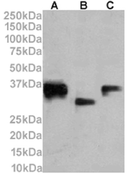


 Immunofluorescence (IF)
(Immunofluorescence staining of fixed Daudi cells with anti-CD74 antibody LN-2 (MBS488743) Immunofluorescence analysis of paraformaldehyde fixed Daudi cells on Shi-fix coverslips stained with the chimeric rabbit IgG version of LN-2 (MBS488743) at 10ug/ml for 1h followed by Alexa Fluor 488 secondary antibody (2ug/ml), showing membrane staining. The nuclear stain is DAPI (blue). Panels show from left-right, top-bottom MBS488743, DAPI, merged channels and an isotype control. The isotype control was an unknown specificity antibody (MBS488140) followed by staining with Alexa Fluor 488 secondary antibody.)
Immunofluorescence (IF)
(Immunofluorescence staining of fixed Daudi cells with anti-CD74 antibody LN-2 (MBS488743) Immunofluorescence analysis of paraformaldehyde fixed Daudi cells on Shi-fix coverslips stained with the chimeric rabbit IgG version of LN-2 (MBS488743) at 10ug/ml for 1h followed by Alexa Fluor 488 secondary antibody (2ug/ml), showing membrane staining. The nuclear stain is DAPI (blue). Panels show from left-right, top-bottom MBS488743, DAPI, merged channels and an isotype control. The isotype control was an unknown specificity antibody (MBS488140) followed by staining with Alexa Fluor 488 secondary antibody.)
CD74, Monoclonal Antibody (Cat# AAA488742)
Full Name
Anti-CD74 [LN-2]
Gene Names
CD74; II; DHLAG; HLADG; Ia-GAMMA
Reactivity
Human, Mouse, Rat
Applications
WB, IHC, IF, FC/FACS, EIA, IP
Purity
Purified antibody. Protein A affinity purified
Pricing
$495/0.2 mg | $2,025/5x0.2 mg


 Western Blot (WB)
(Figure 1. Western blot analysis of CDC27 using anti-CDC27 antibody (MBS1753628).Electrophoresis was performed on a 5-20% SDS-PAGE gel at 70V (Stacking gel) / 90V (Resolving gel) for 2-3 hours. The sample well of each lane was loaded with 50ug of sample under reducing conditions.Lane 1: human Raji whole cell lysatesLane 2: human Jurkat whole cell lysatesLane 3: human Jurkat whole cell lysatesLane 4: mouse thymus tissue lysates.After Electrophoresis, proteins were transferred to a Nitrocellulose membrane at 150mA for 50-90 minutes. Blocked the membrane with 5% Non-fat Milk/ TBS for 1. 5 hour at RT. The membrane was incubated with rabbit anti-CDC27 antigen affinity purified polyclonal antibody (Catalog # MBS1753628) at 0. 5 μg/mL overnight at 4 degree C, then washed with TBS-0. 1%Tween 3 times with 5 minutes each and probed with a goat anti-rabbit IgG-HRP secondary antibody at a dilution of 1:5000 for 1. 5 hour at RT. The signal is developed using an Enhanced Chemiluminescent detection (ECL) kit (Catalog # MBS176460) with Tanon 5200 system. A specific band was detected for CDC27 at approximately 92KD. The expected band size for CDC27 is at 92KD. )
Western Blot (WB)
(Figure 1. Western blot analysis of CDC27 using anti-CDC27 antibody (MBS1753628).Electrophoresis was performed on a 5-20% SDS-PAGE gel at 70V (Stacking gel) / 90V (Resolving gel) for 2-3 hours. The sample well of each lane was loaded with 50ug of sample under reducing conditions.Lane 1: human Raji whole cell lysatesLane 2: human Jurkat whole cell lysatesLane 3: human Jurkat whole cell lysatesLane 4: mouse thymus tissue lysates.After Electrophoresis, proteins were transferred to a Nitrocellulose membrane at 150mA for 50-90 minutes. Blocked the membrane with 5% Non-fat Milk/ TBS for 1. 5 hour at RT. The membrane was incubated with rabbit anti-CDC27 antigen affinity purified polyclonal antibody (Catalog # MBS1753628) at 0. 5 μg/mL overnight at 4 degree C, then washed with TBS-0. 1%Tween 3 times with 5 minutes each and probed with a goat anti-rabbit IgG-HRP secondary antibody at a dilution of 1:5000 for 1. 5 hour at RT. The signal is developed using an Enhanced Chemiluminescent detection (ECL) kit (Catalog # MBS176460) with Tanon 5200 system. A specific band was detected for CDC27 at approximately 92KD. The expected band size for CDC27 is at 92KD. )
CDC27, Polyclonal Antibody (Cat# AAA1753628)
Full Name
Anti-CDC27 Antibody
Gene Names
CDC27; APC3; HNUC; NUC2; ANAPC3; CDC27Hs; D0S1430E; D17S978E
Reactivity
Human, Mouse, Rat
Applications
WB, IHC-P, ICC, IF, FC/FACS/FCM, EIA
Purity
Immunogen affinity purified.
Pricing
$345/0.1 mg | $1,485/5x0.1 mg


 Western Blot (WB)
(Figure 1. Western blot analysis of Cytochrome p450 2C19/CYP2C19 using anti-Cytochrome p450 2C19/CYP2C19 antibody (MBS1754005).Electrophoresis was performed on a 5-20% SDS-PAGE gel at 70V (Stacking gel) / 90V (Resolving gel) for 2-3 hours. The sample well of each lane was loaded with 50ug of sample under reducing conditions.Lane 1: monkey liver tissue lysatesLane 2: rat liver tissue lysatesLane 3: mouse liver tissue lysates.After Electrophoresis, proteins were transferred to a Nitrocellulose membrane at 150mA for 50-90 minutes. Blocked the membrane with 5% Non-fat Milk/ TBS for 1. 5 hour at RT. The membrane was incubated with mouse anti-Cytochrome p450 2C19/CYP2C19 antigen affinity purified monoclonal antibody (Catalog # MBS1754005) at 0. 5 μg/mL overnight at 4 degree C, then washed with TBS-0. 1%Tween 3 times with 5 minutes each and probed with a goat anti-mouse IgG-HRP secondary antibody at a dilution of 1:10000 for 1. 5 hour at RT. The signal is developed using an Enhanced Chemiluminescent detection (ECL) kit (Catalog # MBS176445) with Tanon 5200 system. A specific band was detected for Cytochrome p450 2C19/CYP2C19 at approximately 55KD. The expected band size for Cytochrome p450 2C19/CYP2C19 is at 55KD. )
Western Blot (WB)
(Figure 1. Western blot analysis of Cytochrome p450 2C19/CYP2C19 using anti-Cytochrome p450 2C19/CYP2C19 antibody (MBS1754005).Electrophoresis was performed on a 5-20% SDS-PAGE gel at 70V (Stacking gel) / 90V (Resolving gel) for 2-3 hours. The sample well of each lane was loaded with 50ug of sample under reducing conditions.Lane 1: monkey liver tissue lysatesLane 2: rat liver tissue lysatesLane 3: mouse liver tissue lysates.After Electrophoresis, proteins were transferred to a Nitrocellulose membrane at 150mA for 50-90 minutes. Blocked the membrane with 5% Non-fat Milk/ TBS for 1. 5 hour at RT. The membrane was incubated with mouse anti-Cytochrome p450 2C19/CYP2C19 antigen affinity purified monoclonal antibody (Catalog # MBS1754005) at 0. 5 μg/mL overnight at 4 degree C, then washed with TBS-0. 1%Tween 3 times with 5 minutes each and probed with a goat anti-mouse IgG-HRP secondary antibody at a dilution of 1:10000 for 1. 5 hour at RT. The signal is developed using an Enhanced Chemiluminescent detection (ECL) kit (Catalog # MBS176445) with Tanon 5200 system. A specific band was detected for Cytochrome p450 2C19/CYP2C19 at approximately 55KD. The expected band size for Cytochrome p450 2C19/CYP2C19 is at 55KD. )
Cytochrome p450 2C19/CYP2C19, Monoclonal Antibody (Cat# AAA1754005)
Full Name
Anti-Cytochrome p450 2C19/CYP2C19 Antibody (monoclonal, 10G5)
Gene Names
CYP2C19; CPCJ; CYP2C; P450C2C; P450IIC19
Reactivity
Human, Mouse, Rat, Monkey
Applications
WB, IHC-P, FC/FACS/FCM
Purity
Immunogen affinity purified.
Pricing
$345/0.1 mg | $1,485/5x0.1 mg


 Western Blot (WB)
(Figure 1. Western blot analysis of PCK2 using anti-PCK2 antibody (MBS1754038).Electrophoresis was performed on a 5-20% SDS-PAGE gel at 70V (Stacking gel) / 90V (Resolving gel) for 2-3 hours. The sample well of each lane was loaded with 50ug of sample under reducing conditions.Lane 1: human HEPG2 whole cell lysatesLane 2: human Hela whole cell lysatesLane 3: human HEK293 whole cell lysatesLane 4: human A431 whole cell lysatesLane 5: monkey COS-7 whole cell lysatesLane 6: rat kidney tissue lysatesLane 7: mouse kidney tissue lysatesLane 8: mouse Neuro-2a whole cell lysates.After Electrophoresis, proteins were transferred to a Nitrocellulose membrane at 150mA for 50-90 minutes. Blocked the membrane with 5% Non-fat Milk/ TBS for 1. 5 hour at RT. The membrane was incubated with mouse anti-PCK2 antigen affinity purified monoclonal antibody (Catalog # MBS1754038) at 0. 25 μg/mL overnight at 4 degree C, then washed with TBS-0. 1%Tween 3 times with 5 minutes each and probed with a goat anti-mouse IgG-HRP secondary antibody at a dilution of 1:10000 for 1. 5 hour at RT. The signal is developed using an Enhanced Chemiluminescent detection (ECL) kit (Catalog # MBS176445) with Tanon 5200 system. A specific band was detected for PCK2 at approximately 71KD. The expected band size forPCK2 is at 71KD. )
Western Blot (WB)
(Figure 1. Western blot analysis of PCK2 using anti-PCK2 antibody (MBS1754038).Electrophoresis was performed on a 5-20% SDS-PAGE gel at 70V (Stacking gel) / 90V (Resolving gel) for 2-3 hours. The sample well of each lane was loaded with 50ug of sample under reducing conditions.Lane 1: human HEPG2 whole cell lysatesLane 2: human Hela whole cell lysatesLane 3: human HEK293 whole cell lysatesLane 4: human A431 whole cell lysatesLane 5: monkey COS-7 whole cell lysatesLane 6: rat kidney tissue lysatesLane 7: mouse kidney tissue lysatesLane 8: mouse Neuro-2a whole cell lysates.After Electrophoresis, proteins were transferred to a Nitrocellulose membrane at 150mA for 50-90 minutes. Blocked the membrane with 5% Non-fat Milk/ TBS for 1. 5 hour at RT. The membrane was incubated with mouse anti-PCK2 antigen affinity purified monoclonal antibody (Catalog # MBS1754038) at 0. 25 μg/mL overnight at 4 degree C, then washed with TBS-0. 1%Tween 3 times with 5 minutes each and probed with a goat anti-mouse IgG-HRP secondary antibody at a dilution of 1:10000 for 1. 5 hour at RT. The signal is developed using an Enhanced Chemiluminescent detection (ECL) kit (Catalog # MBS176445) with Tanon 5200 system. A specific band was detected for PCK2 at approximately 71KD. The expected band size forPCK2 is at 71KD. )
PCK2, Monoclonal Antibody (Cat# AAA1754038)
Full Name
Anti-PCK2 Antibody (monoclonal, 3F7)
Gene Names
PCK2; PEPCK; PEPCK2; PEPCK-M
Reactivity
Human, Mouse, Rat, Monkey
Applications
WB, IHC-P, ICC, IF, FC/FACS/FCM
Purity
Immunogen affinity purified.
Pricing
$345/0.1 mg | $1,485/5x0.1 mg


 Western Blot (WB)
(Figure 1. Western blot analysis of ANAPC2 using anti-ANAPC2 antibody (MBS1753725).Electrophoresis was performed on a 5-20% SDS-PAGE gel at 70V (Stacking gel) / 90V (Resolving gel) for 2-3 hours. The sample well of each lane was loaded with 50ug of sample under reducing conditions.Lane 1: human HEK293 whole cell lysatesLane 2: human Jurkat whole cell lysatesLane 3: human HEPG2 whole cell lysatesLane 4: human HELA whole cell lysatesLane 5: monkey COS-7 whole cell lysatesLane 6: human A549 whole cell lysatesLane 7: human PC-3 whole cell lysatesLane 8: rat stomach tissue lysatesLane 9: rat testis tissue lysatesLane 10: rat lung tissue lysatesLane 11: rat PC-12 whole cell lysatesLane 12: mouse stomach tissue lysatesLane 13: mouse lung tissue lysatesLane 14: mouse RAW264. 7 whole cell lysates.After Electrophoresis, proteins were transferred to a Nitrocellulose membrane at 150mA for 50-90 minutes. Blocked the membrane with 5% Non-fat Milk/ TBS for 1. 5 hour at RT. The membrane was incubated with rabbit anti-ANAPC2 antigen affinity purified polyclonal antibody (Catalog # MBS1753725) at 0. 5 μg/mL overnight at 4 degree C, then washed with TBS-0. 1%Tween 3 times with 5 minutes each and probed with a goat anti-rabbit IgG-HRP secondary antibody at a dilution of 1:5000 for 1. 5 hour at RT. The signal is developed using an Enhanced Chemiluminescent detection (ECL) kit (Catalog # MBS176460) with Tanon 5200 system. A specific band was detected for ANAPC2 at approximately 110KD. The expected band size for ANAPC2 is at 94KD. )
Western Blot (WB)
(Figure 1. Western blot analysis of ANAPC2 using anti-ANAPC2 antibody (MBS1753725).Electrophoresis was performed on a 5-20% SDS-PAGE gel at 70V (Stacking gel) / 90V (Resolving gel) for 2-3 hours. The sample well of each lane was loaded with 50ug of sample under reducing conditions.Lane 1: human HEK293 whole cell lysatesLane 2: human Jurkat whole cell lysatesLane 3: human HEPG2 whole cell lysatesLane 4: human HELA whole cell lysatesLane 5: monkey COS-7 whole cell lysatesLane 6: human A549 whole cell lysatesLane 7: human PC-3 whole cell lysatesLane 8: rat stomach tissue lysatesLane 9: rat testis tissue lysatesLane 10: rat lung tissue lysatesLane 11: rat PC-12 whole cell lysatesLane 12: mouse stomach tissue lysatesLane 13: mouse lung tissue lysatesLane 14: mouse RAW264. 7 whole cell lysates.After Electrophoresis, proteins were transferred to a Nitrocellulose membrane at 150mA for 50-90 minutes. Blocked the membrane with 5% Non-fat Milk/ TBS for 1. 5 hour at RT. The membrane was incubated with rabbit anti-ANAPC2 antigen affinity purified polyclonal antibody (Catalog # MBS1753725) at 0. 5 μg/mL overnight at 4 degree C, then washed with TBS-0. 1%Tween 3 times with 5 minutes each and probed with a goat anti-rabbit IgG-HRP secondary antibody at a dilution of 1:5000 for 1. 5 hour at RT. The signal is developed using an Enhanced Chemiluminescent detection (ECL) kit (Catalog # MBS176460) with Tanon 5200 system. A specific band was detected for ANAPC2 at approximately 110KD. The expected band size for ANAPC2 is at 94KD. )
ANAPC2, Polyclonal Antibody (Cat# AAA1753725)
Full Name
Anti-ANAPC2 Antibody
Gene Names
ANAPC2; APC2; RP11-350O14.5
Reactivity
Human, Mouse, Rat, Monkey
Applications
WB, IHC-P, ICC, IF, FC/FACS/FCM, EIA
Purity
Immunogen affinity purified.
Pricing
$345/0.1 mg | $1,485/5x0.1 mg


 Western Blot (WB)
(Figure 1. Western blot analysis of SEZ6L using anti-SEZ6L antibody (MBS1753793).Electrophoresis was performed on a 5-20% SDS-PAGE gel at 70V (Stacking gel) / 90V (Resolving gel) for 2-3 hours. The sample well of each lane was loaded with 50ug of sample under reducing conditions.Lane 1: human HEK293 whole cell lysatesLane 2: human THP-1 whole cell lysatesLane 3: human HL-60 whole cell lysatesLane 4: rat brain tissue lysatesLane 5: mouse brain tissue lysates.After Electrophoresis, proteins were transferred to a Nitrocellulose membrane at 150mA for 50-90 minutes. Blocked the membrane with 5% Non-fat Milk/ TBS for 1. 5 hour at RT. The membrane was incubated with rabbit anti-SEZ6L antigen affinity purified polyclonal antibody (Catalog # MBS1753793) at 0. 5 μg/mL overnight at 4 degree C, then washed with TBS-0. 1%Tween 3 times with 5 minutes each and probed with a goat anti-rabbit IgG-HRP secondary antibody at a dilution of 1:5000 for 1. 5 hour at RT. The signal is developed using an Enhanced Chemiluminescent detection (ECL) kit (Catalog # MBS176460 ) with Tanon 5200 system. A specific band was detected for SEZ6L at approximately 112KD. The expected band size for SEZ6L is at 112KD. )
Western Blot (WB)
(Figure 1. Western blot analysis of SEZ6L using anti-SEZ6L antibody (MBS1753793).Electrophoresis was performed on a 5-20% SDS-PAGE gel at 70V (Stacking gel) / 90V (Resolving gel) for 2-3 hours. The sample well of each lane was loaded with 50ug of sample under reducing conditions.Lane 1: human HEK293 whole cell lysatesLane 2: human THP-1 whole cell lysatesLane 3: human HL-60 whole cell lysatesLane 4: rat brain tissue lysatesLane 5: mouse brain tissue lysates.After Electrophoresis, proteins were transferred to a Nitrocellulose membrane at 150mA for 50-90 minutes. Blocked the membrane with 5% Non-fat Milk/ TBS for 1. 5 hour at RT. The membrane was incubated with rabbit anti-SEZ6L antigen affinity purified polyclonal antibody (Catalog # MBS1753793) at 0. 5 μg/mL overnight at 4 degree C, then washed with TBS-0. 1%Tween 3 times with 5 minutes each and probed with a goat anti-rabbit IgG-HRP secondary antibody at a dilution of 1:5000 for 1. 5 hour at RT. The signal is developed using an Enhanced Chemiluminescent detection (ECL) kit (Catalog # MBS176460 ) with Tanon 5200 system. A specific band was detected for SEZ6L at approximately 112KD. The expected band size for SEZ6L is at 112KD. )
SEZ6L, Polyclonal Antibody (Cat# AAA1753793)
Full Name
Anti-SEZ6L Antibody
Reactivity
Human, Mouse, Rat
Applications
WB, ICC, IF, FC, EIA
Purity
Immunogen affinity purified.
Pricing
$310/0.1 mg | $1,330/5x0.1 mg
 Flow Cytometry (FC/FACS)
(Balb/c mouse splenic T-cells (left) or thymocytes (right) were stained with anti-CD8a (clone: YTS 169.4)(filled histogram) or rat IgG2b isotype control (open histogram). )
Flow Cytometry (FC/FACS)
(Balb/c mouse splenic T-cells (left) or thymocytes (right) were stained with anti-CD8a (clone: YTS 169.4)(filled histogram) or rat IgG2b isotype control (open histogram). )
Type I collagen, Polyclonal Antibody (Cat# AAA524564)
Full Name
Anti-Human Type I collagen, Affinity Purified, (Polyclonal)(Rabbit IgG)
Reactivity
Human
Applications
WB, IHC-P
Purity
Affinity Purified. Affinity Chromatography
Pricing
$440/0.1 mg | $1,760/5x0.1 mg