Loading...

Skip to main content

Call us on + 1 (800) 604-9114 for more information about our products

Looking for specific datasheet Manual/COA/MSDS?
Request a Manual/COA/MSDS

Interested to get a quote about our products?
Request a Quote

Filters


Clonality

Type

Reactivity

Gene Name

Isotype

Host

Application

Clone

2006 results for "Goat IgG Isotype Control" - showing 400-450


Western Blot (WB) (Western blot analysis of lysates from CEM, SH-SY5Y cell line (from left to right), using GATA3 Antibody (Center). It was diluted at 1:1000 at each lane. A goat anti-mouse IgG H&L(HRP) at 1:10000 dilution was used as the secondary antibody. )

GATA3, Monoclonal Antibody (Cat# AAA9231647)

Full Name
GATA3 Antibody (Center)
Gene Names
GATA3; HDR; HDRS
Reactivity
Human
Applications
WB, IF, FC/FACS, EIA
Pricing
$435/0.1 mL | $1,655/5x0.1 mL
Western Blot (WB)

STRN3, Monoclonal Antibody (Cat# AAA435160)

Full Name
STRN3 (617) Mouse mAb
Gene Names
STRN3; SG2NA; S/G2NA; PPP2R6B
Reactivity
Human
Applications
WB, IP, FC/FACS
Pricing
$260/0.02 mL | $405/0.05 mL | $650/0.1 mL | $2,645/5x0.1 mL
Western Blot (WB) (Western blot analysis of lysates from Hela,Jurkat,mouse NIH/3T3 cell line (from left to right), using PPARA Antibody. It was diluted at 1:500 at each lane. A goat anti-mouse IgG H&L(HRP) at 1:10000 dilution was used as the secondary antibody.)

PPARA, Monoclonal Antibody (Cat# AAA9231654)

Full Name
PPARA Antibody
Gene Names
PPARA; PPAR; NR1C1; hPPAR; PPARalpha
Reactivity
Human, Mouse
Applications
WB, IHC, IF, FC/FACS, EIA
Pricing
$435/0.1 mL | $1,655/5x0.1 mL
Flow Cytometry (FC/FACS) (Overlay histogram showing Jurkat cells stained at 37 degree C. The secondary antibody used was Alexa Fluor 488 goat anti-rabbit lgG (H+L) (1583138) at 1/400 dilution for 40 min at 37 degree C. Isotype control antibody (blue line) was rabbit IgG1 (1mug/1x10^6 cells) used under the same conditions. Acquisition of >10, 000 events was performed.)

AVPR2, Polyclonal Antibody (Cat# AAA9232002)

Full Name
AVPR2 Antibody (C-term)
Gene Names
AVPR2; DI1; DIR; NDI; V2R; ADHR; DIR3
Reactivity
Human
Applications
WB, FC/FACS, EIA
Pricing
$425/0.1 mL | $1,615/5x0.1 mL
Western Blot (WB)

MTHFD1, Monoclonal Antibody (Cat# AAA435171)

Full Name
MTHFD1 Mouse mAb
Gene Names
MTHFD1; CIMAH; MTHFC; MTHFD
Reactivity
Human
Applications
WB, IP, FC/FACS
Pricing
$260/0.02 mL | $405/0.05 mL | $650/0.1 mL | $2,645/5x0.1 mL
Western Blot (WB) (Western blot analysis of lysates from Hela, NCI-H1299 cell line (from left to right),using RPL6 Antibody (N-term). It was diluted at 1:1000 at each lane. A goat anti-rabbit IgG H&L(HRP) at 1:10000 dilution was used as the secondary antibody.)

RPL6, Polyclonal Antibody (Cat# AAA9231604)

Full Name
RPL6 Antibody (N-term)
Gene Names
RPL6; L6; TXREB1; SHUJUN-2; TAXREB107
Reactivity
Human
Applications
WB, IHC, IF, FC/FACS, EIA
Pricing
$425/0.1 mL | $1,615/5x0.1 mL
Western Blot (WB) (All lanes : KTN1 Mouse mAb at 1/2000 dilution Lane 1 : Jurkat cells membrane fraction Lane 2 : THP1 cells membrane fraction as negative control Lysates/proteins at 20 ug per lane. Secondary Goat Anti-Mouse IgG-HRP, 5% milk conjugated at 1/10000 dilution Predicted band size : 156 KDa Observed band size : 156 KDa Blocking/Dilution buffer : 1x TBST.)

KTN1, Monoclonal Antibody (Cat# AAA435115)

Full Name
KTN1 (578) Mouse mAb
Gene Names
KTN1; CG1; KNT; MU-RMS-40.19
Reactivity
Human
Applications
WB, IP, FC/FACS
Pricing
$260/0.02 mL | $405/0.05 mL | $650/0.1 mL | $2,645/5x0.1 mL
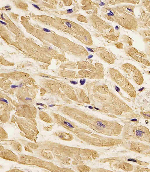
Immunohistochemistry (IHC) (Figure 1. IHC analysis of TAPA1 using anti-TAPA1 antibody (MBS1750607).TAPA1 was detected in paraffin-embedded section of human tonsil tissue. Heat mediated antigen retrieval was performed in citrate buffer (pH6, epitope retrieval solution) for 20 mins. The tissue section was blocked with 10% goat serum. The tissue section was then incubated with 1ug/ml rabbit anti-TAPA1 Antibody (MBS1750607) overnight at 4 degree C. Biotinylated goat anti-rabbit IgG was used as secondary antibody and incubated for 30 minutes at 37 degree C. The tissue section was developed using Strepavidin-Biotin-Complex (SABC) with DAB as the chromogen. )

TAPA1, Polyclonal Antibody (Cat# AAA1750607)

Full Name
Anti-TAPA1 Picoband antibody
Gene Names
CD81; S5.7; CVID6; TAPA1; TSPAN28
Reactivity
Human, Mouse, Rat
No cross reactivity with other proteins.
Applications
EIA, FC/FACS, IHC, ICC
Pricing
$345/0.1 mg | $1,485/5x0.1 mg
Immunocytochemistry/Immunofluorescence (ICC/IF) (Immunocytochemistry/Immunofluorescence analysis using Mouse Anti-Hexanoyl-Lysine adduct Monoclonal Antibody, Clone 5E8. Tissue: Embryonic kidney epithelial cell line (HEK293). Species: Human. Fixation: 5% Formaldehyde for 5 min. Primary Antibody: Mouse Anti-Hexanoyl-Lysine adduct Monoclonal Antibody at 1:50 for 30-60 min at RT. Secondary Antibody: Goat Anti-Mouse Alexa Fluor 488 at 1:1500 for 30-60 min at RT. Counterstain: Phalloidin Alexa Fluor 633 F-Actin stain; DAPI (blue) nuclear stain at 1:250, 1:50000 for 30-60 min at RT. Magnification: 20X (2X Zoom). (A,C,E,G) - Untreated. (B,D,F,H) - Cells cultured overnight with 50 uM H2O2. (A,B) DAPI (blue) nuclear stain. (C,D) Phalloidin Alex Fluor 633 F-Actin stain. (E,F) Hexanoyl-Lysine adduct Antibody. (G,H) Composite. Courtesy of: Dr. Robert Burke, University of Victoria.)

Hexanoyl-Lysine adduct, Monoclonal Antibody (Cat# AAA808675)

Full Name
Hexanoyl-Lysine adduct Antibody, Clone 5E8: Biotin
Reactivity
Species Independent
Applications
WB, ICC, IF, FC/FACS, EIA
Purity
Protein G Purified
Pricing
$555/0.1 mg | $2,255/5x0.1 mg
Immunocytochemistry/Immunofluorescence (ICC/IF) (Immunocytochemistry/Immunofluorescence analysis using Mouse Anti-Dityrosine Monoclonal Antibody, Clone 10A6. Tissue: Embryonic kidney epithelial cell line (HEK293). Species: Human. Fixation: 5% Formaldehyde for 5 min. Primary Antibody: Mouse Anti-Dityrosine Monoclonal Antibody at 1:50 for 30-60 min at RT. Secondary Antibody: Goat Anti-Mouse Alexa Fluor 488 at 1:1500 for 30-60 min at RT. Counterstain: Phalloidin Alexa Fluor 633 F-Actin stain; DAPI (blue) nuclear stain at 1:250, 1:50000 for 30-60 min at RT. Localization: Cytoplasmic. Magnification: 20X (2X Zoom). (A,C,E,G) - Untreated. (B,D,F,H) - Cells cultured overnight with 50 uM H2O2. (A,B) DAPI (blue) nuclear stain. (C,D) Phalloidin Alex Fluor 633 F-Actin stain. (E,F) Dityrosine Antibody. (G,H) Composite. Courtesy of: Dr. Robert Burke, University of Victoria.)

Dityrosine, Monoclonal Antibody (Cat# AAA808891)

Full Name
Dityrosine Antibody, Clone 10A6: Biotin
Reactivity
Species Independent
Applications
WB, ICC, IF, FC/FACS, EIA
Purity
Protein G Purified
Pricing
$555/0.1 mg | $2,255/5x0.1 mg
Immunocytochemistry/Immunofluorescence (ICC/IF) (Immunocytochemistry/Immunofluorescence analysis using Mouse Anti-4-Hydroxy-2-hexenal Monoclonal Antibody, Clone 6F10. Tissue: Embryonic kidney epithelial cell line (HEK293). Species: Human. Fixation: 5% Formaldehyde for 5 min. Primary Antibody: Mouse Anti-4-Hydroxy-2-hexenal Monoclonal Antibody at 1:400 for 30-60 min at RT. Secondary Antibody: Goat Anti-Mouse Alexa Fluor 488 at 1:1500 for 30-60 min at RT. Counterstain: Phalloidin Alexa Fluor 633 F-Actin stain; DAPI (blue) nuclear stain at 1:250, 1:50000 for 30-60 min at RT. Magnification: 20X (2X Zoom). (A,C,E,G) - Untreated. (B,D,F,H) - Cells cultured overnight with 50 uM H2O2. (A,B) DAPI (blue) nuclear stain. (C,D) Phalloidin Alex Fluor 633 F-Actin stain. (E,F) 4-Hydroxy-2-hexenal Antibody. (G,H) Composite. Courtesy of: Dr. Robert Burke, University of Victoria.)

4-Hydroxy-2-hexenal, Monoclonal Antibody (Cat# AAA808686)

Full Name
4-Hydroxy-2-hexenal Antibody, Clone 6F10: ATTO 594
Reactivity
Species Independent
Applications
WB, ICC, IF, EIA, FC/FACS
Purity
Protein G Purified
Pricing
$560/0.1 mg | $2,275/5x0.1 mg
Immunocytochemistry/Immunofluorescence (ICC/IF) (Immunocytochemistry/Immunofluorescence analysis using Mouse Anti-4-Hydroxy-2-hexenal Monoclonal Antibody, Clone 6F10. Tissue: Embryonic kidney epithelial cell line (HEK293). Species: Human. Fixation: 5% Formaldehyde for 5 min. Primary Antibody: Mouse Anti-4-Hydroxy-2-hexenal Monoclonal Antibody at 1:400 for 30-60 min at RT. Secondary Antibody: Goat Anti-Mouse Alexa Fluor 488 at 1:1500 for 30-60 min at RT. Counterstain: Phalloidin Alexa Fluor 633 F-Actin stain; DAPI (blue) nuclear stain at 1:250, 1:50000 for 30-60 min at RT. Magnification: 20X (2X Zoom). (A,C,E,G) - Untreated. (B,D,F,H) - Cells cultured overnight with 50 uM H2O2. (A,B) DAPI (blue) nuclear stain. (C,D) Phalloidin Alex Fluor 633 F-Actin stain. (E,F) 4-Hydroxy-2-hexenal Antibody. (G,H) Composite. Courtesy of: Dr. Robert Burke, University of Victoria.)

4-Hydroxy-2-hexenal, Monoclonal Antibody (Cat# AAA808693)

Full Name
4-Hydroxy-2-hexenal Antibody, Clone 6F10: Biotin
Reactivity
Species Independent
Applications
WB, ICC, IF, EIA, FC/FACS
Purity
Protein G Purified
Pricing
$555/0.1 mg | $2,255/5x0.1 mg
Western Blot (WB) (Figure 1. Western blot analysis of NFIA using anti- NFIA antibody (MBS178836).Electrophoresis was performed on a 5-20% SDS-PAGE gel at 70V (Stacking gel) / 90V (Resolving gel) for 2-3 hours. The sample well of each lane was loaded with 50ug of sample under reducing conditions.Lane 1: JURKAT whole cell lysates,Lane 2: COLO320 whole cell lysates.After Electrophoresis, proteins were transferred to a Nitrocellulose membrane at 150mA for 50-90 minutes. Blocked the membrane with 5% Non-fat Milk/ TBS for 1.5 hour at RT. The membrane was incubated with rabbit anti- NFIA antigen affinity purified polyclonal antibody at 0.5ug/mL overnight at 4 degree C, then washed with TBS-0.1%Tween 3 times with 5 minutes each and probed with a goat anti-rabbit IgG-HRP secondary antibody at a dilution of 1:10000 for 1.5 hour at RT. The signal is developed using an Enhanced Chemiluminescent detection (ECL) kit with Tanon 5200 system. A specific band was detected for NFIA at approximately 69KD. The expected band size for NFIA is at 56KD. )

NFIA, Polyclonal Antibody (Cat# AAA178836)

Full Name
Anti-NFIA Antibody
Gene Names
NFIA; CTF; NF1-A; NFI-A; NFI-L; NF-I/A
Reactivity
Human, Mouse, Rat
Applications
WB, IHC
Purity
Immunogen affinity purified.
Pricing
$345/0.1 mg | $1,485/5x0.1 mg
Western Blot (WB) (All lanes : Anti-MGMT Antibody at1:2000 dilutionLane 1: Jurkat whole cell lysateLane 2: Molt-4 whole cell lysateLysates/proteins at 20 ug per lane. SecondaryGoat Anti-mouse IgG, (H+L), Peroxidase conjugated at 1/10000 dilution. Predicted band size : 22 kDaBlocking/Dilution buffer: 5% NFDM/TBST.)

MGMT, Monoclonal Antibody (Cat# AAA9232196)

Full Name
MGMT Antibody
Reactivity
Human
Predicted: Monkey
Applications
WB, FC/FACS, EIA
Pricing
$435/0.1 mL | $1,655/5x0.1 mL
Western Blot (WB) (Western blot analysis of lysates from A431,KG-1 cell line,mouse kidney,mouse liver tissue lysate,SH-SY5Y cell line (from left to right), using CLN3 Antibody (Center). It was diluted at 1:1000 at each lane. A goat anti-rabbit IgG H&L(HRP) at 1:10000 dilution was used as the secondary antibody.Lysates at 20ug per lane.)

CLN3, Polyclonal Antibody (Cat# AAA9231898)

Full Name
CLN3 Antibody (Center)
Gene Names
CLN3; BTS; BTN1; JNCL
Reactivity
Human, Mouse
Predicted: Monkey
Applications
WB, IHC, FC/FACS, EIA
Pricing
$425/0.1 mL | $1,615/5x0.1 mL
Flow Cytometry (FC/FACS) (Overlay histogram showing Hela cells stained at 37 degree C. The secondary antibody used was Goat-Anti-Rabbit IgG, DyLight 488 Conjugated Highly Cross-Adsorbed(OH191631) at 1/200 dilution for 40 min at 37 degree C. Isotype control antibody (blue line) was rabbit IgG (1mug/1x10^6 cells) used under the same conditions. Acquisition of >10, 000 events was performed.)

RPS7, Polyclonal Antibody (Cat# AAA9232223)

Full Name
RPS7 Antibody (N-Term)
Gene Names
RPS7; S7; eS7; DBA8
Reactivity
Human, Mouse
Predicted: Bovine, Zebrafish, Rat, Xenopus
Applications
WB, EIA
Pricing
$425/0.1 mL | $1,615/5x0.1 mL
Western Blot (WB) (Western blot analysis of lysates from A431,Hela cell line (from left to right), using DFFB Antibody (Center). It was diluted at 1:1000 at each lane. A goat anti-rabbit IgG H&L(HRP) at 1:10000 dilution was used as the secondary antibody.)

DFFB, Polyclonal Antibody (Cat# AAA9231709)

Full Name
DFFB Antibody (Center)
Gene Names
DFFB; CAD; CPAN; DFF2; DFF40; DFF-40
Reactivity
Human
Predicted: Mouse, Rat
Applications
WB, IHC, FC/FACS, EIA
Pricing
$425/0.1 mL | $1,615/5x0.1 mL
Flow Cytometry (FC/FACS) (Flow-cytometry using the anti-IL-18 antibody SAIC-24B-4 Human peripheral blood monocytes (PBMC’s) were fixed using 2% PFA, permeabilised using 0.5% Triton and stained with unimmunized rabbit IgG antibody (MOPC-21; isotype control, black line) or the rabbit IgG-chimeric version of SAIC-24B-4 (MBS488409, blue line) at a dilution of 1:100 for 1h at RT. An additional population of PBMC’s stimulated with PMA and Ionomycin in the presence of Monensin for 4 hours were stained in a similar manner (purple line). After washing, bound antibody was detected using a goat anti-rabbit IgG AlexaFluor 488 antibody at a dilution of 1:1000 and cells analyzed using a FACSCanto flow-cytometer.)

IL-18, Monoclonal Recombinant Antibody (Cat# AAA488409)

Full Name
Anti-IL-18 [SAIC-24B-4]
Gene Names
IL18; IGIF; IL-18; IL-1g; IL1F4
Reactivity
Human
Applications
Immuno-MRM
Purity
Protein A Affinity Purified
Pricing
$495/0.2 mg | $2,025/5x0.2 mg
Western Blot (WB)

MIMIT, Monoclonal Antibody (Cat# AAA435162)

Full Name
MIMIT Mouse mAb
Gene Names
NDUFAF2; MMTN; B17.2L; mimitin; NDUFA12L
Reactivity
Human
Applications
WB, IP, FC/FACS
Pricing
$260/0.02 mL | $405/0.05 mL | $650/0.1 mL | $2,645/5x0.1 mL
Western Blot (WB) (Anti- BMP-5 Picoband antibody, MBS178341, Western blottingAll lanes: Anti BMP-5 (MBS178341) at 0.5ug/mlLane 1: Rat Liver Tissue Lysate at 50ugLane 2: Mouse Liver Tissue Lysate at 50ugLane 3: A549 Whole Cell Lysate at 40ugPredicted bind size: 51KDObserved bind size: 51KD)

BMP5, Polyclonal Antibody (Cat# AAA178341)

Full Name
Anti-BMP5 Antibody
Reactivity
Human, Mouse, Rat
Applications
WB, IHC, EIA
Purity
Immunogen Affinity Purified
Pricing
$345/0.1 mg | $1,485/5x0.1 mg
Immunocytochemistry/Immunofluorescence (ICC/IF) (Immunocytochemistry/Immunofluorescence analysis using Mouse Anti-Nitrotryptophan Monoclonal Antibody, Clone 2D12. Tissue: Embryonic kidney epithelial cell line (HEK293). Species: Human. Fixation: 5% Formaldehyde for 5 min. Primary Antibody: Mouse Anti-Nitrotryptophan Monoclonal Antibody at 1:50 for 30-60 min at RT. Secondary Antibody: Goat Anti-Mouse Alexa Fluor 488 at 1:1500 for 30-60 min at RT. Counterstain: Phalloidin Alexa Fluor 633 F-Actin stain; DAPI (blue) nuclear stain at 1:250, 1:50000 for 30-60 min at RT. Localization: Cytoplasmic. Magnification: 20X (2X Zoom). (A) DAPI (blue) nuclear stain. (B) Phalloidin Alex Fluor 633 F-Actin stain. (C) Nitrotryptophan Antibody. (D) Composite. Courtesy of: Dr. Robert Burke, University of Victoria.)

Nitrotryptophan, Monoclonal Antibody (Cat# AAA808837)

Full Name
Nitrotryptophan Antibody, Clone 2D12: Biotin
Reactivity
Species Independent
Applications
WB, ICC, IF, FC/FACS, EIA
Purity
Protein G Purified
Pricing
$555/0.1 mg | $2,255/5x0.1 mg
Immunocytochemistry/Immunofluorescence (ICC/IF) (Immunocytochemistry/Immunofluorescence analysis using Mouse Anti-Hexanoyl-Lysine adduct Monoclonal Antibody, Clone 5E8. Tissue: Embryonic kidney epithelial cell line (HEK293). Species: Human. Fixation: 5% Formaldehyde for 5 min. Primary Antibody: Mouse Anti-Hexanoyl-Lysine adduct Monoclonal Antibody at 1:50 for 30-60 min at RT. Secondary Antibody: Goat Anti-Mouse Alexa Fluor 488 at 1:1500 for 30-60 min at RT. Counterstain: Phalloidin Alexa Fluor 633 F-Actin stain; DAPI (blue) nuclear stain at 1:250, 1:50000 for 30-60 min at RT. Magnification: 20X (2X Zoom). (A,C,E,G) - Untreated. (B,D,F,H) - Cells cultured overnight with 50 uM H2O2. (A,B) DAPI (blue) nuclear stain. (C,D) Phalloidin Alex Fluor 633 F-Actin stain. (E,F) Hexanoyl-Lysine adduct Antibody. (G,H) Composite. Courtesy of: Dr. Robert Burke, University of Victoria.)

Hexanoyl-Lysine adduct, Monoclonal Antibody (Cat# AAA808668)

Full Name
Hexanoyl-Lysine adduct Antibody, Clone 5E8: ATTO 594
Reactivity
Species Independent
Applications
WB, ICC, IF, FC/FACS, EIA
Purity
Protein G Purified
Pricing
$560/0.1 mg | $2,275/5x0.1 mg
Immunocytochemistry/Immunofluorescence (ICC/IF) (Immunocytochemistry/Immunofluorescence analysis using Mouse Anti-Hexanoyl-Lysine adduct Monoclonal Antibody, Clone 5E8. Tissue: Embryonic kidney epithelial cell line (HEK293). Species: Human. Fixation: 5% Formaldehyde for 5 min. Primary Antibody: Mouse Anti-Hexanoyl-Lysine adduct Monoclonal Antibody at 1:50 for 30-60 min at RT. Secondary Antibody: Goat Anti-Mouse Alexa Fluor 488 at 1:1500 for 30-60 min at RT. Counterstain: Phalloidin Alexa Fluor 633 F-Actin stain; DAPI (blue) nuclear stain at 1:250, 1:50000 for 30-60 min at RT. Magnification: 20X (2X Zoom). (A,C,E,G) - Untreated. (B,D,F,H) - Cells cultured overnight with 50 uM H2O2. (A,B) DAPI (blue) nuclear stain. (C,D) Phalloidin Alex Fluor 633 F-Actin stain. (E,F) Hexanoyl-Lysine adduct Antibody. (G,H) Composite. Courtesy of: Dr. Robert Burke, University of Victoria.)

Hexanoyl-Lysine adduct, Monoclonal Antibody (Cat# AAA808679)

Full Name
Hexanoyl-Lysine adduct Antibody, Clone 5E8: PerCP
Reactivity
Species Independent
Applications
WB, ICC, IF, FC/FACS, EIA
Purity
Protein G Purified
Pricing
$560/0.1 mg | $2,270/5x0.1 mg
Immunofluorescence (IF) (Immunofluorescence staining of fixed mouse splenocytes with anti-CD27 antibody LG.3A10 Immunofluorescence analysis of paraformaldehyde fixed mouse (Mus musculus) splenocytes on Shi-fix coverslips, permeabilized with 0.15% Triton stained with the chimeric rabbit version of LG.3A10 at 10 ug/ml for 1h followed by Alexa Fluor 488 secondary antibody (1 ug/ml), showing membrane and some cytoplasmic staining in a subset of cells. The nuclear stain is DAPI (blue). Panels show from left-right, top-bottom, DAPI, merged channels and an isotype control. The isotype control was stained with an anti-Fluorescein antibody followed by Alexa Fluor 488 secondary antibody.)

CD27, Monoclonal Recombinant Antibody (Cat# AAA488454)

Full Name
Anti-CD27 [LG.3A10]
Gene Names
Cd27; S152; Tp55; Tnfrsf7
Reactivity
Mouse
Applications
IP, IF, FC/FACS
Purity
Protein A Affinity Purified
Pricing
$495/0.2 mg | $2,025/5x0.2 mg
Western Blot (WB) (Western blot analysis of lysates from Hela,A431,MCF-7,mouse C2C12,rat C6 cell line (from left to right), using ACTB Antibody. It was diluted at 1:1000 at each lane. A goat anti-mouse IgG H&L(HRP) at 1:10000 dilution was used as the secondary antibody.)

Beta-Actin, Monoclonal Antibody (Cat# AAA9231852)

Full Name
Beta-Actin Antibody
Gene Names
ACTB; BRWS1; PS1TP5BP1
Reactivity
Human, Rat
Predicted: Xenopus, Bovine, Chicken, Horse, Monkey, Mouse, Pig, Sheep
Applications
WB, IHC, FC/FACS, EIA
Pricing
$435/0.1 mL | $1,655/5x0.1 mL
Testing Data (Figure A. RPE conjugated Mouse anti Human CD19 and FITC conjugated Rat IgG2b isotype control . Figure B. RPE conjugated Mouse anti Human CD19 and FITC conjugated Rat anti Human CD18 . All experiments performed on human peripheral blood lymphocytes in the presence of human SeroBlock (BUF070A).)

IgG2b, Negative Control (Cat# AAA225676)

Full Name
Rat IgG2b Negative Control: Pacific Blue
Reactivity
Rat
Applications
FC/FACS
Purity
Purified IgG prepared by affinity chromatography on Protein G from tissue culture supernatant
Pricing
$300/100 Tests | $1,200/5x100 Tests
Testing Data (1:1000 dilution of this antibody detected SMC1A on 40ug of cell lysates)

SMC1A, Monoclonal Antibody (Cat# AAA9400360)

Full Name
SMC1A(N-term) Monoclonal Antibody
Gene Names
SMC1A; SMC1; SMCB; CDLS2; SB1.8; SMC1L1; DXS423E; SMC1alpha
Reactivity
Human, Porcine, Mouse, Rat
Applications
WB, IHC, FC/FACS
Purity
Purified by antigen-affinity chromatography.
Pricing
$305/0.1 mL | $1,230/5x0.1 mL
Western Blot (WB) (Western blot analysis of lysates from Daudi,Raji cell line (from left to right), using BCL10 Antibody. It was diluted at 1:1000 at each lane. A goat anti-mouse IgG H&L(HRP) at 1:10000 dilution was used as the secondary antibody.Lysates at 20ug per lane.)

BCL10, Monoclonal Antibody (Cat# AAA9231645)

Full Name
BCL10 Antibody
Gene Names
BCL10; CLAP; mE10; CIPER; IMD37; c-E10; CARMEN
Reactivity
Human
Applications
WB, IHC, FC/FACS, EIA
Pricing
$435/0.1 mL | $1,655/5x0.1 mL
Flow Cytometry (FC/FACS) (Overlay histogram showing U-2OS cells stained at 37 degree C. The secondary antibody used was Goat-Anti-Rabbit IgG, DyLight 488 Conjugated Highly Cross-Adsorbed(NA168821) at 1/400 dilution for 40 min at 37 degree C. Isotype control antibody (blue line) was rabbit IgG (1mug/1x10^6 cells) used under the same conditions. Acquisition of >10, 000 events was performed.)

CF150, Polyclonal Antibody (Cat# AAA9231966)

Full Name
CF150 Antibody (Center)
Gene Names
CGAS; MB21D1; h-cGAS; C6orf150
Reactivity
Human
Applications
WB, EIA
Pricing
$425/0.1 mL | $1,615/5x0.1 mL
Immunohistochemistry (IHC) (Formalin-fixed, paraffin-embedded human spleen stained with CD43 Mouse Monoclonal Antibody (SPN/3388).)

CD43, Monoclonal Antibody (Cat# AAA4381264)

Full Name
CD43 (T-Cell Marker)
Gene Names
SPN; LSN; CD43; GALGP; GPL115
Reactivity
Human
Applications
FC/FACS, IF, WB, IHC
Purity
Purified Ab with BSA and Azide at 200ug/ml OR Purified Ab WITHOUT BSA and Azide at 1.0mg/ml
Pricing
$390/0.1 mg (With BSA & Azide at 0.2mg/mL) | $390/0.1 mg (Without BSA & Azide at 1mg/mL) | $215/0.02 mg (With BSA & Azide at 0.2mg/mL) | $1,710/5x0.1 mg (With BSA & Azide at 0.2mg/mL) | $1,710/5x0.1 mg (Without BSA & Azide at 1mg/mL)
SDS-Page (SDS-PAGE Analysis Purified Estrogen Receptor alpha Mouse Monoclonal Antibody (ESR1/3559). Confirmation of Integrity and Purity of Antibody.)

Estrogen Receptor, alpha, Monoclonal Antibody (Cat# AAA4380880)

Full Name
Estrogen Receptor, alpha (Marker of Estrogen Dependence)
Gene Names
ESR1; ER; ESR; Era; ESRA; ESTRR; NR3A1
Reactivity
Human
Applications
FC/FACS, IHC
Purity
Purified Ab with BSA and Azide at 200ug/ml OR Purified Ab WITHOUT BSA and Azide at 1.0mg/ml
Pricing
$390/0.1 mg (With BSA & Azide at 0.2mg/mL) | $390/0.1 mg (Without BSA & Azide at 1mg/mL) | $215/0.02 mg (With BSA & Azide at 0.2mg/mL) | $1,710/5x0.1 mg (With BSA & Azide at 0.2mg/mL) | $1,710/5x0.1 mg (Without BSA & Azide at 1mg/mL)
Immunohistochemistry (IHC) (Formalin-fixed, paraffin-embedded human Tonsil stained with CD73 Mouse Monoclonal Antibody (NT5E/2646).)

CD73, Monoclonal Antibody (Cat# AAA4381133)

Full Name
CD73 (Immuno-Oncology Target)
Gene Names
NT5E; NT; eN; NT5; NTE; eNT; CD73; E5NT; CALJA
Reactivity
Human
Applications
EIA, FC/FACS, IF, WB, IHC
Purity
Purified Ab with BSA and Azide at 200ug/ml OR Purified Ab WITHOUT BSA and Azide at 1.0mg/ml
Pricing
$390/0.1 mg (With BSA & Azide at 0.2mg/mL) | $390/0.1 mg (Without BSA & Azide at 1mg/mL) | $215/0.02 mg (With BSA & Azide at 0.2mg/mL) | $1,710/5x0.1 mg (With BSA & Azide at 0.2mg/mL) | $1,710/5x0.1 mg (Without BSA & Azide at 1mg/mL)
Quality Control (Western blot analysis of immunized recombinant protein, using anti-DHX29 monoclonal antibody. )

DHX29, Monoclonal Antibody (Cat# AAA120200)

Full Name
Mouse monoclonal antibody Anti-Human DHX29
Gene Names
DHX29; DDX29
Reactivity
Human
Applications
DB, ICC, IP, WB, FC/FACS
Pricing
$460/0.1 mg | $2,070/5x0.1 mg
Testing Data (Figure A. FITC conjugated Mouse anti Human CD19 and RPE-Alexa Fluor 647 conjugated Mouse IgG1 isotype control . Figure B. FITC conjugated Mouse anti Human CD19 and RPE-Alexa Fluor 647 conjugated Mouse anti Human CD81 . All experiments performed on red cell lysed human blood gated on lymphoid cells in the presence of 10% human serum. Data acquired on the ZE5 Cell Analyzer.)

CD81, Monoclonal Antibody (Cat# AAA225635)

Full Name
Mouse Anti Human CD81: RPE
Gene Names
CD81; S5.7; CVID6; TAPA1; TSPAN28
Reactivity
Human, Chimpanzee, Sheep, Goat
Applications
FC/FACS
Purity
Purified IgG prepared by affinity chromatography on Protein A from tissue culture supernatant
Pricing
$500/100 Tests | $2,090/5x100 Tests
Western Blot (WB) (Western blot analysis of lysates from mouse kidney and liver tissue lysate (from left to right), using Mouse Csnk1g3 Antibody (C-term). It was diluted at 1:1000 at each lane. A goat anti-rabbit IgG H&L(HRP) at 1:5000 dilution was used as the secondary antibody.)

Csnk1g3, Polyclonal Antibody (Cat# AAA9231587)

Full Name
Mouse Csnk1g3 Antibody (C-term)
Gene Names
Csnk1g3; 3300002K07Rik; C330049O21Rik
Reactivity
Mouse
Predicted: Rat
Applications
WB, IHC, FC/FACS, EIA
Pricing
$425/0.1 mL | $1,615/5x0.1 mL
Western Blot (WB) (All lanes : CPSF1 Mouse mAb at 1/2000 dilution Lane 1 : THP1 cells membrane fraction Lane 2 : Jurkat cells membrane fraction Lysates/proteins at 20 ug per lane. Secondary Goat Anti-Mouse IgG-HRP, 5% milk conjugated at 1/10000 dilution Predicted band size : 160 KDa Observed band size : 160 KDa Blocking/Dilution buffer : 1x TBST.)

CPSF1, Monoclonal Antibody (Cat# AAA435129)

Full Name
CPSF1 (514) Mouse mAb
Reactivity
Human
Applications
WB, IP, FC/FACS
Pricing
$260/0.02 mL | $405/0.05 mL | $650/0.1 mL | $2,645/5x0.1 mL
Western Blot (WB) (All lanes : Anti-GARS Antibody at 1:1000 dilutionLane 1: HT-1080 whole cell lysateLane 2: Jurkat whole cell lysateLane 3: Ramos whole cell lysateLane 4: PC-3 whole cell lysateLane 4: mouse brain lysateLysates/proteins at 15 ug per lane. SecondaryGoat Anti-mouse IgG, (H+L), Peroxidase conjugated at 1/10000 dilution. Predicted band size : 83 kDaBlocking/Dilution buffer: 5% NFDM/TBST.)

GARS, Monoclonal Antibody (Cat# AAA9232203)

Full Name
GARS Antibody
Gene Names
GARS1; GARS; HMN5; CMT2D; DSMAV; GlyRS; SMAD1
Reactivity
Human, Mouse
Applications
WB, FC/FACS, EIA
Pricing
$435/0.1 mL | $1,655/5x0.1 mL
Western Blot (WB) (All lanes : ROA1 Mouse mAb at 1/2000 dilution Lane 1 : THP1 cells membrane fraction Lane 2 : Jurkat cells membrane fraction Lysates/proteins at 20 ug per lane. Secondary Goat Anti-Mouse IgG-HRP, 5% milk conjugated at 1/10000 dilution Predicted band size : 38 KDa Observed band size : 38 KDa Blocking/Dilution buffer : 1x TBST.)

ROA1, Monoclonal Antibody (Cat# AAA435122)

Full Name
ROA1 (592) Mouse mAb
Reactivity
Human
Applications
WB, IP, FC/FACS
Pricing
$260/0.02 mL | $405/0.05 mL | $650/0.1 mL | $2,645/5x0.1 mL
Immunocytochemistry/Immunofluorescence (ICC/IF) (Immunocytochemistry/Immunofluorescence analysis using Mouse Anti-Nitrotryptophan Monoclonal Antibody, Clone 2D12. Tissue: Embryonic kidney epithelial cell line (HEK293). Species: Human. Fixation: 5% Formaldehyde for 5 min. Primary Antibody: Mouse Anti-Nitrotryptophan Monoclonal Antibody at 1:50 for 30-60 min at RT. Secondary Antibody: Goat Anti-Mouse Alexa Fluor 488 at 1:1500 for 30-60 min at RT. Counterstain: Phalloidin Alexa Fluor 633 F-Actin stain; DAPI (blue) nuclear stain at 1:250, 1:50000 for 30-60 min at RT. Localization: Cytoplasmic. Magnification: 20X (2X Zoom). (A) DAPI (blue) nuclear stain. (B) Phalloidin Alex Fluor 633 F-Actin stain. (C) Nitrotryptophan Antibody. (D) Composite. Courtesy of: Dr. Robert Burke, University of Victoria.)

Nitrotryptophan, Monoclonal Antibody (Cat# AAA808838)

Full Name
Nitrotryptophan Antibody, Clone 2D12: FITC
Reactivity
Species Independent
Applications
WB, ICC, IF, FC/FACS, EIA
Purity
Protein G Purified
Pricing
$550/0.1 mg | $2,235/5x0.1 mg
Immunocytochemistry/Immunofluorescence (ICC/IF) (Immunocytochemistry/Immunofluorescence analysis using Mouse Anti-Nitrotryptophan Monoclonal Antibody, Clone 2D12. Tissue: Embryonic kidney epithelial cell line (HEK293). Species: Human. Fixation: 5% Formaldehyde for 5 min. Primary Antibody: Mouse Anti-Nitrotryptophan Monoclonal Antibody at 1:50 for 30-60 min at RT. Secondary Antibody: Goat Anti-Mouse Alexa Fluor 488 at 1:1500 for 30-60 min at RT. Counterstain: Phalloidin Alexa Fluor 633 F-Actin stain; DAPI (blue) nuclear stain at 1:250, 1:50000 for 30-60 min at RT. Localization: Cytoplasmic. Magnification: 20X (2X Zoom). (A) DAPI (blue) nuclear stain. (B) Phalloidin Alex Fluor 633 F-Actin stain. (C) Nitrotryptophan Antibody. (D) Composite. Courtesy of: Dr. Robert Burke, University of Victoria.)

Nitrotryptophan, Monoclonal Antibody (Cat# AAA808826)

Full Name
Nitrotryptophan Antibody, Clone 2D12
Reactivity
Species Independent
Applications
WB, ICC, IF, FC/FACS, EIA
Purity
Protein G Purified
Pricing
$505/0.1 mg | $2,050/5x0.1 mg
Immunocytochemistry/Immunofluorescence (ICC/IF) (Immunocytochemistry/Immunofluorescence analysis using Mouse Anti-Hexanoyl-Lysine adduct Monoclonal Antibody, Clone 5E8. Tissue: Embryonic kidney epithelial cell line (HEK293). Species: Human. Fixation: 5% Formaldehyde for 5 min. Primary Antibody: Mouse Anti-Hexanoyl-Lysine adduct Monoclonal Antibody at 1:50 for 30-60 min at RT. Secondary Antibody: Goat Anti-Mouse Alexa Fluor 488 at 1:1500 for 30-60 min at RT. Counterstain: Phalloidin Alexa Fluor 633 F-Actin stain; DAPI (blue) nuclear stain at 1:250, 1:50000 for 30-60 min at RT. Magnification: 20X (2X Zoom). (A,C,E,G) - Untreated. (B,D,F,H) - Cells cultured overnight with 50 uM H2O2. (A,B) DAPI (blue) nuclear stain. (C,D) Phalloidin Alex Fluor 633 F-Actin stain. (E,F) Hexanoyl-Lysine adduct Antibody. (G,H) Composite. Courtesy of: Dr. Robert Burke, University of Victoria.)

Hexanoyl-Lysine adduct, Monoclonal Antibody (Cat# AAA808677)

Full Name
Hexanoyl-Lysine adduct Antibody, Clone 5E8: HRP
Reactivity
Species Independent
Applications
WB, ICC, IF, FC/FACS, EIA
Purity
Protein G Purified
Pricing
$545/0.1 mg | $2,215/5x0.1 mg
Immunocytochemistry/Immunofluorescence (ICC/IF) (Immunocytochemistry/Immunofluorescence analysis using Mouse Anti-Dityrosine Monoclonal Antibody, Clone 10A6. Tissue: Embryonic kidney epithelial cell line (HEK293). Species: Human. Fixation: 5% Formaldehyde for 5 min. Primary Antibody: Mouse Anti-Dityrosine Monoclonal Antibody at 1:50 for 30-60 min at RT. Secondary Antibody: Goat Anti-Mouse Alexa Fluor 488 at 1:1500 for 30-60 min at RT. Counterstain: Phalloidin Alexa Fluor 633 F-Actin stain; DAPI (blue) nuclear stain at 1:250, 1:50000 for 30-60 min at RT. Localization: Cytoplasmic. Magnification: 20X (2X Zoom). (A,C,E,G) - Untreated. (B,D,F,H) - Cells cultured overnight with 50 uM H2O2. (A,B) DAPI (blue) nuclear stain. (C,D) Phalloidin Alex Fluor 633 F-Actin stain. (E,F) Dityrosine Antibody. (G,H) Composite. Courtesy of: Dr. Robert Burke, University of Victoria.)

Dityrosine, Monoclonal Antibody (Cat# AAA808893)

Full Name
Dityrosine Antibody, Clone 10A6: HRP
Reactivity
Species Independent
Applications
WB, ICC, IF, FC/FACS, EIA
Purity
Protein G Purified
Pricing
$545/0.1 mg | $2,215/5x0.1 mg
Immunocytochemistry/Immunofluorescence (ICC/IF) (Immunocytochemistry/Immunofluorescence analysis using Mouse Anti-4-Hydroxy-2-hexenal Monoclonal Antibody, Clone 6F10. Tissue: Embryonic kidney epithelial cell line (HEK293). Species: Human. Fixation: 5% Formaldehyde for 5 min. Primary Antibody: Mouse Anti-4-Hydroxy-2-hexenal Monoclonal Antibody at 1:400 for 30-60 min at RT. Secondary Antibody: Goat Anti-Mouse Alexa Fluor 488 at 1:1500 for 30-60 min at RT. Counterstain: Phalloidin Alexa Fluor 633 F-Actin stain; DAPI (blue) nuclear stain at 1:250, 1:50000 for 30-60 min at RT. Magnification: 20X (2X Zoom). (A,C,E,G) - Untreated. (B,D,F,H) - Cells cultured overnight with 50 uM H2O2. (A,B) DAPI (blue) nuclear stain. (C,D) Phalloidin Alex Fluor 633 F-Actin stain. (E,F) 4-Hydroxy-2-hexenal Antibody. (G,H) Composite. Courtesy of: Dr. Robert Burke, University of Victoria.)

4-Hydroxy-2-hexenal, Monoclonal Antibody (Cat# AAA808695)

Full Name
4-Hydroxy-2-hexenal Antibody, Clone 6F10: HRP
Reactivity
Species Independent
Applications
WB, ICC, IF, EIA, FC/FACS
Purity
Protein G Purified
Pricing
$545/0.1 mg | $2,215/5x0.1 mg
Immunocytochemistry/Immunofluorescence (ICC/IF) (Immunocytochemistry/Immunofluorescence analysis using Mouse Anti-Acrolein Monoclonal Antibody, Clone 10A10. Tissue: Embryonic kidney epithelial cell line (HEK293). Species: Human. Fixation: 5% Formaldehyde for 5 min. Primary Antibody: Mouse Anti-Acrolein Monoclonal Antibody at 1:50 for 30-60 min at RT. Secondary Antibody: Goat Anti-Mouse Alexa Fluor 488 at 1:1500 for 30-60 min at RT. Counterstain: Phalloidin Alexa Fluor 633 F-Actin stain; DAPI (blue) nuclear stain at 1:250, 1:50000 for 30-60 min at RT. Magnification: 20X (2X Zoom). (A,C,E,G) - Untreated. (B,D,F,H) - Cells cultured overnight with 50 uM H2O2. (A,B) DAPI (blue) nuclear stain. (C,D) Phalloidin Alex Fluor 633 F-Actin stain. (E,F) Acrolein Antibody. (G,H) Composite. Courtesy of: Dr. Robert Burke, University of Victoria.)

Acrolein, Monoclonal Antibody (Cat# AAA808608)

Full Name
Acrolein Antibody, Clone 10A10: RPE
Reactivity
Species Independent
Applications
WB, ICC, IF, FC/FACS, EIA
Purity
Protein G Purified
Pricing
$555/0.1 mg | $2,260/5x0.1 mg
Western Blot (WB) (Figure 1. Western blot analysis of C-Kit using anti-C-Kit antibody (MBS177665).Electrophoresis was performed on a 5-20% SDS-PAGE gel at 70V (Stacking gel) / 90V (Resolving gel) for 2-3 hours.Lane 1: Recombinan Human C-Kit Protein 0.5ngAfter Electrophoresis, proteins were transferred to a Nitrocellulose membrane at 150mA for 50-90 minutes. Blocked the membrane with 5% Non-fat Milk/ TBS for 1.5 hour at RT. The membrane was incubated with rabbit anti-C-Kit antigen affinity purified polyclonal antibody at 0.5ug/mL overnight at 4 degree C, then washed with TBS-0.1%Tween 3 times with 5 minutes each and probed with a goat anti-rabbit IgG-HRP secondary antibody at a dilution of 1:10000 for 1.5 hour at RT. The signal is developed using an Enhanced Chemiluminescent detection (ECL) kit with Tanon 5200 system. A specific band was detected for C-Kit at approximately49KD. The expected band size for C-Kit is at 49KD. )

c-Kit, Polyclonal Antibody (Cat# AAA177665)

Full Name
Anti-c-Kit Antibody
Gene Names
KIT; PBT; SCFR; C-Kit; CD117
Reactivity
Human
Applications
WB, IHC
Purity
Immunogen Affinity Purified
Pricing
$345/0.1 mg | $1,485/5x0.1 mg
Western Blot (WB) (Figure 1. Western blot analysis of NFATC4 using anti-NFATC4 antibody (MBS1750778).Electrophoresis was performed on a 5-20% SDS-PAGE gel at 70V (Stacking gel) / 90V (Resolving gel) for 2-3 hours. The sample well of each lane was loaded with 50ug of sample under reducing conditions.Lane 1: rat testis tissue lysates,Lane 2: mouse testis tissue lysates,Lane 3: human Hela whole cell lysates,Lane 4: human placenta tissue lysates,Lane 5: human A549 whole cell lysates,Lane 6: human SK-OV-3 whole cell lysates.After Electrophoresis, proteins were transferred to a Nitrocellulose membrane at 150mA for 50-90 minutes. Blocked the membrane with 5% Non-fat Milk/ TBS for 1.5 hour at RT. The membrane was incubated with rabbit anti-NFATC4 antigen affinity purified polyclonal antibody at 0.5 ug/mL overnight at 4 degree C, then washed with TBS-0.1%Tween 3 times with 5 minutes each and probed with a goat anti-rabbit IgG-HRP secondary antibody at a dilution of 1:10000 for 1.5 hour at RT. The signal is developed using an Enhanced Chemiluminescent detection (ECL) kit with Tanon 5200 system. A specific band was detected for NFATC4 at approximately 95120KD. The expected band size for NFATC4 is at 95KD. )

NFATC4, Polyclonal Antibody (Cat# AAA1750778)

Full Name
Anti-NFATC4 Picoband Antibody
Gene Names
NFATC4; NFAT3; NF-AT3; NF-ATC4
Reactivity
Human, Mouse, Rat
No cross reactivity with other proteins.
Applications
EIA, WB
Purity
Immunogen affinity purified
Pricing
$345/0.1 mg | $1,485/5x0.1 mg
Western Blot (WB) (Figure 1. Western blot analysis of ATF2 using anti-ATF2 antibody (MBS177474).Electrophoresis was performed on a 5-20% SDS-PAGE gel at 70V (Stacking gel) / 90V (Resolving gel) for 2-3 hours.Lane 1: Recombinant Human ATF2 Protein 0.5ng.After Electrophoresis, proteins were transferred to a Nitrocellulose membrane at 150mA for 50-90 minutes. Blocked the membrane with 5% Non-fat Milk/ TBS for 1.5 hour at RT. The membrane was incubated with rabbit anti-ATF2 antigen affinity purified polyclonal antibody at 0.5ug/mL overnight at 4 degree C, then washed with TBS-0.1%Tween 3 times with 5 minutes each and probed with a goat anti-rabbit IgG-HRP secondary antibody at a dilution of 1:10000 for 1.5 hour at RT. The signal is developed using an Enhanced Chemiluminescent detection (ECL) kit with Tanon 5200 system. A specific band was detected for ATF2 at approximately 49KD. The expected band size for ATF2 is at 49KD. )

Cyclic AMP-dependent transcription factor ATF-2, Polyclonal Antibody (Cat# AAA177474)

Full Name
Anti-ATF2 Antibody
Gene Names
ATF2; HB16; CREB2; TREB7; CREB-2; CRE-BP1
Reactivity
Human, Mouse, Rat. No cross reactivity with other proteins.
Applications
WB, IHC-P, IHC-F
Purity
Immunogen affinity purified.
Pricing
$345/0.1 mg | $1,485/5x0.1 mg
Western Blot (WB) (Western blot analysis of lysates from MCF-7,A549,A431 cell line (from left to right), using CAPN1 Antibody. It was diluted at 1:1000 at each lane. A goat anti-mouse IgG H&L(HRP) at 1:10000 dilution was used as the secondary antibody.)

CAPN1, Monoclonal Antibody (Cat# AAA9231801)

Full Name
CAPN1 Antibody
Gene Names
CAPN1; CANP; muCL; CANP1; SPG76; CANPL1; muCANP
Reactivity
Human
Applications
WB, IHC, FC/FACS, EIA
Pricing
$435/0.1 mL | $1,655/5x0.1 mL
Western Blot (WB) (Western blot analysis of lysates from A431,mouse NIH/3T3,L929,rat C6,Hela cell line (from left to right), using HIST1H4A Antibody (C-term). It was diluted at 1:1000 at each lane. A goat anti-rabbit IgG H&L(HRP) at 1:10000 dilution was used as the secondary antibody.)

HIST1H4A, Polyclonal Antibody (Cat# AAA9231704)

Full Name
HIST1H4A Antibody (C-term)
Reactivity
Human, Mouse, Rat
Predicted: Bovine, C.Elegans, Chicken, D, Monkey, Pig, Xenopus, Yeast
Applications
WB, IHC, FC/FACS, EIA
Pricing
$425/0.1 mL | $1,615/5x0.1 mL
Western Blot (WB) (All lanes : ESYT1 Mouse mAb at 1/2000 dilution Lane 1 : THP1 cells membrane fraction Lane 2 : Jurkat cells membrane fraction Lysates/proteins at 20 ug per lane. Secondary Goat Anti-Mouse IgG-HRP, 5% milk conjugated at 1/10000 dilution Predicted band size : 124 KDa Observed band size : 124 KDa Blocking/Dilution buffer : 1x TBST.)

FAM62A, Monoclonal Antibody (Cat# AAA435145)

Full Name
FAM62A Mouse mAb
Gene Names
ESYT1; MBC2; FAM62A
Reactivity
Human
Applications
WB, IP, FC/FACS
Pricing
$260/0.02 mL | $405/0.05 mL | $650/0.1 mL | $2,645/5x0.1 mL
Looking for a specific manual?
Request a Manual

Request more Information

Please complete the form below and a representative will contact you as soon as possible.

Request a Manual

Please complete the form below and a representative will contact you as soon as possible.

Request a Quote

Please complete the form below and a representative will contact you as soon as possible.