Filters
Clonality
Type
Reactivity
Gene Name
Isotype
Host
Application
Clone
2006 results for "Goat IgG Isotype Control" - showing 550-600


 Immunocytochemistry/Immunofluorescence (ICC/IF)
(Immunocytochemistry/Immunofluorescence analysis using Mouse Anti-Hexanoyl-Lysine adduct Monoclonal Antibody, Clone 5D9. Tissue: Embryonic kidney epithelial cell line (HEK293). Species: Human. Fixation: 5% Formaldehyde for 5 min. Primary Antibody: Mouse Anti-Hexanoyl-Lysine adduct Monoclonal Antibody at 1:50 for 30-60 min at RT. Secondary Antibody: Goat Anti-Mouse Alexa Fluor 488 at 1:1500 for 30-60 min at RT. Counterstain: Phalloidin Alexa Fluor 633 F-Actin stain; DAPI (blue) nuclear stain at 1:250, 1:50000 for 30-60 min at RT. Magnification: 20X (2X Zoom). (A,C,E,G) - Untreated. (B,D,F,H) - Cells cultured overnight with 50 uM H2O2. (A,B) DAPI (blue) nuclear stain. (C,D) Phalloidin Alex Fluor 633 F-Actin stain. (E,F) Hexanoyl-Lysine adduct Antibody. (G,H) Composite. Courtesy of: Dr. Robert Burke, University of Victoria.)
Immunocytochemistry/Immunofluorescence (ICC/IF)
(Immunocytochemistry/Immunofluorescence analysis using Mouse Anti-Hexanoyl-Lysine adduct Monoclonal Antibody, Clone 5D9. Tissue: Embryonic kidney epithelial cell line (HEK293). Species: Human. Fixation: 5% Formaldehyde for 5 min. Primary Antibody: Mouse Anti-Hexanoyl-Lysine adduct Monoclonal Antibody at 1:50 for 30-60 min at RT. Secondary Antibody: Goat Anti-Mouse Alexa Fluor 488 at 1:1500 for 30-60 min at RT. Counterstain: Phalloidin Alexa Fluor 633 F-Actin stain; DAPI (blue) nuclear stain at 1:250, 1:50000 for 30-60 min at RT. Magnification: 20X (2X Zoom). (A,C,E,G) - Untreated. (B,D,F,H) - Cells cultured overnight with 50 uM H2O2. (A,B) DAPI (blue) nuclear stain. (C,D) Phalloidin Alex Fluor 633 F-Actin stain. (E,F) Hexanoyl-Lysine adduct Antibody. (G,H) Composite. Courtesy of: Dr. Robert Burke, University of Victoria.)
Hexanoyl-Lysine adduct, Monoclonal Antibody (Cat# AAA808662)
Full Name
Hexanoyl-Lysine adduct Antibody, Clone 5D9: RPE
Reactivity
Species Independent
Applications
WB, ICC, IF, FC/FACS, EIA
Purity
Protein G Purified
Pricing
$555/0.1 mg | $2,260/5x0.1 mg


 Immunocytochemistry/Immunofluorescence (ICC/IF)
(Immunocytochemistry/Immunofluorescence analysis using Mouse Anti-Malondialdehyde Monoclonal Antibody, Clone 6H6. Tissue: Embryonic kidney epithelial cell line (HEK293). Species: Human. Fixation: 5% Formaldehyde for 5 min. Primary Antibody: Mouse Anti-Malondialdehyde Monoclonal Antibody at 1:50 for 30-60 min at RT. Secondary Antibody: Goat Anti-Mouse Alexa Fluor 488 at 1:1500 for 30-60 min at RT. Counterstain: Phalloidin Alexa Fluor 633 F-Actin stain; DAPI (blue) nuclear stain at 1:250, 1:50000 for 30-60 min at RT. Magnification: 20X (2X Zoom). (A,C,E,G) - Untreated. (B,D,F,H) - Cells cultured overnight with 50 uM H2O2. (A,B) DAPI (blue) nuclear stain. (C,D) Phalloidin Alex Fluor 633 F-Actin stain. (E,F) Malondialdehyde Antibody. (G,H) Composite. Courtesy of: Dr. Robert Burke, University of Victoria.)
Immunocytochemistry/Immunofluorescence (ICC/IF)
(Immunocytochemistry/Immunofluorescence analysis using Mouse Anti-Malondialdehyde Monoclonal Antibody, Clone 6H6. Tissue: Embryonic kidney epithelial cell line (HEK293). Species: Human. Fixation: 5% Formaldehyde for 5 min. Primary Antibody: Mouse Anti-Malondialdehyde Monoclonal Antibody at 1:50 for 30-60 min at RT. Secondary Antibody: Goat Anti-Mouse Alexa Fluor 488 at 1:1500 for 30-60 min at RT. Counterstain: Phalloidin Alexa Fluor 633 F-Actin stain; DAPI (blue) nuclear stain at 1:250, 1:50000 for 30-60 min at RT. Magnification: 20X (2X Zoom). (A,C,E,G) - Untreated. (B,D,F,H) - Cells cultured overnight with 50 uM H2O2. (A,B) DAPI (blue) nuclear stain. (C,D) Phalloidin Alex Fluor 633 F-Actin stain. (E,F) Malondialdehyde Antibody. (G,H) Composite. Courtesy of: Dr. Robert Burke, University of Victoria.)
Malondialdehyde, Monoclonal Antibody (Cat# AAA808756)
Full Name
Malondialdehyde Antibody, Clone 6H6: ATTO 488
Reactivity
Species Independent
Applications
WB, ICC, IF, FC/FACS, EIA
Purity
Protein G Purified
Pricing
$560/0.1 mg | $2,275/5x0.1 mg


 Immunocytochemistry/Immunofluorescence (ICC/IF)
(Immunocytochemistry/Immunofluorescence analysis using Mouse Anti-Acrolein Monoclonal Antibody, Clone 10A10. Tissue: Embryonic kidney epithelial cell line (HEK293). Species: Human. Fixation: 5% Formaldehyde for 5 min. Primary Antibody: Mouse Anti-Acrolein Monoclonal Antibody at 1:50 for 30-60 min at RT. Secondary Antibody: Goat Anti-Mouse Alexa Fluor 488 at 1:1500 for 30-60 min at RT. Counterstain: Phalloidin Alexa Fluor 633 F-Actin stain; DAPI (blue) nuclear stain at 1:250, 1:50000 for 30-60 min at RT. Magnification: 20X (2X Zoom). (A,C,E,G) - Untreated. (B,D,F,H) - Cells cultured overnight with 50 uM H2O2. (A,B) DAPI (blue) nuclear stain. (C,D) Phalloidin Alex Fluor 633 F-Actin stain. (E,F) Acrolein Antibody. (G,H) Composite. Courtesy of: Dr. Robert Burke, University of Victoria.)
Immunocytochemistry/Immunofluorescence (ICC/IF)
(Immunocytochemistry/Immunofluorescence analysis using Mouse Anti-Acrolein Monoclonal Antibody, Clone 10A10. Tissue: Embryonic kidney epithelial cell line (HEK293). Species: Human. Fixation: 5% Formaldehyde for 5 min. Primary Antibody: Mouse Anti-Acrolein Monoclonal Antibody at 1:50 for 30-60 min at RT. Secondary Antibody: Goat Anti-Mouse Alexa Fluor 488 at 1:1500 for 30-60 min at RT. Counterstain: Phalloidin Alexa Fluor 633 F-Actin stain; DAPI (blue) nuclear stain at 1:250, 1:50000 for 30-60 min at RT. Magnification: 20X (2X Zoom). (A,C,E,G) - Untreated. (B,D,F,H) - Cells cultured overnight with 50 uM H2O2. (A,B) DAPI (blue) nuclear stain. (C,D) Phalloidin Alex Fluor 633 F-Actin stain. (E,F) Acrolein Antibody. (G,H) Composite. Courtesy of: Dr. Robert Burke, University of Victoria.)
Acrolein, Monoclonal Antibody (Cat# AAA808603)
Full Name
Acrolein Antibody, Clone 10A10: Biotin
Reactivity
Species Independent
Applications
WB, ICC, IF, FC/FACS, EIA
Purity
Protein G Purified
Pricing
$555/0.1 mg | $2,255/5x0.1 mg


 Immunocytochemistry/Immunofluorescence (ICC/IF)
(Immunocytochemistry/Immunofluorescence analysis using Mouse Anti-Malondialdehyde Monoclonal Antibody, Clone 6H6. Tissue: Embryonic kidney epithelial cell line (HEK293). Species: Human. Fixation: 5% Formaldehyde for 5 min. Primary Antibody: Mouse Anti-Malondialdehyde Monoclonal Antibody at 1:50 for 30-60 min at RT. Secondary Antibody: Goat Anti-Mouse Alexa Fluor 488 at 1:1500 for 30-60 min at RT. Counterstain: Phalloidin Alexa Fluor 633 F-Actin stain; DAPI (blue) nuclear stain at 1:250, 1:50000 for 30-60 min at RT. Magnification: 20X (2X Zoom). (A,C,E,G) - Untreated. (B,D,F,H) - Cells cultured overnight with 50 uM H2O2. (A,B) DAPI (blue) nuclear stain. (C,D) Phalloidin Alex Fluor 633 F-Actin stain. (E,F) Malondialdehyde Antibody. (G,H) Composite. Courtesy of: Dr. Robert Burke, University of Victoria.)
Immunocytochemistry/Immunofluorescence (ICC/IF)
(Immunocytochemistry/Immunofluorescence analysis using Mouse Anti-Malondialdehyde Monoclonal Antibody, Clone 6H6. Tissue: Embryonic kidney epithelial cell line (HEK293). Species: Human. Fixation: 5% Formaldehyde for 5 min. Primary Antibody: Mouse Anti-Malondialdehyde Monoclonal Antibody at 1:50 for 30-60 min at RT. Secondary Antibody: Goat Anti-Mouse Alexa Fluor 488 at 1:1500 for 30-60 min at RT. Counterstain: Phalloidin Alexa Fluor 633 F-Actin stain; DAPI (blue) nuclear stain at 1:250, 1:50000 for 30-60 min at RT. Magnification: 20X (2X Zoom). (A,C,E,G) - Untreated. (B,D,F,H) - Cells cultured overnight with 50 uM H2O2. (A,B) DAPI (blue) nuclear stain. (C,D) Phalloidin Alex Fluor 633 F-Actin stain. (E,F) Malondialdehyde Antibody. (G,H) Composite. Courtesy of: Dr. Robert Burke, University of Victoria.)
Malondialdehyde, Monoclonal Antibody (Cat# AAA808755)
Full Name
Malondialdehyde Antibody, Clone 6H6: ATTO 390
Reactivity
Species Independent
Applications
WB, ICC, IF, FC/FACS, EIA
Purity
Protein G Purified
Pricing
$560/0.1 mg | $2,280/5x0.1 mg


 Western Blot (WB)
(Figure 1. Western blot analysis of PLK2/Snk using anti- PLK2/Snk antibody (MBS176157). Electrophoresis was performed on a 5-20% SDS-PAGE gel at 70V (Stacking gel) / 90V (Resolving gel) for 2-3 hours. The sample well of each lane was loaded with 50ug of sample under reducing conditions. Lane 1: A431 Cell Lysate Lane 2: 293T Cell Lysate Lane 3: COLO-320 Cell Lysate After Electrophoresis, proteins were transferred to a Nitrocellulose membrane at 150mA for 50-90 minutes. Blocked the membrane with 5% Non-fat Milk/ TBS for 1.5 hour at RT. The membrane was incubated with rabbit anti- PLK2/Snk antigen affinity purified polyclonal antibody at 0.5ug/mL overnight at 4 degree C, then washed with TBS-0.1%Tween 3 times with 5 minutes each and probed with a goat anti-rabbit IgG-HRP secondary antibody at a dilution of 1:10000 for 1.5 hour at RT. The signal is developed using an Enhanced Chemiluminescent detection (ECL) kit with Tanon 5200 system. A specific band was detected for PLK2/Snk at approximately 78KD. The expected band size for PLK2/Snk is at 77KD. )
Western Blot (WB)
(Figure 1. Western blot analysis of PLK2/Snk using anti- PLK2/Snk antibody (MBS176157). Electrophoresis was performed on a 5-20% SDS-PAGE gel at 70V (Stacking gel) / 90V (Resolving gel) for 2-3 hours. The sample well of each lane was loaded with 50ug of sample under reducing conditions. Lane 1: A431 Cell Lysate Lane 2: 293T Cell Lysate Lane 3: COLO-320 Cell Lysate After Electrophoresis, proteins were transferred to a Nitrocellulose membrane at 150mA for 50-90 minutes. Blocked the membrane with 5% Non-fat Milk/ TBS for 1.5 hour at RT. The membrane was incubated with rabbit anti- PLK2/Snk antigen affinity purified polyclonal antibody at 0.5ug/mL overnight at 4 degree C, then washed with TBS-0.1%Tween 3 times with 5 minutes each and probed with a goat anti-rabbit IgG-HRP secondary antibody at a dilution of 1:10000 for 1.5 hour at RT. The signal is developed using an Enhanced Chemiluminescent detection (ECL) kit with Tanon 5200 system. A specific band was detected for PLK2/Snk at approximately 78KD. The expected band size for PLK2/Snk is at 77KD. )
PLK2, Polyclonal Antibody (Cat# AAA176157)
Full Name
Anti-PLK2 antibody
Gene Names
PLK2; SNK; hSNK; hPlk2
Reactivity
Human, Mouse, Rat
Applications
WB, IHC, ICC
Purity
Immunogen affinity purified.
Pricing
$345/0.1 mg | $1,485/5x0.1 mg


 Testing Data
(Published customer image: Ex vivo recognition of yeast particles and live fungi by inflammatory cells. A) Representative flow-cytometric analysis, gated on Ly-6G+ neutrophils, after coincubation of BIOgel-elicited inflammatory cells from wild type or dectin-1-deficient (Clec7a-/-) 129S6/SvEv interaction with serum-opsonized or non-opsonized zymosan. Positive staining for the A405-labelled zymosan identifies the neutrophils that are associated zymosan and only these cells exhibit conversion of APF, the ROS reporter. B) Representsative flow-cytometric analysis of CD11b and dectin-1 expression by inflammatory neutrophils and monocyte/M˜. Data represents specific receptor staining (shaded histograms) and isotype control staining (bold lines). Data representative of 2 independent experiments and consistent with previous experiments with thioglycollate. C) Inflammatory cells were loaded with APF and then incubated with serum-opsonized or non-opsonized A405-labelled zymosan or Pacific Orange-labelled C. albicans for 15 minutes. After this time the association of the inflammatory cells with zymosan was measured by flow-cytometry (upper panels) and in those cells that were interacting with zymosan the evidence for fluorescent conversion of APF was also quantified (lower panels). Data is derived from three independent experiments and the data derived from the use of dectin-1-deficient cells is shown relative to wild type cells (100%) as mean+/-95% confidence interval (raw representative data from one of the 3 independent experiments are shown in the Figure S1). The impact of complement osponization (˜C') and the use of different fungal particle used (˜F') were assessed by Two-way ANOVA (˜I' = Interaction statistic). Samples in which the 95% confidence intervals do not overlap with the mean wildtype are specific indicated with a # symbol. Differences in impairment of response observed with dectin-1-deficient cells were further analysed by Bonferroni post-tests. P values derived from individual Bonferroni post-tests are indicated with bracketed pairs of samples.From: McDonald JU, Rosas M, Brown GD, Jones SA, Taylor PR (2012) Differential Dependencies of Monocytes and Neutrophils on Dectin-1, Dectin-2 and Complement for the Recognition of Fungal Particles in Inflammation. PLoS ONE 7(9): e45781.)
Testing Data
(Published customer image: Ex vivo recognition of yeast particles and live fungi by inflammatory cells. A) Representative flow-cytometric analysis, gated on Ly-6G+ neutrophils, after coincubation of BIOgel-elicited inflammatory cells from wild type or dectin-1-deficient (Clec7a-/-) 129S6/SvEv interaction with serum-opsonized or non-opsonized zymosan. Positive staining for the A405-labelled zymosan identifies the neutrophils that are associated zymosan and only these cells exhibit conversion of APF, the ROS reporter. B) Representsative flow-cytometric analysis of CD11b and dectin-1 expression by inflammatory neutrophils and monocyte/M˜. Data represents specific receptor staining (shaded histograms) and isotype control staining (bold lines). Data representative of 2 independent experiments and consistent with previous experiments with thioglycollate. C) Inflammatory cells were loaded with APF and then incubated with serum-opsonized or non-opsonized A405-labelled zymosan or Pacific Orange-labelled C. albicans for 15 minutes. After this time the association of the inflammatory cells with zymosan was measured by flow-cytometry (upper panels) and in those cells that were interacting with zymosan the evidence for fluorescent conversion of APF was also quantified (lower panels). Data is derived from three independent experiments and the data derived from the use of dectin-1-deficient cells is shown relative to wild type cells (100%) as mean+/-95% confidence interval (raw representative data from one of the 3 independent experiments are shown in the Figure S1). The impact of complement osponization (˜C') and the use of different fungal particle used (˜F') were assessed by Two-way ANOVA (˜I' = Interaction statistic). Samples in which the 95% confidence intervals do not overlap with the mean wildtype are specific indicated with a # symbol. Differences in impairment of response observed with dectin-1-deficient cells were further analysed by Bonferroni post-tests. P values derived from individual Bonferroni post-tests are indicated with bracketed pairs of samples.From: McDonald JU, Rosas M, Brown GD, Jones SA, Taylor PR (2012) Differential Dependencies of Monocytes and Neutrophils on Dectin-1, Dectin-2 and Complement for the Recognition of Fungal Particles in Inflammation. PLoS ONE 7(9): e45781.)
DECTIN-1, Monoclonal Antibody (Cat# AAA215790)
Full Name
RAT ANTI MOUSE DECTIN-1:Biotin
Gene Names
Clec7a; BGR; beta-GR; Clecsf12
Applications
FC/FACS
Pricing
$460/0.1 mg | $1,910/5x0.1 mg


 Quality Control
(Western blot analysis of immunized recombinant protein, using anti-EIF2S1 monoclonal antibody. )
Quality Control
(Western blot analysis of immunized recombinant protein, using anti-EIF2S1 monoclonal antibody. )
EIF2S1, Monoclonal Antibody (Cat# AAA120093)
Full Name
Mouse monoclonal antibody Anti-Human EIF2S1
Gene Names
EIF2S1; EIF2; EIF-2; EIF2A; EIF-2A; EIF-2alpha
Reactivity
Human
Applications
DB, ICC, WB, FC/FACS
Pricing
$460/0.1 mg | $2,070/5x0.1 mg


 Western Blot (WB)
(All lanes : RPN1 Mouse mAb at 1/2000 dilution Lane 1 : Jurkat cells membrane fraction Lane 2 : THP1 cells membrane fraction Lysates/proteins at 20 ug per lane. Secondary Goat Anti-Mouse IgG-HRP, 5% milk conjugated at 1/10000 dilution Predicted band size : 68 KDa Observed band size : 68 KDa Blocking/Dilution buffer : 1x TBST.)
Western Blot (WB)
(All lanes : RPN1 Mouse mAb at 1/2000 dilution Lane 1 : Jurkat cells membrane fraction Lane 2 : THP1 cells membrane fraction Lysates/proteins at 20 ug per lane. Secondary Goat Anti-Mouse IgG-HRP, 5% milk conjugated at 1/10000 dilution Predicted band size : 68 KDa Observed band size : 68 KDa Blocking/Dilution buffer : 1x TBST.)
RPN1, Monoclonal Antibody (Cat# AAA435181)
Full Name
RPN1 Mouse mAb
Gene Names
RPN1; OST1; RBPH1
Reactivity
Human
Applications
WB, IP, FC/FACS
Pricing
$260/0.02 mL | $405/0.05 mL | $650/0.1 mL | $2,645/5x0.1 mL


 Western Blot (WB)
(Western blot analysis of lysates from K562 cell line,mouse brain,rat brain tissue,Jurkat cell line (from left to right), using USP14 Antibody (N-term). It was diluted at 1:1000 at each lane. A goat anti-mouse IgG H&L(HRP) at 1:10000 dilution was used as the secondary antibody.Lysates at 20ug per lane.)
Western Blot (WB)
(Western blot analysis of lysates from K562 cell line,mouse brain,rat brain tissue,Jurkat cell line (from left to right), using USP14 Antibody (N-term). It was diluted at 1:1000 at each lane. A goat anti-mouse IgG H&L(HRP) at 1:10000 dilution was used as the secondary antibody.Lysates at 20ug per lane.)
USP14, Monoclonal Antibody (Cat# AAA9231760)
Full Name
USP14 Antibody (N-term)
Gene Names
USP14; TGT
Reactivity
Human, Mouse, Rat
Applications
WB, EIA
Pricing
$435/0.1 mL | $1,655/5x0.1 mL


 Immunocytochemistry/Immunofluorescence (ICC/IF)
(Immunocytochemistry/Immunofluorescence analysis using Mouse Anti-Nitrotryptophan Monoclonal Antibody, Clone 2D12. Tissue: Embryonic kidney epithelial cell line (HEK293). Species: Human. Fixation: 5% Formaldehyde for 5 min. Primary Antibody: Mouse Anti-Nitrotryptophan Monoclonal Antibody at 1:50 for 30-60 min at RT. Secondary Antibody: Goat Anti-Mouse Alexa Fluor 488 at 1:1500 for 30-60 min at RT. Counterstain: Phalloidin Alexa Fluor 633 F-Actin stain; DAPI (blue) nuclear stain at 1:250, 1:50000 for 30-60 min at RT. Localization: Cytoplasmic. Magnification: 20X (2X Zoom). (A) DAPI (blue) nuclear stain. (B) Phalloidin Alex Fluor 633 F-Actin stain. (C) Nitrotryptophan Antibody. (D) Composite. Courtesy of: Dr. Robert Burke, University of Victoria.)
Immunocytochemistry/Immunofluorescence (ICC/IF)
(Immunocytochemistry/Immunofluorescence analysis using Mouse Anti-Nitrotryptophan Monoclonal Antibody, Clone 2D12. Tissue: Embryonic kidney epithelial cell line (HEK293). Species: Human. Fixation: 5% Formaldehyde for 5 min. Primary Antibody: Mouse Anti-Nitrotryptophan Monoclonal Antibody at 1:50 for 30-60 min at RT. Secondary Antibody: Goat Anti-Mouse Alexa Fluor 488 at 1:1500 for 30-60 min at RT. Counterstain: Phalloidin Alexa Fluor 633 F-Actin stain; DAPI (blue) nuclear stain at 1:250, 1:50000 for 30-60 min at RT. Localization: Cytoplasmic. Magnification: 20X (2X Zoom). (A) DAPI (blue) nuclear stain. (B) Phalloidin Alex Fluor 633 F-Actin stain. (C) Nitrotryptophan Antibody. (D) Composite. Courtesy of: Dr. Robert Burke, University of Victoria.)
Nitrotryptophan, Monoclonal Antibody (Cat# AAA808842)
Full Name
Nitrotryptophan Antibody, Clone 2D12: RPE
Reactivity
Species Independent
Applications
WB, ICC, IF, FC/FACS, EIA
Purity
Protein G Purified
Pricing
$555/0.1 mg | $2,260/5x0.1 mg


 Immunocytochemistry/Immunofluorescence (ICC/IF)
(Immunocytochemistry/Immunofluorescence analysis using Mouse Anti-Methylglyoxal Monoclonal Antibody, Clone 9F11. Tissue: Embryonic kidney epithelial cell line (HEK293). Species: Human. Fixation: 5% Formaldehyde for 5 min. Primary Antibody: Mouse Anti-Methylglyoxal Monoclonal Antibody at 1:50 for 30-60 min at RT. Secondary Antibody: Goat Anti-Mouse Alexa Fluor 488 at 1:1500 for 30-60 min at RT. Counterstain: Phalloidin Alexa Fluor 633 F-Actin stain; DAPI (blue) nuclear stain at 1:250, 1:50000 for 30-60 min at RT. Magnification: 20X (2X Zoom). (A,C,E,G) - Untreated. (B,D,F,H) - Cells cultured overnight with 50 uM H2O2. (A,B) DAPI (blue) nuclear stain. (C,D) Phalloidin Alex Fluor 633 F-Actin stain. (E,F) Methylglyoxal Antibody. (G,H) Composite. Courtesy of: Dr. Robert Burke, University of Victoria.)
Immunocytochemistry/Immunofluorescence (ICC/IF)
(Immunocytochemistry/Immunofluorescence analysis using Mouse Anti-Methylglyoxal Monoclonal Antibody, Clone 9F11. Tissue: Embryonic kidney epithelial cell line (HEK293). Species: Human. Fixation: 5% Formaldehyde for 5 min. Primary Antibody: Mouse Anti-Methylglyoxal Monoclonal Antibody at 1:50 for 30-60 min at RT. Secondary Antibody: Goat Anti-Mouse Alexa Fluor 488 at 1:1500 for 30-60 min at RT. Counterstain: Phalloidin Alexa Fluor 633 F-Actin stain; DAPI (blue) nuclear stain at 1:250, 1:50000 for 30-60 min at RT. Magnification: 20X (2X Zoom). (A,C,E,G) - Untreated. (B,D,F,H) - Cells cultured overnight with 50 uM H2O2. (A,B) DAPI (blue) nuclear stain. (C,D) Phalloidin Alex Fluor 633 F-Actin stain. (E,F) Methylglyoxal Antibody. (G,H) Composite. Courtesy of: Dr. Robert Burke, University of Victoria.)
Methylglyoxal, Monoclonal Antibody (Cat# AAA808817)
Full Name
Methylglyoxal Antibody, Clone 9F11: Alkaline Phosphatase
Reactivity
Species Independent
Applications
WB, ICC, IF, FC/FACS, EIA
Purity
Protein G Purified
Pricing
$550/0.1 mg | $2,245/5x0.1 mg


 Immunocytochemistry/Immunofluorescence (ICC/IF)
(Immunocytochemistry/Immunofluorescence analysis using Mouse Anti-Methylglyoxal Monoclonal Antibody, Clone 9F11. Tissue: Embryonic kidney epithelial cell line (HEK293). Species: Human. Fixation: 5% Formaldehyde for 5 min. Primary Antibody: Mouse Anti-Methylglyoxal Monoclonal Antibody at 1:50 for 30-60 min at RT. Secondary Antibody: Goat Anti-Mouse Alexa Fluor 488 at 1:1500 for 30-60 min at RT. Counterstain: Phalloidin Alexa Fluor 633 F-Actin stain; DAPI (blue) nuclear stain at 1:250, 1:50000 for 30-60 min at RT. Magnification: 20X (2X Zoom). (A,C,E,G) - Untreated. (B,D,F,H) - Cells cultured overnight with 50 uM H2O2. (A,B) DAPI (blue) nuclear stain. (C,D) Phalloidin Alex Fluor 633 F-Actin stain. (E,F) Methylglyoxal Antibody. (G,H) Composite. Courtesy of: Dr. Robert Burke, University of Victoria.)
Immunocytochemistry/Immunofluorescence (ICC/IF)
(Immunocytochemistry/Immunofluorescence analysis using Mouse Anti-Methylglyoxal Monoclonal Antibody, Clone 9F11. Tissue: Embryonic kidney epithelial cell line (HEK293). Species: Human. Fixation: 5% Formaldehyde for 5 min. Primary Antibody: Mouse Anti-Methylglyoxal Monoclonal Antibody at 1:50 for 30-60 min at RT. Secondary Antibody: Goat Anti-Mouse Alexa Fluor 488 at 1:1500 for 30-60 min at RT. Counterstain: Phalloidin Alexa Fluor 633 F-Actin stain; DAPI (blue) nuclear stain at 1:250, 1:50000 for 30-60 min at RT. Magnification: 20X (2X Zoom). (A,C,E,G) - Untreated. (B,D,F,H) - Cells cultured overnight with 50 uM H2O2. (A,B) DAPI (blue) nuclear stain. (C,D) Phalloidin Alex Fluor 633 F-Actin stain. (E,F) Methylglyoxal Antibody. (G,H) Composite. Courtesy of: Dr. Robert Burke, University of Victoria.)
Methylglyoxal, Monoclonal Antibody (Cat# AAA808819)
Full Name
Methylglyoxal Antibody, Clone 9F11: Biotin
Reactivity
Species Independent
Applications
WB, ICC, IF, FC/FACS, EIA
Purity
Protein G Purified
Pricing
$555/0.1 mg | $2,255/5x0.1 mg


 Immunocytochemistry/Immunofluorescence (ICC/IF)
(Immunocytochemistry/Immunofluorescence analysis using Mouse Anti-4-Hydroxy-2-hexenal Monoclonal Antibody, Clone 6F10. Tissue: Embryonic kidney epithelial cell line (HEK293). Species: Human. Fixation: 5% Formaldehyde for 5 min. Primary Antibody: Mouse Anti-4-Hydroxy-2-hexenal Monoclonal Antibody at 1:400 for 30-60 min at RT. Secondary Antibody: Goat Anti-Mouse Alexa Fluor 488 at 1:1500 for 30-60 min at RT. Counterstain: Phalloidin Alexa Fluor 633 F-Actin stain; DAPI (blue) nuclear stain at 1:250, 1:50000 for 30-60 min at RT. Magnification: 20X (2X Zoom). (A,C,E,G) - Untreated. (B,D,F,H) - Cells cultured overnight with 50 uM H2O2. (A,B) DAPI (blue) nuclear stain. (C,D) Phalloidin Alex Fluor 633 F-Actin stain. (E,F) 4-Hydroxy-2-hexenal Antibody. (G,H) Composite. Courtesy of: Dr. Robert Burke, University of Victoria.)
Immunocytochemistry/Immunofluorescence (ICC/IF)
(Immunocytochemistry/Immunofluorescence analysis using Mouse Anti-4-Hydroxy-2-hexenal Monoclonal Antibody, Clone 6F10. Tissue: Embryonic kidney epithelial cell line (HEK293). Species: Human. Fixation: 5% Formaldehyde for 5 min. Primary Antibody: Mouse Anti-4-Hydroxy-2-hexenal Monoclonal Antibody at 1:400 for 30-60 min at RT. Secondary Antibody: Goat Anti-Mouse Alexa Fluor 488 at 1:1500 for 30-60 min at RT. Counterstain: Phalloidin Alexa Fluor 633 F-Actin stain; DAPI (blue) nuclear stain at 1:250, 1:50000 for 30-60 min at RT. Magnification: 20X (2X Zoom). (A,C,E,G) - Untreated. (B,D,F,H) - Cells cultured overnight with 50 uM H2O2. (A,B) DAPI (blue) nuclear stain. (C,D) Phalloidin Alex Fluor 633 F-Actin stain. (E,F) 4-Hydroxy-2-hexenal Antibody. (G,H) Composite. Courtesy of: Dr. Robert Burke, University of Victoria.)
4-Hydroxy-2-hexenal, Monoclonal Antibody (Cat# AAA808683)
Full Name
4-Hydroxy-2-hexenal Antibody, Clone 6F10: ATTO 390
Reactivity
Species Independent
Applications
WB, ICC, IF, EIA, FC/FACS
Purity
Protein G Purified
Pricing
$560/0.1 mg | $2,280/5x0.1 mg


 Immunocytochemistry/Immunofluorescence (ICC/IF)
(Immunocytochemistry/Immunofluorescence analysis using Mouse Anti-Hexanoyl-Lysine adduct Monoclonal Antibody, Clone 5D9. Tissue: Embryonic kidney epithelial cell line (HEK293). Species: Human. Fixation: 5% Formaldehyde for 5 min. Primary Antibody: Mouse Anti-Hexanoyl-Lysine adduct Monoclonal Antibody at 1:50 for 30-60 min at RT. Secondary Antibody: Goat Anti-Mouse Alexa Fluor 488 at 1:1500 for 30-60 min at RT. Counterstain: Phalloidin Alexa Fluor 633 F-Actin stain; DAPI (blue) nuclear stain at 1:250, 1:50000 for 30-60 min at RT. Magnification: 20X (2X Zoom). (A,C,E,G) - Untreated. (B,D,F,H) - Cells cultured overnight with 50 uM H2O2. (A,B) DAPI (blue) nuclear stain. (C,D) Phalloidin Alex Fluor 633 F-Actin stain. (E,F) Hexanoyl-Lysine adduct Antibody. (G,H) Composite. Courtesy of: Dr. Robert Burke, University of Victoria.)
Immunocytochemistry/Immunofluorescence (ICC/IF)
(Immunocytochemistry/Immunofluorescence analysis using Mouse Anti-Hexanoyl-Lysine adduct Monoclonal Antibody, Clone 5D9. Tissue: Embryonic kidney epithelial cell line (HEK293). Species: Human. Fixation: 5% Formaldehyde for 5 min. Primary Antibody: Mouse Anti-Hexanoyl-Lysine adduct Monoclonal Antibody at 1:50 for 30-60 min at RT. Secondary Antibody: Goat Anti-Mouse Alexa Fluor 488 at 1:1500 for 30-60 min at RT. Counterstain: Phalloidin Alexa Fluor 633 F-Actin stain; DAPI (blue) nuclear stain at 1:250, 1:50000 for 30-60 min at RT. Magnification: 20X (2X Zoom). (A,C,E,G) - Untreated. (B,D,F,H) - Cells cultured overnight with 50 uM H2O2. (A,B) DAPI (blue) nuclear stain. (C,D) Phalloidin Alex Fluor 633 F-Actin stain. (E,F) Hexanoyl-Lysine adduct Antibody. (G,H) Composite. Courtesy of: Dr. Robert Burke, University of Victoria.)
Hexanoyl-Lysine adduct, Monoclonal Antibody (Cat# AAA808659)
Full Name
Hexanoyl-Lysine adduct Antibody, Clone 5D9: HRP
Reactivity
Species Independent
Applications
WB, ICC, IF, FC/FACS, EIA
Purity
Protein G Purified
Pricing
$545/0.1 mg | $2,215/5x0.1 mg


 Western Blot (WB)
(Figure 1. Western blot analysis of ARSA using anti-ARSA antibody (MBS177695).Electrophoresis was performed on a 5-20% SDS-PAGE gel at 70V (Stacking gel) / 90V (Resolving gel) for 2-3 hours. The sample well of each lane was loaded with 50ug of sample under reducing conditions.Lane 1: Rat Testis Tissue Lysate,Lane 2: Rat Pancreas Tissue Lysate,Lane 3: Rat Skeletal Muscle Tissue Lysate,Lane 4: Mouse Kidney Tissue Lysate,Lane 5: MCF-7 Whole Cell Lysate.After Electrophoresis, proteins were transferred to a Nitrocellulose membrane at 150mA for 50-90 minutes. Blocked the membrane with 5% Non-fat Milk/ TBS for 1.5 hour at RT. The membrane was incubated with rabbit anti-ARSA antigen affinity purified polyclonal antibody at 0.5ug/mL overnight at 4 degree C, then washed with TBS-0.1%Tween 3 times with 5 minutes each and probed with a goat anti-rabbit IgG-HRP secondary antibody at a dilution of 1:10000 for 1.5 hour at RT. The signal is developed using an Enhanced Chemiluminescent detection (ECL) kit with Tanon 5200 system. A specific band was detected for ARSA at approximately 54KD. The expected band size for ARSA is at 54KD. )
Western Blot (WB)
(Figure 1. Western blot analysis of ARSA using anti-ARSA antibody (MBS177695).Electrophoresis was performed on a 5-20% SDS-PAGE gel at 70V (Stacking gel) / 90V (Resolving gel) for 2-3 hours. The sample well of each lane was loaded with 50ug of sample under reducing conditions.Lane 1: Rat Testis Tissue Lysate,Lane 2: Rat Pancreas Tissue Lysate,Lane 3: Rat Skeletal Muscle Tissue Lysate,Lane 4: Mouse Kidney Tissue Lysate,Lane 5: MCF-7 Whole Cell Lysate.After Electrophoresis, proteins were transferred to a Nitrocellulose membrane at 150mA for 50-90 minutes. Blocked the membrane with 5% Non-fat Milk/ TBS for 1.5 hour at RT. The membrane was incubated with rabbit anti-ARSA antigen affinity purified polyclonal antibody at 0.5ug/mL overnight at 4 degree C, then washed with TBS-0.1%Tween 3 times with 5 minutes each and probed with a goat anti-rabbit IgG-HRP secondary antibody at a dilution of 1:10000 for 1.5 hour at RT. The signal is developed using an Enhanced Chemiluminescent detection (ECL) kit with Tanon 5200 system. A specific band was detected for ARSA at approximately 54KD. The expected band size for ARSA is at 54KD. )
ARSA, Polyclonal Antibody (Cat# AAA177695)
Full Name
Anti-ARSA Antibody
Gene Names
ARSA; MLD
Reactivity
Human, Mouse, Rat
Applications
WB, IHC
Purity
Immunogen Affinity Purified
Pricing
$345/0.1 mg | $1,485/5x0.1 mg


 Immunohistochemistry (IHC)
(Formalin-fixed, paraffin-embedded human Renal Cell Carcinoma stained with CD137-Monospecific Mouse Monoclonal Antibody (4-1BB/3201).)
Immunohistochemistry (IHC)
(Formalin-fixed, paraffin-embedded human Renal Cell Carcinoma stained with CD137-Monospecific Mouse Monoclonal Antibody (4-1BB/3201).)
CD137/4-1BB/TNFRSF9, Monoclonal Antibody (Cat# AAA4381020)
Full Name
CD137/4-1BB/TNFRSF9
Gene Names
TNFRSF9; ILA; 4-1BB; CD137; CDw137
Reactivity
Human
Applications
EIA, IF, FC/FACS, IHC
Purity
Purified Ab with BSA and Azide at 200ug/ml OR Purified Ab WITHOUT BSA and Azide at 1.0mg/ml
Pricing
$390/0.1 mg (With BSA & Azide at 0.2mg/mL) | $390/0.1 mg (Without BSA & Azide at 1mg/mL) | $215/0.02 mg (With BSA & Azide at 0.2mg/mL) | $1,710/5x0.1 mg (With BSA & Azide at 0.2mg/mL) | $1,710/5x0.1 mg (Without BSA & Azide at 1mg/mL)


 Western Blot (WB)
(Figure 1. Western blot analysis of EIF2C1/AGO1 using anti- EIF2C1/AGO1 antibody (MBS1750490).Electrophoresis was performed on a 5-20% SDS-PAGE gel at 70V (Stacking gel) / 90V (Resolving gel) for 2-3 hours. The sample well of each lane was loaded with 50ug of sample under reducing conditions.Lane 1: rat brain tissue lysates,Lane 2: rat kidney tissue lysates,Lane 3: NRK whole Cell lysates,Lane 4: mouse brain tissue lysates,Lane 5: mouse kidney tissue lysates,Lane 6: HELA whole cell lysates,Lane 7: JURKAT whole cell lysates,Lane 8: K562 whole cell lysates,After Electrophoresis, proteins were transferred to a Nitrocellulose membrane at 150mA for 50-90 minutes. Blocked the membrane with 5% Non-fat Milk/ TBS for 1.5 hour at RT. The membrane was incubated with rabbit anti- EIF2C1/AGO1 antigen affinity purified polyclonal antibody at 0.5ug/mL overnight at 4 degree C, then washed with TBS-0.1%Tween 3 times with 5 minutes each and probed with a goat anti-rabbit IgG-HRP secondary antibody at a dilution of 1:10000 for 1.5 hour at RT. The signal is developed using an Enhanced Chemiluminescent detection (ECL) kit with Tanon 5200 system. A specific band was detected for EIF2C1/AGO1 at approximately 97KD. The expected band size for EIF2C1/AGO1 is at 97KD.)
Western Blot (WB)
(Figure 1. Western blot analysis of EIF2C1/AGO1 using anti- EIF2C1/AGO1 antibody (MBS1750490).Electrophoresis was performed on a 5-20% SDS-PAGE gel at 70V (Stacking gel) / 90V (Resolving gel) for 2-3 hours. The sample well of each lane was loaded with 50ug of sample under reducing conditions.Lane 1: rat brain tissue lysates,Lane 2: rat kidney tissue lysates,Lane 3: NRK whole Cell lysates,Lane 4: mouse brain tissue lysates,Lane 5: mouse kidney tissue lysates,Lane 6: HELA whole cell lysates,Lane 7: JURKAT whole cell lysates,Lane 8: K562 whole cell lysates,After Electrophoresis, proteins were transferred to a Nitrocellulose membrane at 150mA for 50-90 minutes. Blocked the membrane with 5% Non-fat Milk/ TBS for 1.5 hour at RT. The membrane was incubated with rabbit anti- EIF2C1/AGO1 antigen affinity purified polyclonal antibody at 0.5ug/mL overnight at 4 degree C, then washed with TBS-0.1%Tween 3 times with 5 minutes each and probed with a goat anti-rabbit IgG-HRP secondary antibody at a dilution of 1:10000 for 1.5 hour at RT. The signal is developed using an Enhanced Chemiluminescent detection (ECL) kit with Tanon 5200 system. A specific band was detected for EIF2C1/AGO1 at approximately 97KD. The expected band size for EIF2C1/AGO1 is at 97KD.)
EIF2C1/AGO1, Polyclonal Antibody (Cat# AAA1750490)
Full Name
Anti-EIF2C1/AGO1 Picoband Antibody
Gene Names
AGO1; Q99; EIF2C; hAgo1; EIF2C1; GERP95
Reactivity
Human, Mouse, Rat
No cross reactivity with other proteins
No cross reactivity with other proteins
Applications
FC/FACS, IHC, ICC, WB
Purity
Immunogen affinity purified
Pricing
$345/0.1 mg | $1,485/5x0.1 mg

 Western Blot (WB)
(Western blot analysis of lysates from mouse brain,mouse testis,rat brain tissue (from left to right), using TUBA1C Antibody (N-term). It was diluted at 1:1000 at each lane. A goat anti-rabbit IgG H&L(HRP) at 1:10000 dilution was used as the secondary antibody.)
Western Blot (WB)
(Western blot analysis of lysates from mouse brain,mouse testis,rat brain tissue (from left to right), using TUBA1C Antibody (N-term). It was diluted at 1:1000 at each lane. A goat anti-rabbit IgG H&L(HRP) at 1:10000 dilution was used as the secondary antibody.)
TUBA1C, Polyclonal Antibody (Cat# AAA9231706)
Full Name
TUBA1C Antibody (N-term)
Gene Names
TUBA1C; TUBA6; bcm948
Reactivity
Human, Mouse, Rat
Predicted: Hamster, Pig, Bovine, Monkey, Chicken, Drosophila, Xenopus
Predicted: Hamster, Pig, Bovine, Monkey, Chicken, Drosophila, Xenopus
Applications
WB, FC/FACS, EIA
Pricing
$425/0.1 mL | $1,615/5x0.1 mL


 Western Blot (WB)
(All lanes : SFXN1 Mouse mAb at 1/2000 dilution Lane 1 : Jurkat cells membrane fraction Lane 2 : THP1 cells membrane fraction Lysates/proteins at 20 ug per lane. Secondary Goat Anti-Mouse IgG-HRP, 5% milk conjugated at 1/10000 dilution Predicted band size : 36 KDa Observed band size : 36 KDa Blocking/Dilution buffer : 1x TBST.)
Western Blot (WB)
(All lanes : SFXN1 Mouse mAb at 1/2000 dilution Lane 1 : Jurkat cells membrane fraction Lane 2 : THP1 cells membrane fraction Lysates/proteins at 20 ug per lane. Secondary Goat Anti-Mouse IgG-HRP, 5% milk conjugated at 1/10000 dilution Predicted band size : 36 KDa Observed band size : 36 KDa Blocking/Dilution buffer : 1x TBST.)
SFXN1, Monoclonal Antibody (Cat# AAA435114)
Full Name
SFXN1 (319) Mouse mAb
Gene Names
SFXN1; TCC; SLC56A1
Reactivity
Human
Applications
WB, IP, FC/FACS
Pricing
$260/0.02 mL | $405/0.05 mL | $650/0.1 mL | $2,645/5x0.1 mL


 Western Blot (WB)
(All lanes : ROA2 Mouse mAb at 1/2000 dilution Lane 1 : THP1 cells membrane fraction Lane 2 : Jurkat cells membrane fraction Lysates/proteins at 20 ug per lane. Secondary Goat Anti-Mouse IgG-HRP, 5% milk conjugated at 1/10000 dilution Predicted band size : 37 KDa Observed band size : 37 KDa Blocking/Dilution buffer : 1x TBST.)
Western Blot (WB)
(All lanes : ROA2 Mouse mAb at 1/2000 dilution Lane 1 : THP1 cells membrane fraction Lane 2 : Jurkat cells membrane fraction Lysates/proteins at 20 ug per lane. Secondary Goat Anti-Mouse IgG-HRP, 5% milk conjugated at 1/10000 dilution Predicted band size : 37 KDa Observed band size : 37 KDa Blocking/Dilution buffer : 1x TBST.)
HNRPA2B1, Monoclonal Antibody (Cat# AAA435120)
Full Name
HNRPA2B1 Mouse mAb
Gene Names
HNRNPA2B1; RNPA2; HNRPA2; HNRPB1; SNRPB1; HNRNPA2; HNRNPB1; IBMPFD2; HNRPA2B1
Reactivity
Human
Applications
WB, IP, FC/FACS
Pricing
$260/0.02 mL | $405/0.05 mL | $650/0.1 mL | $2,645/5x0.1 mL


 Immunocytochemistry/Immunofluorescence (ICC/IF)
(Immunocytochemistry/Immunofluorescence analysis using Mouse Anti-Methylglyoxal Monoclonal Antibody, Clone 9E7. Tissue: Embryonic kidney epithelial cell line (HEK293). Species: Human. Fixation: 5% Formaldehyde for 5 min. Primary Antibody: Mouse Anti-Methylglyoxal Monoclonal Antibody at 1:50 for 30-60 min at RT. Secondary Antibody: Goat Anti-Mouse Alexa Fluor 488 at 1:1500 for 30-60 min at RT. Counterstain: Phalloidin Alexa Fluor 633 F-Actin stain; DAPI (blue) nuclear stain at 1:250, 1:50000 for 30-60 min at RT. Magnification: 20X (2X Zoom). (A,C,E,G) - Untreated. (B,D,F,H) - Cells cultured overnight with 50 uM H2O2. (A,B) DAPI (blue) nuclear stain. (C,D) Phalloidin Alex Fluor 633 F-Actin stain. (E,F) Methylglyoxal Antibody. (G,H) Composite. Courtesy of: Dr. Robert Burke, University of Victoria.)
Immunocytochemistry/Immunofluorescence (ICC/IF)
(Immunocytochemistry/Immunofluorescence analysis using Mouse Anti-Methylglyoxal Monoclonal Antibody, Clone 9E7. Tissue: Embryonic kidney epithelial cell line (HEK293). Species: Human. Fixation: 5% Formaldehyde for 5 min. Primary Antibody: Mouse Anti-Methylglyoxal Monoclonal Antibody at 1:50 for 30-60 min at RT. Secondary Antibody: Goat Anti-Mouse Alexa Fluor 488 at 1:1500 for 30-60 min at RT. Counterstain: Phalloidin Alexa Fluor 633 F-Actin stain; DAPI (blue) nuclear stain at 1:250, 1:50000 for 30-60 min at RT. Magnification: 20X (2X Zoom). (A,C,E,G) - Untreated. (B,D,F,H) - Cells cultured overnight with 50 uM H2O2. (A,B) DAPI (blue) nuclear stain. (C,D) Phalloidin Alex Fluor 633 F-Actin stain. (E,F) Methylglyoxal Antibody. (G,H) Composite. Courtesy of: Dr. Robert Burke, University of Victoria.)
Methylglyoxal, Monoclonal Antibody (Cat# AAA808791)
Full Name
Methylglyoxal Antibody, Clone 9E7: ATTO 390
Reactivity
Species Independent
Applications
WB, ICC, IF, FC/FACS, EIA
Purity
Protein G Purified
Pricing
$560/0.1 mg | $2,280/5x0.1 mg


 Western Blot (WB)
(Figure 1. Western blot analysis of CLPX using anti-CLPX antibody (MBS1750529).Electrophoresis was performed on a 5-20% SDS-PAGE gel at 70V (Stacking gel) / 90V (Resolving gel) for 2-3 hours. The sample well of each lane was loaded with 50ug of sample under reducing conditions.Lane 1: human Hela whole cell lysates,Lane 2: human HepG2 whole cell lysates.After Electrophoresis, proteins were transferred to a Nitrocellulose membrane at 150mA for 50-90 minutes. Blocked the membrane with 5% Non-fat Milk/ TBS for 1.5 hour at RT. The membrane was incubated with rabbit anti-CLPX antigen affinity purified polyclonal antibody at 0.5ug/mL overnight at 4 degree C, then washed with TBS-0.1%Tween 3 times with 5 minutes each and probed with a goat anti-rabbit IgG-HRP secondary antibody at a dilution of 1:10000 for 1.5 hour at RT. The signal is developed using an Enhanced Chemiluminescent detection (ECL) kit with Tanon 5200 system. A specific band was detected for CLPX at approximately 69KD. The expected band size for CLPX is at 69KD. )
Western Blot (WB)
(Figure 1. Western blot analysis of CLPX using anti-CLPX antibody (MBS1750529).Electrophoresis was performed on a 5-20% SDS-PAGE gel at 70V (Stacking gel) / 90V (Resolving gel) for 2-3 hours. The sample well of each lane was loaded with 50ug of sample under reducing conditions.Lane 1: human Hela whole cell lysates,Lane 2: human HepG2 whole cell lysates.After Electrophoresis, proteins were transferred to a Nitrocellulose membrane at 150mA for 50-90 minutes. Blocked the membrane with 5% Non-fat Milk/ TBS for 1.5 hour at RT. The membrane was incubated with rabbit anti-CLPX antigen affinity purified polyclonal antibody at 0.5ug/mL overnight at 4 degree C, then washed with TBS-0.1%Tween 3 times with 5 minutes each and probed with a goat anti-rabbit IgG-HRP secondary antibody at a dilution of 1:10000 for 1.5 hour at RT. The signal is developed using an Enhanced Chemiluminescent detection (ECL) kit with Tanon 5200 system. A specific band was detected for CLPX at approximately 69KD. The expected band size for CLPX is at 69KD. )
CLPX, Polyclonal Antibody (Cat# AAA1750529)
Full Name
Anti-CLPX Picoband antibody
Reactivity
Human, Mouse, Rat
No cross reactivity with other proteins.
No cross reactivity with other proteins.
Applications
EIA, IHC, WB
Pricing
$345/0.1 mg | $1,485/5x0.1 mg


 Testing Data
(Published customer image: Ex vivo recognition of yeast particles and live fungi by inflammatory cells. A) Representative flow-cytometric analysis, gated on Ly-6G+ neutrophils, after coincubation of BIOgel-elicited inflammatory cells from wild type or dectin-1-deficient (Clec7a-/-) 129S6/SvEv interaction with serum-opsonized or non-opsonized zymosan. Positive staining for the A405-labelled zymosan identifies the neutrophils that are associated zymosan and only these cells exhibit conversion of APF, the ROS reporter. B) Representsative flow-cytometric analysis of CD11b and dectin-1 expression by inflammatory neutrophils and monocyte/M˜. Data represents specific receptor staining (shaded histograms) and isotype control staining (bold lines). Data representative of 2 independent experiments and consistent with previous experiments with thioglycollate. C) Inflammatory cells were loaded with APF and then incubated with serum-opsonized or non-opsonized A405-labelled zymosan or Pacific Orange-labelled C. albicans for 15 minutes. After this time the association of the inflammatory cells with zymosan was measured by flow-cytometry (upper panels) and in those cells that were interacting with zymosan the evidence for fluorescent conversion of APF was also quantified (lower panels). Data is derived from three independent experiments and the data derived from the use of dectin-1-deficient cells is shown relative to wild type cells (100%) as mean+/-95% confidence interval (raw representative data from one of the 3 independent experiments are shown in the Figure S1). The impact of complement osponization (˜C') and the use of different fungal particle used (˜F') were assessed by Two-way ANOVA (˜I' = Interaction statistic). Samples in which the 95% confidence intervals do not overlap with the mean wildtype are specific indicated with a # symbol. Differences in impairment of response observed with dectin-1-deficient cells were further analysed by Bonferroni post-tests. P values derived from individual Bonferroni post-tests are indicated with bracketed pairs of samples.From: McDonald JU, Rosas M, Brown GD, Jones SA, Taylor PR (2012) Differential Dependencies of Monocytes and Neutrophils on Dectin-1, Dectin-2 and Complement for the Recognition of Fungal Particles in Inflammation. PLoS ONE 7(9): e45781.)
Testing Data
(Published customer image: Ex vivo recognition of yeast particles and live fungi by inflammatory cells. A) Representative flow-cytometric analysis, gated on Ly-6G+ neutrophils, after coincubation of BIOgel-elicited inflammatory cells from wild type or dectin-1-deficient (Clec7a-/-) 129S6/SvEv interaction with serum-opsonized or non-opsonized zymosan. Positive staining for the A405-labelled zymosan identifies the neutrophils that are associated zymosan and only these cells exhibit conversion of APF, the ROS reporter. B) Representsative flow-cytometric analysis of CD11b and dectin-1 expression by inflammatory neutrophils and monocyte/M˜. Data represents specific receptor staining (shaded histograms) and isotype control staining (bold lines). Data representative of 2 independent experiments and consistent with previous experiments with thioglycollate. C) Inflammatory cells were loaded with APF and then incubated with serum-opsonized or non-opsonized A405-labelled zymosan or Pacific Orange-labelled C. albicans for 15 minutes. After this time the association of the inflammatory cells with zymosan was measured by flow-cytometry (upper panels) and in those cells that were interacting with zymosan the evidence for fluorescent conversion of APF was also quantified (lower panels). Data is derived from three independent experiments and the data derived from the use of dectin-1-deficient cells is shown relative to wild type cells (100%) as mean+/-95% confidence interval (raw representative data from one of the 3 independent experiments are shown in the Figure S1). The impact of complement osponization (˜C') and the use of different fungal particle used (˜F') were assessed by Two-way ANOVA (˜I' = Interaction statistic). Samples in which the 95% confidence intervals do not overlap with the mean wildtype are specific indicated with a # symbol. Differences in impairment of response observed with dectin-1-deficient cells were further analysed by Bonferroni post-tests. P values derived from individual Bonferroni post-tests are indicated with bracketed pairs of samples.From: McDonald JU, Rosas M, Brown GD, Jones SA, Taylor PR (2012) Differential Dependencies of Monocytes and Neutrophils on Dectin-1, Dectin-2 and Complement for the Recognition of Fungal Particles in Inflammation. PLoS ONE 7(9): e45781.)
DECTIN-1, Monoclonal Antibody (Cat# AAA215796)
Full Name
RAT ANTI MOUSE DECTIN-1:FITC
Gene Names
Clec7a; BGR; beta-GR; Clecsf12
Applications
FC/FACS
Pricing
$210/0.025 mg | $800/5x0.025 mg


 Testing Data
(Published customer image: Ex vivo recognition of yeast particles and live fungi by inflammatory cells. A) Representative flow-cytometric analysis, gated on Ly-6G+ neutrophils, after coincubation of BIOgel-elicited inflammatory cells from wild type or dectin-1-deficient (Clec7a-/-) 129S6/SvEv interaction with serum-opsonized or non-opsonized zymosan. Positive staining for the A405-labelled zymosan identifies the neutrophils that are associated zymosan and only these cells exhibit conversion of APF, the ROS reporter. B) Representsative flow-cytometric analysis of CD11b and dectin-1 expression by inflammatory neutrophils and monocyte/M˜. Data represents specific receptor staining (shaded histograms) and isotype control staining (bold lines). Data representative of 2 independent experiments and consistent with previous experiments with thioglycollate. C) Inflammatory cells were loaded with APF and then incubated with serum-opsonized or non-opsonized A405-labelled zymosan or Pacific Orange-labelled C. albicans for 15 minutes. After this time the association of the inflammatory cells with zymosan was measured by flow-cytometry (upper panels) and in those cells that were interacting with zymosan the evidence for fluorescent conversion of APF was also quantified (lower panels). Data is derived from three independent experiments and the data derived from the use of dectin-1-deficient cells is shown relative to wild type cells (100%) as mean+/-95% confidence interval (raw representative data from one of the 3 independent experiments are shown in the Figure S1). The impact of complement osponization (˜C') and the use of different fungal particle used (˜F') were assessed by Two-way ANOVA (˜I' = Interaction statistic). Samples in which the 95% confidence intervals do not overlap with the mean wildtype are specific indicated with a # symbol. Differences in impairment of response observed with dectin-1-deficient cells were further analysed by Bonferroni post-tests. P values derived from individual Bonferroni post-tests are indicated with bracketed pairs of samples.From: McDonald JU, Rosas M, Brown GD, Jones SA, Taylor PR (2012) Differential Dependencies of Monocytes and Neutrophils on Dectin-1, Dectin-2 and Complement for the Recognition of Fungal Particles in Inflammation. PLoS ONE 7(9): e45781.)
Testing Data
(Published customer image: Ex vivo recognition of yeast particles and live fungi by inflammatory cells. A) Representative flow-cytometric analysis, gated on Ly-6G+ neutrophils, after coincubation of BIOgel-elicited inflammatory cells from wild type or dectin-1-deficient (Clec7a-/-) 129S6/SvEv interaction with serum-opsonized or non-opsonized zymosan. Positive staining for the A405-labelled zymosan identifies the neutrophils that are associated zymosan and only these cells exhibit conversion of APF, the ROS reporter. B) Representsative flow-cytometric analysis of CD11b and dectin-1 expression by inflammatory neutrophils and monocyte/M˜. Data represents specific receptor staining (shaded histograms) and isotype control staining (bold lines). Data representative of 2 independent experiments and consistent with previous experiments with thioglycollate. C) Inflammatory cells were loaded with APF and then incubated with serum-opsonized or non-opsonized A405-labelled zymosan or Pacific Orange-labelled C. albicans for 15 minutes. After this time the association of the inflammatory cells with zymosan was measured by flow-cytometry (upper panels) and in those cells that were interacting with zymosan the evidence for fluorescent conversion of APF was also quantified (lower panels). Data is derived from three independent experiments and the data derived from the use of dectin-1-deficient cells is shown relative to wild type cells (100%) as mean+/-95% confidence interval (raw representative data from one of the 3 independent experiments are shown in the Figure S1). The impact of complement osponization (˜C') and the use of different fungal particle used (˜F') were assessed by Two-way ANOVA (˜I' = Interaction statistic). Samples in which the 95% confidence intervals do not overlap with the mean wildtype are specific indicated with a # symbol. Differences in impairment of response observed with dectin-1-deficient cells were further analysed by Bonferroni post-tests. P values derived from individual Bonferroni post-tests are indicated with bracketed pairs of samples.From: McDonald JU, Rosas M, Brown GD, Jones SA, Taylor PR (2012) Differential Dependencies of Monocytes and Neutrophils on Dectin-1, Dectin-2 and Complement for the Recognition of Fungal Particles in Inflammation. PLoS ONE 7(9): e45781.)
DECTIN-1, Monoclonal Antibody (Cat# AAA215794)
Full Name
RAT ANTI MOUSE DECTIN-1:FITC
Gene Names
Clec7a; BGR; beta-GR; Clecsf12
Applications
FC/FACS
Pricing
$280/0.05 mg | $1,115/5x0.05 mg

 Western Blot (WB)
(Western blot analysis of lysates from Jurkat,Hela cell line (from left to right), using USP11 Antibody (C-term R565). It was diluted at 1:1000 at each lane. A goat anti-mouse IgG H&L(HRP) at 1:10000 dilution was used as the secondary antibody.Lysates at 20ug per lane.)
Western Blot (WB)
(Western blot analysis of lysates from Jurkat,Hela cell line (from left to right), using USP11 Antibody (C-term R565). It was diluted at 1:1000 at each lane. A goat anti-mouse IgG H&L(HRP) at 1:10000 dilution was used as the secondary antibody.Lysates at 20ug per lane.)
USP11, Monoclonal Antibody (Cat# AAA9231646)
Full Name
USP11 Antibody (N-term)
Gene Names
USP11; UHX1
Reactivity
Human
Applications
WB, FC/FACS, EIA
Pricing
$435/0.1 mL | $1,655/5x0.1 mL


 Western Blot (WB)
STRN3, Monoclonal Antibody (Cat# AAA435159)
Full Name
STRN3 (804) Mouse mAb
Gene Names
STRN3; SG2NA; S/G2NA; PPP2R6B
Reactivity
Human
Applications
WB, IP, FC/FACS
Pricing
$260/0.02 mL | $405/0.05 mL | $650/0.1 mL | $2,645/5x0.1 mL
 Flow Cytometry (FC/FACS)
(Flow-cytometry using the anti-CD28 antibody 1C6 Dog peripheral blood leukocytes were stained with anti-Fluorescein IgG antibody (4-4-20; isotype control, black line) or the mouse IgG1 version of 1C6 (MBS488426, blue line) at a dilution of 1:100 for 1h at RT. After washing, bound antibody was detected using a goat anti-mouse IgG AlexaFluor 488 antibody at a dilution of 1:1000 and cells analyzed using a FACSCanto flow-cytometer.)
Flow Cytometry (FC/FACS)
(Flow-cytometry using the anti-CD28 antibody 1C6 Dog peripheral blood leukocytes were stained with anti-Fluorescein IgG antibody (4-4-20; isotype control, black line) or the mouse IgG1 version of 1C6 (MBS488426, blue line) at a dilution of 1:100 for 1h at RT. After washing, bound antibody was detected using a goat anti-mouse IgG AlexaFluor 488 antibody at a dilution of 1:1000 and cells analyzed using a FACSCanto flow-cytometer.)
CD28, Monoclonal Recombinant Antibody (Cat# AAA488426)
Full Name
Anti-CD28 [1C6]
Reactivity
Dog
Applications
FC/FACS, BL, EIA
Purity
Protein A Affinity Purified
Pricing
$495/0.2 mg | $2,025/5x0.2 mg


 Western Blot (WB)
(Western blot analysis of lysates from human heart tissue, HepG2, PC-3, Ramos cell line (from left to right), using NDUFB4 Antibody (N-term). It was diluted at 1:1000 at each lane. A goat anti-rabbit IgG H&L(HRP) at 1:10000 dilution was used as the secondary antibody.)
Western Blot (WB)
(Western blot analysis of lysates from human heart tissue, HepG2, PC-3, Ramos cell line (from left to right), using NDUFB4 Antibody (N-term). It was diluted at 1:1000 at each lane. A goat anti-rabbit IgG H&L(HRP) at 1:10000 dilution was used as the secondary antibody.)
NDUFB4, Polyclonal Antibody (Cat# AAA9231612)
Full Name
NDUFB4 Antibody (N-term)
Gene Names
NDUFB4; B15; CI-B15
Reactivity
Human
Applications
WB, IHC, IF, FC/FACS, EIA
Pricing
$425/0.1 mL | $1,615/5x0.1 mL


 Western Blot (WB)
(All lanes : FUBP2 Mouse mAb at 1/2000 dilution Lane 1 : THP1 cells membrane fraction Lane 2 : Jurkat cells membrane fraction Lysates/proteins at 20 ug per lane. Secondary Goat Anti-Mouse IgG-HRP, 5% milk conjugated at 1/10000 dilution Predicted band size : 73 KDa Observed band size : 73 KDa Blocking/Dilution buffer : 1x TBST.)
Western Blot (WB)
(All lanes : FUBP2 Mouse mAb at 1/2000 dilution Lane 1 : THP1 cells membrane fraction Lane 2 : Jurkat cells membrane fraction Lysates/proteins at 20 ug per lane. Secondary Goat Anti-Mouse IgG-HRP, 5% milk conjugated at 1/10000 dilution Predicted band size : 73 KDa Observed band size : 73 KDa Blocking/Dilution buffer : 1x TBST.)
KHSRP, Monoclonal Antibody (Cat# AAA435128)
Full Name
KHSRP Mouse mAb
Gene Names
KHSRP; p75; FBP2; KSRP; FUBP2
Reactivity
Human
Applications
WB, IP, FC/FACS
Pricing
$260/0.02 mL | $405/0.05 mL | $650/0.1 mL | $2,645/5x0.1 mL


 Immunocytochemistry/Immunofluorescence (ICC/IF)
(Immunocytochemistry/Immunofluorescence analysis using Mouse Anti-Dityrosine Monoclonal Antibody, Clone 10A6. Tissue: Embryonic kidney epithelial cell line (HEK293). Species: Human. Fixation: 5% Formaldehyde for 5 min. Primary Antibody: Mouse Anti-Dityrosine Monoclonal Antibody at 1:50 for 30-60 min at RT. Secondary Antibody: Goat Anti-Mouse Alexa Fluor 488 at 1:1500 for 30-60 min at RT. Counterstain: Phalloidin Alexa Fluor 633 F-Actin stain; DAPI (blue) nuclear stain at 1:250, 1:50000 for 30-60 min at RT. Localization: Cytoplasmic. Magnification: 20X (2X Zoom). (A,C,E,G) - Untreated. (B,D,F,H) - Cells cultured overnight with 50 uM H2O2. (A,B) DAPI (blue) nuclear stain. (C,D) Phalloidin Alex Fluor 633 F-Actin stain. (E,F) Dityrosine Antibody. (G,H) Composite. Courtesy of: Dr. Robert Burke, University of Victoria.)
Immunocytochemistry/Immunofluorescence (ICC/IF)
(Immunocytochemistry/Immunofluorescence analysis using Mouse Anti-Dityrosine Monoclonal Antibody, Clone 10A6. Tissue: Embryonic kidney epithelial cell line (HEK293). Species: Human. Fixation: 5% Formaldehyde for 5 min. Primary Antibody: Mouse Anti-Dityrosine Monoclonal Antibody at 1:50 for 30-60 min at RT. Secondary Antibody: Goat Anti-Mouse Alexa Fluor 488 at 1:1500 for 30-60 min at RT. Counterstain: Phalloidin Alexa Fluor 633 F-Actin stain; DAPI (blue) nuclear stain at 1:250, 1:50000 for 30-60 min at RT. Localization: Cytoplasmic. Magnification: 20X (2X Zoom). (A,C,E,G) - Untreated. (B,D,F,H) - Cells cultured overnight with 50 uM H2O2. (A,B) DAPI (blue) nuclear stain. (C,D) Phalloidin Alex Fluor 633 F-Actin stain. (E,F) Dityrosine Antibody. (G,H) Composite. Courtesy of: Dr. Robert Burke, University of Victoria.)
Dityrosine, Monoclonal Antibody (Cat# AAA808880)
Full Name
Dityrosine Antibody, Clone 10A6
Reactivity
Species Independent
Applications
WB, ICC, IF, FC/FACS, EIA
Purity
Protein G Purified
Pricing
$505/0.1 mg | $2,050/5x0.1 mg


 Immunocytochemistry/Immunofluorescence (ICC/IF)
(Immunocytochemistry/Immunofluorescence analysis using Mouse Anti-Dityrosine Monoclonal Antibody, Clone 10A6. Tissue: Embryonic kidney epithelial cell line (HEK293). Species: Human. Fixation: 5% Formaldehyde for 5 min. Primary Antibody: Mouse Anti-Dityrosine Monoclonal Antibody at 1:50 for 30-60 min at RT. Secondary Antibody: Goat Anti-Mouse Alexa Fluor 488 at 1:1500 for 30-60 min at RT. Counterstain: Phalloidin Alexa Fluor 633 F-Actin stain; DAPI (blue) nuclear stain at 1:250, 1:50000 for 30-60 min at RT. Localization: Cytoplasmic. Magnification: 20X (2X Zoom). (A,C,E,G) - Untreated. (B,D,F,H) - Cells cultured overnight with 50 uM H2O2. (A,B) DAPI (blue) nuclear stain. (C,D) Phalloidin Alex Fluor 633 F-Actin stain. (E,F) Dityrosine Antibody. (G,H) Composite. Courtesy of: Dr. Robert Burke, University of Victoria.)
Immunocytochemistry/Immunofluorescence (ICC/IF)
(Immunocytochemistry/Immunofluorescence analysis using Mouse Anti-Dityrosine Monoclonal Antibody, Clone 10A6. Tissue: Embryonic kidney epithelial cell line (HEK293). Species: Human. Fixation: 5% Formaldehyde for 5 min. Primary Antibody: Mouse Anti-Dityrosine Monoclonal Antibody at 1:50 for 30-60 min at RT. Secondary Antibody: Goat Anti-Mouse Alexa Fluor 488 at 1:1500 for 30-60 min at RT. Counterstain: Phalloidin Alexa Fluor 633 F-Actin stain; DAPI (blue) nuclear stain at 1:250, 1:50000 for 30-60 min at RT. Localization: Cytoplasmic. Magnification: 20X (2X Zoom). (A,C,E,G) - Untreated. (B,D,F,H) - Cells cultured overnight with 50 uM H2O2. (A,B) DAPI (blue) nuclear stain. (C,D) Phalloidin Alex Fluor 633 F-Actin stain. (E,F) Dityrosine Antibody. (G,H) Composite. Courtesy of: Dr. Robert Burke, University of Victoria.)
Dityrosine, Monoclonal Antibody (Cat# AAA808882)
Full Name
Dityrosine Antibody, Clone 10A6: ATTO 488
Reactivity
Species Independent
Applications
WB, ICC, IF, FC/FACS, EIA
Purity
Protein G Purified
Pricing
$560/0.1 mg | $2,275/5x0.1 mg


 Immunofluorescence (IF)
(Immunofluorescent analysis of 4% paraformaldehyde-fixed, 0. 1% Triton X-100 permeabilized U-2 OS (human bone osteosarcoma cell line) cells labeling Pdx1 at 1/25 dilution, followed by Dylight 488-conjugated goat anti-mouse IgG (NA166821) secondary antibody at 1/200 dilution (green). Immunofluorescence image showing cytoplasm staining on U-2 OS cell line. Cytoplasmic actin is detected with Dylight 554 Phalloidin (PD18466410) at 1/100 dilution (red). The nuclear counter stain is DAPI (blue).)
Immunofluorescence (IF)
(Immunofluorescent analysis of 4% paraformaldehyde-fixed, 0. 1% Triton X-100 permeabilized U-2 OS (human bone osteosarcoma cell line) cells labeling Pdx1 at 1/25 dilution, followed by Dylight 488-conjugated goat anti-mouse IgG (NA166821) secondary antibody at 1/200 dilution (green). Immunofluorescence image showing cytoplasm staining on U-2 OS cell line. Cytoplasmic actin is detected with Dylight 554 Phalloidin (PD18466410) at 1/100 dilution (red). The nuclear counter stain is DAPI (blue).)
PPP2R1B, Monoclonal Antibody (Cat# AAA9232165)
Full Name
PPP2R1B Antibody
Gene Names
PPP2R1B; PR65B; PP2A-Abeta
Reactivity
Human, Mouse, Rat
Applications
WB, EIA
Pricing
$435/0.1 mL | $1,655/5x0.1 mL


 Immunofluorescence (IF)
(Immunofluorescent analysis of 4% paraformaldehyde-fixed, 0. 1% Triton X-100 permeabilized HepG2 (human liver hepatocellular carcinoma cell line) cells labeling Pdx1 with AP22330a at 1/25 dilution, followed by Dylight 488-conjugated goat anti-rabbit IgG (NK179883) secondary antibody at 1/200 dilution (green). Immunofluorescence image showing cytoplasm staining on HepG2 cell line. Cytoplasmic actin is detected with Dylight 554 Phalloidin (PD18466410) at 1/100 dilution (red). The nuclear counter stain is DAPI (blue).)
Immunofluorescence (IF)
(Immunofluorescent analysis of 4% paraformaldehyde-fixed, 0. 1% Triton X-100 permeabilized HepG2 (human liver hepatocellular carcinoma cell line) cells labeling Pdx1 with AP22330a at 1/25 dilution, followed by Dylight 488-conjugated goat anti-rabbit IgG (NK179883) secondary antibody at 1/200 dilution (green). Immunofluorescence image showing cytoplasm staining on HepG2 cell line. Cytoplasmic actin is detected with Dylight 554 Phalloidin (PD18466410) at 1/100 dilution (red). The nuclear counter stain is DAPI (blue).)
MKS1, Polyclonal Antibody (Cat# AAA9231277)
Full Name
MKS1 Antibody (N-Term)
Gene Names
MKS1; MES; MKS; BBS13; POC12; JBTS28
Reactivity
Human
Applications
IF, FC/FACS, WB, EIA
Purity
Purified through a protein A column, followed by peptide affinity purification.
Pricing
$185/0.05 mL | $405/0.2 mL | $1,585/5x0.2 mL


 Western Blot (WB)
(Figure 1. Western blot analysis of CCT3 using anti- CCT3 antibody (MBS178426). Electrophoresis was performed on a 5-20% SDS-PAGE gel at 70V (Stacking gel) / 90V (Resolving gel) for 2-3 hours. The sample well of each lane was loaded with 50ug of sample under reducing conditions. Lane 1: rat kidney tissue lysates, Lane 2: HELA whole cell lysates. After Electrophoresis, proteins were transferred to a Nitrocellulose membrane at 150mA for 50-90 minutes. Blocked the membrane with 5% Non-fat Milk/ TBS for 1.5 hour at RT. The membrane was incubated with rabbit anti- CCT3 antigen affinity purified polyclonal antibody at 0.5ug/mL overnight at 4 degree C, then washed with TBS-0.1%Tween 3 times with 5 minutes each and probed with a goat anti-rabbit IgG-HRP secondary antibody at a dilution of 1:10000 for 1.5 hour at RT. The signal is developed using an Enhanced Chemiluminescent detection (ECL) kit with Tanon 5200 system. A specific band was detected for CCT3 at approximately 61KD. The expected band size for CCT3 is at 61KD. )
Western Blot (WB)
(Figure 1. Western blot analysis of CCT3 using anti- CCT3 antibody (MBS178426). Electrophoresis was performed on a 5-20% SDS-PAGE gel at 70V (Stacking gel) / 90V (Resolving gel) for 2-3 hours. The sample well of each lane was loaded with 50ug of sample under reducing conditions. Lane 1: rat kidney tissue lysates, Lane 2: HELA whole cell lysates. After Electrophoresis, proteins were transferred to a Nitrocellulose membrane at 150mA for 50-90 minutes. Blocked the membrane with 5% Non-fat Milk/ TBS for 1.5 hour at RT. The membrane was incubated with rabbit anti- CCT3 antigen affinity purified polyclonal antibody at 0.5ug/mL overnight at 4 degree C, then washed with TBS-0.1%Tween 3 times with 5 minutes each and probed with a goat anti-rabbit IgG-HRP secondary antibody at a dilution of 1:10000 for 1.5 hour at RT. The signal is developed using an Enhanced Chemiluminescent detection (ECL) kit with Tanon 5200 system. A specific band was detected for CCT3 at approximately 61KD. The expected band size for CCT3 is at 61KD. )
CCT3, Polyclonal Antibody (Cat# AAA178426)
Full Name
Anti-CCT3 Antibody
Gene Names
CCT3; CCTG; PIG48; TRIC5; CCT-gamma; TCP-1-gamma
Reactivity
Human, Rat
No cross reactivity with other proteins.
No cross reactivity with other proteins.
Applications
WB
Purity
Immunogen affinity purified.
Pricing
$345/0.1 mg | $1,485/5x0.1 mg


 Immunohistochemistry (IHC)
(Formalin-fixed, paraffin-embedded human Tonsil stained with CD20 Mouse Monoclonal Antibody (L26).)
Immunohistochemistry (IHC)
(Formalin-fixed, paraffin-embedded human Tonsil stained with CD20 Mouse Monoclonal Antibody (L26).)
CD20/MS4A1, Monoclonal Antibody (Cat# AAA4381412)
Full Name
CD20/MS4A1 (B-Cell Marker)
Gene Names
MS4A1; B1; S7; Bp35; CD20; CVID5; MS4A2; LEU-16
Reactivity
Human
Applications
FC/FACS, IF, WB, IHC
Purity
Purified Ab with BSA and Azide at 200ug/ml OR Purified Ab WITHOUT BSA and Azide at 1.0mg/ml
Pricing
$390/0.1 mg (With BSA & Azide at 0.2mg/mL) | $390/0.1 mg (Without BSA & Azide at 1mg/mL) | $215/0.02 mg (With BSA & Azide at 0.2mg/mL) | $1,710/5x0.1 mg (With BSA & Azide at 0.2mg/mL) | $1,710/5x0.1 mg (Without BSA & Azide at 1mg/mL)

 Western Blot (WB)
(Western blot analysis of lysates from Ramos,Raji,Daudi cell line (from left to right), using TCL1A Antibody (Center). It was diluted at 1:1000 at each lane. A goat anti-rabbit IgG H&L(HRP) at 1:10000 dilution was used as the secondary antibody.Lysates at 20ug per lane.)
Western Blot (WB)
(Western blot analysis of lysates from Ramos,Raji,Daudi cell line (from left to right), using TCL1A Antibody (Center). It was diluted at 1:1000 at each lane. A goat anti-rabbit IgG H&L(HRP) at 1:10000 dilution was used as the secondary antibody.Lysates at 20ug per lane.)
TCL1A, Polyclonal Antibody (Cat# AAA9231916)
Full Name
TCL1A Antibody (Center)
Gene Names
TCL1A; TCL1
Reactivity
Human
Applications
WB, FC/FACS, EIA
Pricing
$425/0.1 mL | $1,615/5x0.1 mL

 Flow Cytometry (FC/FACS)
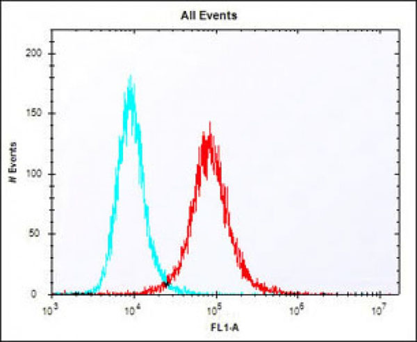
(Overlay histogram showing NIH/3T3 cells stained at 37 degree C. The secondary antibody used was Alexa Fluor 488 goat anti-rabbit lgG (H+L) (1583138) at 1/400 dilution for 40 min at 37 degree C. Isotype control antibody (blue line) was rabbit IgG1 (1mug/1x10^6 cells) used under the same conditions. Acquisition of >10, 000 events was performed.)
Flow Cytometry (FC/FACS)
(Overlay histogram showing NIH/3T3 cells stained at 37 degree C. The secondary antibody used was Alexa Fluor 488 goat anti-rabbit lgG (H+L) (1583138) at 1/400 dilution for 40 min at 37 degree C. Isotype control antibody (blue line) was rabbit IgG1 (1mug/1x10^6 cells) used under the same conditions. Acquisition of >10, 000 events was performed.)
Klf4, Polyclonal Antibody (Cat# AAA9232000)
Full Name
Mouse Klf4 Antibody (Center)
Gene Names
Klf4; EZF; Zie; Gklf
Reactivity
Mouse
Applications
WB, FC/FACS, EIA
Pricing
$425/0.1 mL | $1,615/5x0.1 mL


 Western Blot (WB)
(All lanes : CD5 Mouse mAb at 1/2000 dilution Lane 1 : Jurkat cells membrane fraction Lane 2 : THP1 cells membrane fraction Lysates/proteins at 20 ug per lane. Secondary Goat Anti-Mouse IgG-HRP, 5% milk conjugated at 1/10000 dilution Predicted band size : 55 KDa Observed band size : 55 KDa Blocking/Dilution buffer : 1x TBST.)
Western Blot (WB)
(All lanes : CD5 Mouse mAb at 1/2000 dilution Lane 1 : Jurkat cells membrane fraction Lane 2 : THP1 cells membrane fraction Lysates/proteins at 20 ug per lane. Secondary Goat Anti-Mouse IgG-HRP, 5% milk conjugated at 1/10000 dilution Predicted band size : 55 KDa Observed band size : 55 KDa Blocking/Dilution buffer : 1x TBST.)
CD5, Monoclonal Antibody (Cat# AAA435110)
Full Name
CD5 Mouse mAb
Gene Names
CD5; T1; LEU1
Reactivity
Human
Applications
WB, IP, FC/FACS
Pricing
$260/0.02 mL | $405/0.05 mL | $650/0.1 mL | $2,645/5x0.1 mL


 Western Blot (WB)
TIM50, Monoclonal Antibody (Cat# AAA435157)
Full Name
TIM50 (680) Mouse mAb
Gene Names
TIMM50; MGCA9; TIM50; TIM50L
Reactivity
Human
Applications
WB, IP, FC/FACS
Pricing
$260/0.02 mL | $405/0.05 mL | $650/0.1 mL | $2,645/5x0.1 mL


 Quality Control
(Western blot analysis of immunized recombinant protein, using anti-POLR2G monoclonal antibody. )
Quality Control
(Western blot analysis of immunized recombinant protein, using anti-POLR2G monoclonal antibody. )
POLR2G, Monoclonal Antibody (Cat# AAA120241)
Full Name
Mouse monoclonal antibody Anti-Human POLR2G
Gene Names
POLR2G; RPB7; RPB19; hRPB19; hsRPB7
Reactivity
Human
Applications
DB, WB, FC/FACS
Pricing
$460/0.1 mg | $2,070/5x0.1 mg

 Flow Cytometry (FC/FACS)
(Flow cytometric analysis of Raji cells using CD74(green) compared to an isotype control of mouse IgG2b(blue). MBS9200181 was diluted at 1:25 dilution. An Alexa Fluor 488 goat anti-mouse lgG at 1:400 dilution was used as the secondary antibody.)
Flow Cytometry (FC/FACS)
(Flow cytometric analysis of Raji cells using CD74(green) compared to an isotype control of mouse IgG2b(blue). MBS9200181 was diluted at 1:25 dilution. An Alexa Fluor 488 goat anti-mouse lgG at 1:400 dilution was used as the secondary antibody.)
CD74, Monoclonal Antibody (Cat# AAA9200181)
Full Name
CD74 Antibody
Gene Names
CD74; II; DHLAG; HLADG; Ia-GAMMA
Reactivity
Human
Applications
WB, EIA, FC/FACS
Purity
Purified Mouse Monoclonal Antibody (Mab)
Pricing
$425/0.4 mL | $190/0.08 mL | $1,665/5x0.4 mL

 SDS-Page
(SDS-PAGE Analysis Purified CD71 Mouse Monoclonal Antibody (TFRC/1396). Confirmation of Integrity and Purity of Antibody.)
SDS-Page
(SDS-PAGE Analysis Purified CD71 Mouse Monoclonal Antibody (TFRC/1396). Confirmation of Integrity and Purity of Antibody.)
CD71/Transferrin Receptor (TFRC), Monoclonal Antibody (Cat# AAA4381286)
Full Name
CD71/Transferrin Receptor (TFRC)
Gene Names
TFRC; T9; TR; TFR; p90; CD71; TFR1; TRFR; IMD46
Reactivity
Human
Applications
FC/FACS, IF, IHC
Purity
Purified Ab with BSA and Azide at 200ug/ml OR Purified Ab WITHOUT BSA and Azide at 1.0mg/ml
Pricing
$390/0.1 mg (With BSA & Azide at 0.2mg/mL) | $390/0.1 mg (Without BSA & Azide at 1mg/mL) | $215/0.02 mg (With BSA & Azide at 0.2mg/mL) | $1,710/5x0.1 mg (With BSA & Azide at 0.2mg/mL) | $1,710/5x0.1 mg (Without BSA & Azide at 1mg/mL)


 Western Blot (WB)
(All lanes : Anti-EEF1E1 Antibody at1:2000 dilutionLane 1: HepG2 whole cell lysateLane 2: A431 whole cell lysateLane 3: A549 whole cell lysateLane 4: Jurkat whole cell lysateLysates/proteins at 20 ug per lane. SecondaryGoat Anti-mouse IgG, (H+L), Peroxidase conjugated at 1/10000 dilution. Predicted band size : 20 kDaBlocking/Dilution buffer: 5% NFDM/TBST.)
Western Blot (WB)
(All lanes : Anti-EEF1E1 Antibody at1:2000 dilutionLane 1: HepG2 whole cell lysateLane 2: A431 whole cell lysateLane 3: A549 whole cell lysateLane 4: Jurkat whole cell lysateLysates/proteins at 20 ug per lane. SecondaryGoat Anti-mouse IgG, (H+L), Peroxidase conjugated at 1/10000 dilution. Predicted band size : 20 kDaBlocking/Dilution buffer: 5% NFDM/TBST.)
EEF1E1, Monoclonal Antibody (Cat# AAA9232191)
Full Name
EEF1E1 Antibody
Gene Names
EEF1E1; P18; AIMP3
Reactivity
Human
Applications
WB, IF, FC/FACS, EIA
Pricing
$435/0.1 mL | $1,655/5x0.1 mL


 Immunohistochemistry (IHC)-Paraffin
(Staining Epcam in Mouse colon tissue sections by Immunohistochemistry (IHC-P - paraformaldehyde-fixed, paraffin-embedded sections). Tissue was fixed with formaldehyde and blocked with 3% BSA for 0. 5 hour at room temperature; antigen retrieval was by heat mediation with a citrate buffer (pH6). Samples were incubated with primary antibody (1/25) for 1 hours at 37 degree C. A undiluted biotinylated goat polyvalent antibody was used as the secondary antibody.)
Immunohistochemistry (IHC)-Paraffin
(Staining Epcam in Mouse colon tissue sections by Immunohistochemistry (IHC-P - paraformaldehyde-fixed, paraffin-embedded sections). Tissue was fixed with formaldehyde and blocked with 3% BSA for 0. 5 hour at room temperature; antigen retrieval was by heat mediation with a citrate buffer (pH6). Samples were incubated with primary antibody (1/25) for 1 hours at 37 degree C. A undiluted biotinylated goat polyvalent antibody was used as the secondary antibody.)
Epcam, Polyclonal Antibody (Cat# AAA9232060)
Full Name
(Mouse) Epcam Antibody (C-term)
Gene Names
Epcam; EGP; Ly74; gp40; CD326; EGP-2; TROP1; Egp314; Ep-CAM; EpCAM1; Tacsd1; GA733-2; Tacstd1
Reactivity
Mouse
Applications
WB, IHC, FC/FACS, EIA
Pricing
$425/0.1 mL | $1,615/5x0.1 mL


 Western Blot (WB)
(Western blot analysis of CD27 expression in HELA whole cell lysates (lane 1). CD27 at 51KD was detected using rabbit anti- CD27 Antigen Affinity purified polyclonal antibody at 0.5 ug/mL. The blot was developed using chemiluminescence (ECL) method. )
Western Blot (WB)
(Western blot analysis of CD27 expression in HELA whole cell lysates (lane 1). CD27 at 51KD was detected using rabbit anti- CD27 Antigen Affinity purified polyclonal antibody at 0.5 ug/mL. The blot was developed using chemiluminescence (ECL) method. )
CD27, Polyclonal Antibody (Cat# AAA178431)
Full Name
Anti-CD27 Antibody
Gene Names
CD27; T14; S152; Tp55; TNFRSF7; S152. LPFS2
Reactivity
Human
No cross reactivity with other proteins.
No cross reactivity with other proteins.
Applications
WB, IHC
Purity
Immunogen affinity purified.
Pricing
$345/0.1 mg | $1,485/5x0.1 mg


 Immunocytochemistry/Immunofluorescence (ICC/IF)
(Immunocytochemistry/Immunofluorescence analysis using Mouse Anti-Methylglyoxal Monoclonal Antibody, Clone 9F11. Tissue: Embryonic kidney epithelial cell line (HEK293). Species: Human. Fixation: 5% Formaldehyde for 5 min. Primary Antibody: Mouse Anti-Methylglyoxal Monoclonal Antibody at 1:50 for 30-60 min at RT. Secondary Antibody: Goat Anti-Mouse Alexa Fluor 488 at 1:1500 for 30-60 min at RT. Counterstain: Phalloidin Alexa Fluor 633 F-Actin stain; DAPI (blue) nuclear stain at 1:250, 1:50000 for 30-60 min at RT. Magnification: 20X (2X Zoom). (A,C,E,G) - Untreated. (B,D,F,H) - Cells cultured overnight with 50 uM H2O2. (A,B) DAPI (blue) nuclear stain. (C,D) Phalloidin Alex Fluor 633 F-Actin stain. (E,F) Methylglyoxal Antibody. (G,H) Composite. Courtesy of: Dr. Robert Burke, University of Victoria.)
Immunocytochemistry/Immunofluorescence (ICC/IF)
(Immunocytochemistry/Immunofluorescence analysis using Mouse Anti-Methylglyoxal Monoclonal Antibody, Clone 9F11. Tissue: Embryonic kidney epithelial cell line (HEK293). Species: Human. Fixation: 5% Formaldehyde for 5 min. Primary Antibody: Mouse Anti-Methylglyoxal Monoclonal Antibody at 1:50 for 30-60 min at RT. Secondary Antibody: Goat Anti-Mouse Alexa Fluor 488 at 1:1500 for 30-60 min at RT. Counterstain: Phalloidin Alexa Fluor 633 F-Actin stain; DAPI (blue) nuclear stain at 1:250, 1:50000 for 30-60 min at RT. Magnification: 20X (2X Zoom). (A,C,E,G) - Untreated. (B,D,F,H) - Cells cultured overnight with 50 uM H2O2. (A,B) DAPI (blue) nuclear stain. (C,D) Phalloidin Alex Fluor 633 F-Actin stain. (E,F) Methylglyoxal Antibody. (G,H) Composite. Courtesy of: Dr. Robert Burke, University of Victoria.)
Methylglyoxal, Monoclonal Antibody (Cat# AAA808810)
Full Name
Methylglyoxal Antibody, Clone 9F11: ATTO 488
Reactivity
Species Independent
Applications
WB, ICC, IF, FC/FACS, EIA
Purity
Protein G Purified
Pricing
$560/0.1 mg | $2,275/5x0.1 mg


 Immunocytochemistry/Immunofluorescence (ICC/IF)
(Immunocytochemistry/Immunofluorescence analysis using Mouse Anti-Acrolein Monoclonal Antibody, Clone 2H2. Tissue: Embryonic kidney epithelial cell line (HEK293). Species: Human. Fixation: 5% Formaldehyde for 5 min. Primary Antibody: Mouse Anti-Acrolein Monoclonal Antibody at 1:50 for 30-60 min at RT. Secondary Antibody: Goat Anti-Mouse Alexa Fluor 488 at 1:1500 for 30-60 min at RT. Counterstain: Phalloidin Alexa Fluor 633 F-Actin stain; DAPI (blue) nuclear stain at 1:250, 1:50000 for 30-60 min at RT. Magnification: 20X (2X Zoom). (A,C,E,G) - Untreated. (B,D,F,H) - Cells cultured overnight with 50 uM H2O2. (A,B) DAPI (blue) nuclear stain. (C,D) Phalloidin Alex Fluor 633 F-Actin stain. (E,F) Acrolein Antibody. (G,H) Composite. Courtesy of: Dr. Robert Burke, University of Victoria.)
Immunocytochemistry/Immunofluorescence (ICC/IF)
(Immunocytochemistry/Immunofluorescence analysis using Mouse Anti-Acrolein Monoclonal Antibody, Clone 2H2. Tissue: Embryonic kidney epithelial cell line (HEK293). Species: Human. Fixation: 5% Formaldehyde for 5 min. Primary Antibody: Mouse Anti-Acrolein Monoclonal Antibody at 1:50 for 30-60 min at RT. Secondary Antibody: Goat Anti-Mouse Alexa Fluor 488 at 1:1500 for 30-60 min at RT. Counterstain: Phalloidin Alexa Fluor 633 F-Actin stain; DAPI (blue) nuclear stain at 1:250, 1:50000 for 30-60 min at RT. Magnification: 20X (2X Zoom). (A,C,E,G) - Untreated. (B,D,F,H) - Cells cultured overnight with 50 uM H2O2. (A,B) DAPI (blue) nuclear stain. (C,D) Phalloidin Alex Fluor 633 F-Actin stain. (E,F) Acrolein Antibody. (G,H) Composite. Courtesy of: Dr. Robert Burke, University of Victoria.)
Acrolein, Monoclonal Antibody (Cat# AAA808583)
Full Name
Acrolein Antibody, Clone 2H2: Alkaline Phosphatase
Reactivity
Species Independent
Applications
WB, ICC, IF, FC/FACS, EIA
Purity
Protein G Purified
Pricing
$550/0.1 mg | $2,245/5x0.1 mg


 Immunocytochemistry/Immunofluorescence (ICC/IF)
(Immunocytochemistry/Immunofluorescence analysis using Mouse Anti-Hexanoyl-Lysine adduct Monoclonal Antibody, Clone 5D9. Tissue: Embryonic kidney epithelial cell line (HEK293). Species: Human. Fixation: 5% Formaldehyde for 5 min. Primary Antibody: Mouse Anti-Hexanoyl-Lysine adduct Monoclonal Antibody at 1:50 for 30-60 min at RT. Secondary Antibody: Goat Anti-Mouse Alexa Fluor 488 at 1:1500 for 30-60 min at RT. Counterstain: Phalloidin Alexa Fluor 633 F-Actin stain; DAPI (blue) nuclear stain at 1:250, 1:50000 for 30-60 min at RT. Magnification: 20X (2X Zoom). (A,C,E,G) - Untreated. (B,D,F,H) - Cells cultured overnight with 50 uM H2O2. (A,B) DAPI (blue) nuclear stain. (C,D) Phalloidin Alex Fluor 633 F-Actin stain. (E,F) Hexanoyl-Lysine adduct Antibody. (G,H) Composite. Courtesy of: Dr. Robert Burke, University of Victoria.)
Immunocytochemistry/Immunofluorescence (ICC/IF)
(Immunocytochemistry/Immunofluorescence analysis using Mouse Anti-Hexanoyl-Lysine adduct Monoclonal Antibody, Clone 5D9. Tissue: Embryonic kidney epithelial cell line (HEK293). Species: Human. Fixation: 5% Formaldehyde for 5 min. Primary Antibody: Mouse Anti-Hexanoyl-Lysine adduct Monoclonal Antibody at 1:50 for 30-60 min at RT. Secondary Antibody: Goat Anti-Mouse Alexa Fluor 488 at 1:1500 for 30-60 min at RT. Counterstain: Phalloidin Alexa Fluor 633 F-Actin stain; DAPI (blue) nuclear stain at 1:250, 1:50000 for 30-60 min at RT. Magnification: 20X (2X Zoom). (A,C,E,G) - Untreated. (B,D,F,H) - Cells cultured overnight with 50 uM H2O2. (A,B) DAPI (blue) nuclear stain. (C,D) Phalloidin Alex Fluor 633 F-Actin stain. (E,F) Hexanoyl-Lysine adduct Antibody. (G,H) Composite. Courtesy of: Dr. Robert Burke, University of Victoria.)
Hexanoyl-Lysine adduct, Monoclonal Antibody (Cat# AAA808661)
Full Name
Hexanoyl-Lysine adduct Antibody, Clone 5D9: PerCP
Reactivity
Species Independent
Applications
WB, ICC, IF, FC/FACS, EIA
Purity
Protein G Purified
Pricing
$560/0.1 mg | $2,270/5x0.1 mg


 Immunocytochemistry/Immunofluorescence (ICC/IF)
(Immunocytochemistry/Immunofluorescence analysis using Mouse Anti-Dityrosine Monoclonal Antibody, Clone 10A6. Tissue: Embryonic kidney epithelial cell line (HEK293). Species: Human. Fixation: 5% Formaldehyde for 5 min. Primary Antibody: Mouse Anti-Dityrosine Monoclonal Antibody at 1:50 for 30-60 min at RT. Secondary Antibody: Goat Anti-Mouse Alexa Fluor 488 at 1:1500 for 30-60 min at RT. Counterstain: Phalloidin Alexa Fluor 633 F-Actin stain; DAPI (blue) nuclear stain at 1:250, 1:50000 for 30-60 min at RT. Localization: Cytoplasmic. Magnification: 20X (2X Zoom). (A,C,E,G) - Untreated. (B,D,F,H) - Cells cultured overnight with 50 uM H2O2. (A,B) DAPI (blue) nuclear stain. (C,D) Phalloidin Alex Fluor 633 F-Actin stain. (E,F) Dityrosine Antibody. (G,H) Composite. Courtesy of: Dr. Robert Burke, University of Victoria.)
Immunocytochemistry/Immunofluorescence (ICC/IF)
(Immunocytochemistry/Immunofluorescence analysis using Mouse Anti-Dityrosine Monoclonal Antibody, Clone 10A6. Tissue: Embryonic kidney epithelial cell line (HEK293). Species: Human. Fixation: 5% Formaldehyde for 5 min. Primary Antibody: Mouse Anti-Dityrosine Monoclonal Antibody at 1:50 for 30-60 min at RT. Secondary Antibody: Goat Anti-Mouse Alexa Fluor 488 at 1:1500 for 30-60 min at RT. Counterstain: Phalloidin Alexa Fluor 633 F-Actin stain; DAPI (blue) nuclear stain at 1:250, 1:50000 for 30-60 min at RT. Localization: Cytoplasmic. Magnification: 20X (2X Zoom). (A,C,E,G) - Untreated. (B,D,F,H) - Cells cultured overnight with 50 uM H2O2. (A,B) DAPI (blue) nuclear stain. (C,D) Phalloidin Alex Fluor 633 F-Actin stain. (E,F) Dityrosine Antibody. (G,H) Composite. Courtesy of: Dr. Robert Burke, University of Victoria.)
Dityrosine, Monoclonal Antibody (Cat# AAA808884)
Full Name
Dityrosine Antibody, Clone 10A6: ATTO 594
Reactivity
Species Independent
Applications
WB, ICC, IF, FC/FACS, EIA
Purity
Protein G Purified
Pricing
$560/0.1 mg | $2,275/5x0.1 mg


 Western Blot (WB)
(Figure 1. Western blot analysis of APRT using anti-APRT antibody (MBS178668). Electrophoresis was performed on a 5-20% SDS-PAGE gel at 70V (Stacking gel) / 90V (Resolving gel) for 2-3 hours. The sample well of each lane was loaded with 50ug of sample under reducing conditions. lane 1: K562 whole cell lysates. After Electrophoresis, proteins were transferred to a Nitrocellulose membrane at 150mA for 50-90 minutes. Blocked the membrane with 5% Non-fat Milk/ TBS for 1.5 hour at RT. The membrane was incubated with rabbit anti-APRT antigen affinity purified polyclonal antibody at 0.5ug/mL overnight at 4 degree C, then washed with TBS-0.1%Tween 3 times with 5 minutes each and probed with a goat anti-rabbit IgG-HRP secondary antibody at a dilution of 1:10000 for 1.5 hour at RT. The signal is developed using an Enhanced Chemiluminescent detection (ECL) kit with Tanon 5200 system. A specific band was detected for APRT at approximately 20KD. The expected band size for APRT is at 20KD. )
Western Blot (WB)
(Figure 1. Western blot analysis of APRT using anti-APRT antibody (MBS178668). Electrophoresis was performed on a 5-20% SDS-PAGE gel at 70V (Stacking gel) / 90V (Resolving gel) for 2-3 hours. The sample well of each lane was loaded with 50ug of sample under reducing conditions. lane 1: K562 whole cell lysates. After Electrophoresis, proteins were transferred to a Nitrocellulose membrane at 150mA for 50-90 minutes. Blocked the membrane with 5% Non-fat Milk/ TBS for 1.5 hour at RT. The membrane was incubated with rabbit anti-APRT antigen affinity purified polyclonal antibody at 0.5ug/mL overnight at 4 degree C, then washed with TBS-0.1%Tween 3 times with 5 minutes each and probed with a goat anti-rabbit IgG-HRP secondary antibody at a dilution of 1:10000 for 1.5 hour at RT. The signal is developed using an Enhanced Chemiluminescent detection (ECL) kit with Tanon 5200 system. A specific band was detected for APRT at approximately 20KD. The expected band size for APRT is at 20KD. )
APRT, Polyclonal Antibody (Cat# AAA178668)
Full Name
Anti-APRT Antibody
Gene Names
APRT; AMP; APRTD
Reactivity
Human
Applications
WB
Purity
Immunogen affinity purified.
Pricing
$345/0.1 mg | $1,485/5x0.1 mg