Filters
Clonality
Type
Reactivity
Gene Name
Isotype
Host
Application
Clone
1194 results for " E" - showing 1050-1100


 Application Data
(Publised customer image:Mouse anti Human CD163 antibody, clone EDHu-1 used for the identification of perivascular macrophages in human brain by immunofluorescence.Image caption:Images demonstrating immunohistological stainings of amylin and double immunofluorescence staining against NG2/amylin, laminin/amylin and CD163/amylin in the hippocampus of the patient with AD and T2D.Amylin cell inclusions are indicated with arrows in (a) and shown in a higher magnification in (b). Pericytes with round cell bodies and NG2-positive coverage of the microvessel surface (green in c), without amylin cell inclusions (red in d), displayed round DAPI-positive cell nuclei (blue in e). The images in (c), (d) and (e) are merged in (f). Cells with more diffuse and weak NG2 staining (indicated by the arrowhead, green in g) and cytosolic amylin cell inclusions (red in h) showed altered cell nuclei (indicated with arrow, blue in i).The adjacent unaffected NG2-positive cell is indicated with an arrowhead in (i). The images in (g), (h) and (i) are merged in (j). Cells enclosed by laminin (green in K) contained amylin grains (red in l) and fragmented DAPI-positive cell nuclei (indicated with an arrow blue in m). The images in (k), (l) and (m) are merged in (n).Loss of NG2 coverage (green in o) was associated with polarized amylin cell inclusion (red in p) and fragmented DAPI-positive cell nuclei (indicated with an arrow, blue in q). The images in (o), (p) and (q) are merged in (r). Staining against macrophage marker CD163 (green in s) did not co-localize with amylin cell inclusions (red in t). The cell nucleus was stained with DAPI (blue in U). The images in (s), (t) and (u) are merged in (v). Scale bars: (a) 50 mum, (b), (c) to (v) 5 mum.From: Schultz, N. et al. (2016).Amylin alters human brain pericyte viability and NG2 expression.J Cereb Blood Flow & Metab. Jun 28 [Epub ahead of print]This is from an open access article distributed under the terms of the Creative Commons Attribution License.)
Application Data
(Publised customer image:Mouse anti Human CD163 antibody, clone EDHu-1 used for the identification of perivascular macrophages in human brain by immunofluorescence.Image caption:Images demonstrating immunohistological stainings of amylin and double immunofluorescence staining against NG2/amylin, laminin/amylin and CD163/amylin in the hippocampus of the patient with AD and T2D.Amylin cell inclusions are indicated with arrows in (a) and shown in a higher magnification in (b). Pericytes with round cell bodies and NG2-positive coverage of the microvessel surface (green in c), without amylin cell inclusions (red in d), displayed round DAPI-positive cell nuclei (blue in e). The images in (c), (d) and (e) are merged in (f). Cells with more diffuse and weak NG2 staining (indicated by the arrowhead, green in g) and cytosolic amylin cell inclusions (red in h) showed altered cell nuclei (indicated with arrow, blue in i).The adjacent unaffected NG2-positive cell is indicated with an arrowhead in (i). The images in (g), (h) and (i) are merged in (j). Cells enclosed by laminin (green in K) contained amylin grains (red in l) and fragmented DAPI-positive cell nuclei (indicated with an arrow blue in m). The images in (k), (l) and (m) are merged in (n).Loss of NG2 coverage (green in o) was associated with polarized amylin cell inclusion (red in p) and fragmented DAPI-positive cell nuclei (indicated with an arrow, blue in q). The images in (o), (p) and (q) are merged in (r). Staining against macrophage marker CD163 (green in s) did not co-localize with amylin cell inclusions (red in t). The cell nucleus was stained with DAPI (blue in U). The images in (s), (t) and (u) are merged in (v). Scale bars: (a) 50 mum, (b), (c) to (v) 5 mum.From: Schultz, N. et al. (2016).Amylin alters human brain pericyte viability and NG2 expression.J Cereb Blood Flow & Metab. Jun 28 [Epub ahead of print]This is from an open access article distributed under the terms of the Creative Commons Attribution License.)
CD163, Monoclonal Antibody (Cat# AAA12256)
Full Name
Mouse Anti Human CD163: RPE
Gene Names
CD163; M130; MM130; SCARI1
Reactivity
Human
Applications
FC/FACS


 FCM (Flow Cytometry)
(Figure 11. Flow Cytometry analysis of U937 cells using anti-Coronin 1a/TACO/CORO1A antibody (AAA19291).Overlay histogram showing U937 cells stained with AAA19291 (Blue line). The cells were blocked with 10% normal goat serum. And then incubated with rabbit anti-Coronin 1a/TACO/CORO1A Antibody (AAA19291, 1μg/1x106 cells) for 30 min at 20 degree C. DyLight®488 conjugated goat anti-rabbit IgG (5-10μg/1x106 cells) was used as secondary antibody for 30 minutes at 20 degree C. Isotype control antibody (Green line) was rabbit IgG (1μg/1x106) used under the same conditions. Unlabelled sample (Red line) was also used as a control.)
FCM (Flow Cytometry)
(Figure 11. Flow Cytometry analysis of U937 cells using anti-Coronin 1a/TACO/CORO1A antibody (AAA19291).Overlay histogram showing U937 cells stained with AAA19291 (Blue line). The cells were blocked with 10% normal goat serum. And then incubated with rabbit anti-Coronin 1a/TACO/CORO1A Antibody (AAA19291, 1μg/1x106 cells) for 30 min at 20 degree C. DyLight®488 conjugated goat anti-rabbit IgG (5-10μg/1x106 cells) was used as secondary antibody for 30 minutes at 20 degree C. Isotype control antibody (Green line) was rabbit IgG (1μg/1x106) used under the same conditions. Unlabelled sample (Red line) was also used as a control.)
Coronin 1a/TACO/CORO1A, Polyclonal Antibody (Cat# AAA19291)
Full Name
Anti-Coronin 1a/TACO/CORO1A Antibody
Gene Names
CORO1A; p57; TACO; CLABP; HCORO1; CLIPINA
Reactivity
Human, Mouse, Rat
Applications
WB, IHC-P, FC/FACS/FCM, EIA
Purity
Immunogen affinity purified.


 IF (Immunofluorescence)
(Figure 6. IF analysis of SCG10/STMN2 using anti- SCG10/STMN2 antibody (AAA19299).SCG10/STMN2 was detected in immunocytochemical section of U20S cells. Enzyme antigen retrieval was performed using IHC enzyme antigen retrieval reagent for 15 mins. The cells were blocked with 10% goat serum. And then incubated with 5μg/mL rabbit anti- SCG10/STMN2 Antibody (AAA19299) overnight at 4 degree C. DyLight®488 Conjugated Goat Anti-Rabbit IgG was used as secondary antibody at 1:100 dilution and incubated for 30 minutes at 37 degree C. The section was counterstained with DAPI. Visualize using a fluorescence microscope and filter sets appropriate for the label used.)
IF (Immunofluorescence)
(Figure 6. IF analysis of SCG10/STMN2 using anti- SCG10/STMN2 antibody (AAA19299).SCG10/STMN2 was detected in immunocytochemical section of U20S cells. Enzyme antigen retrieval was performed using IHC enzyme antigen retrieval reagent for 15 mins. The cells were blocked with 10% goat serum. And then incubated with 5μg/mL rabbit anti- SCG10/STMN2 Antibody (AAA19299) overnight at 4 degree C. DyLight®488 Conjugated Goat Anti-Rabbit IgG was used as secondary antibody at 1:100 dilution and incubated for 30 minutes at 37 degree C. The section was counterstained with DAPI. Visualize using a fluorescence microscope and filter sets appropriate for the label used.)
SCG10/STMN2, Polyclonal Antibody (Cat# AAA19299)
Full Name
Anti-SCG10/STMN2 Antibody
Gene Names
STMN2; SCG10; SCGN10
Reactivity
Human, Mouse, Rat
Applications
WB, IHC-P, ICC, IF, FC/FACS/FCM, EIA
Purity
Immunogen affinity purified.


 FCM (Flow Cytometry)
(Figure 7. Flow Cytometry analysis of CACO-2 cells using anti-Glycine decarboxylase/GLDC antibody (AAA19300).Overlay histogram showing CACO-2 cells stained with AAA19300 (Blue line). The cells were blocked with 10% normal goat serum. And then incubated with rabbit anti-Glycine decarboxylase/GLDC Antibody (AAA19300, 1μg/1x106 cells) for 30 min at 20 degree C. DyLight®488 conjugated goat anti-rabbit IgG (5-10μg/1x106 cells) was used as secondary antibody for 30 minutes at 20 degree C. Isotype control antibody (Green line) was rabbit IgG (1μg/1x106) used under the same conditions. Unlabelled sample (Red line) was also used as a control.)
FCM (Flow Cytometry)
(Figure 7. Flow Cytometry analysis of CACO-2 cells using anti-Glycine decarboxylase/GLDC antibody (AAA19300).Overlay histogram showing CACO-2 cells stained with AAA19300 (Blue line). The cells were blocked with 10% normal goat serum. And then incubated with rabbit anti-Glycine decarboxylase/GLDC Antibody (AAA19300, 1μg/1x106 cells) for 30 min at 20 degree C. DyLight®488 conjugated goat anti-rabbit IgG (5-10μg/1x106 cells) was used as secondary antibody for 30 minutes at 20 degree C. Isotype control antibody (Green line) was rabbit IgG (1μg/1x106) used under the same conditions. Unlabelled sample (Red line) was also used as a control.)
Glycine decarboxylase/GLDC, Polyclonal Antibody (Cat# AAA19300)
Full Name
Anti-Glycine decarboxylase/GLDC Antibody
Gene Names
GLDC; GCE; GCSP; HYGN1
Reactivity
Human, Mouse, Rat
Applications
WB, IHC-P, FC/FACS/FCM, EIA
Purity
Immunogen affinity purified.


 FCM (Flow Cytometry)
(Figure 7. Flow Cytometry analysis of U20S cells using anti-VDAC3 antibody (AAA19301).Overlay histogram showing U20S cells stained with AAA19301 (Blue line). The cells were blocked with 10% normal goat serum. And then incubated with rabbit anti-VDAC3 Antibody (AAA19301, 1μg/1x106 cells) for 30 min at 20 degree C. DyLight®488 conjugated goat anti-rabbit IgG (5-10μg/1x106 cells) was used as secondary antibody for 30 minutes at 20 degree C. Isotype control antibody (Green line) was rabbit IgG (1μg/1x106) used under the same conditions. Unlabelled sample (Red line) was also used as a control.)
FCM (Flow Cytometry)
(Figure 7. Flow Cytometry analysis of U20S cells using anti-VDAC3 antibody (AAA19301).Overlay histogram showing U20S cells stained with AAA19301 (Blue line). The cells were blocked with 10% normal goat serum. And then incubated with rabbit anti-VDAC3 Antibody (AAA19301, 1μg/1x106 cells) for 30 min at 20 degree C. DyLight®488 conjugated goat anti-rabbit IgG (5-10μg/1x106 cells) was used as secondary antibody for 30 minutes at 20 degree C. Isotype control antibody (Green line) was rabbit IgG (1μg/1x106) used under the same conditions. Unlabelled sample (Red line) was also used as a control.)
VDAC3, Polyclonal Antibody (Cat# AAA19301)
Full Name
Anti-VDAC3 Antibody
Gene Names
VDAC3; VDAC-3; HD-VDAC3
Reactivity
Human, Mouse, Rat
Applications
WB, IHC-P, FC/FACS/FCM, EIA
Purity
Immunogen affinity purified.


 FCM (Flow Cytometry)
(Figure 9. Flow Cytometry analysis of 293T cells using anti-CDC45L antibody (AAA19360).Overlay histogram showing 293T cells stained with AAA19360 (Blue line). The cells were blocked with 10% normal goat serum. And then incubated with mouse anti- CDC45L Antibody (AAA19360, 1μg/1x106 cells) for 30 min at 20 degree C. DyLight®488 conjugated goat anti-mouse IgG (BA1126, 5-10μg/1x106 cells) was used as secondary antibody for 30 minutes at 20 degree C. Isotype control antibody (Green line) was mouse IgG (1μg/1x106) used under the same conditions. Unlabelled sample (Red line) was also used as a control.)
FCM (Flow Cytometry)
(Figure 9. Flow Cytometry analysis of 293T cells using anti-CDC45L antibody (AAA19360).Overlay histogram showing 293T cells stained with AAA19360 (Blue line). The cells were blocked with 10% normal goat serum. And then incubated with mouse anti- CDC45L Antibody (AAA19360, 1μg/1x106 cells) for 30 min at 20 degree C. DyLight®488 conjugated goat anti-mouse IgG (BA1126, 5-10μg/1x106 cells) was used as secondary antibody for 30 minutes at 20 degree C. Isotype control antibody (Green line) was mouse IgG (1μg/1x106) used under the same conditions. Unlabelled sample (Red line) was also used as a control.)
CDC45L, Monoclonal Antibody (Cat# AAA19360)
Full Name
Anti-CDC45L Antibody (monoclonal, 6H6)
Gene Names
CDC45; CDC45L; CDC45L2; PORC-PI-1
Reactivity
Human, Mouse, Rat
Applications
WB, IHC-P, FC/FACS/FCM
Purity
Immunogen affinity purified.


 FCM (Flow Cytometry)
(Figure 8. Flow Cytometry analysis of SiHa cells using anti- ASS1 antibody (AAA19369).Overlay histogram showing SiHa cells stained with AAA19369 (Blue line). The cells were blocked with 10% normal goat serum. And then incubated with mouse anti-ASS1 Antibody (AAA19369, 1μg/1x106 cells) for 30 min at 20 degree C. DyLight®488 conjugated goat anti-mouse IgG (BA1126, 5-10μg/1x106 cells) was used as secondary antibody for 30 minutes at 20 degree C. Isotype control antibody (Green line) was mouse IgG (1μg/1x106) used under the same conditions. Unlabelled sample (Red line) was also used as a control.)
FCM (Flow Cytometry)
(Figure 8. Flow Cytometry analysis of SiHa cells using anti- ASS1 antibody (AAA19369).Overlay histogram showing SiHa cells stained with AAA19369 (Blue line). The cells were blocked with 10% normal goat serum. And then incubated with mouse anti-ASS1 Antibody (AAA19369, 1μg/1x106 cells) for 30 min at 20 degree C. DyLight®488 conjugated goat anti-mouse IgG (BA1126, 5-10μg/1x106 cells) was used as secondary antibody for 30 minutes at 20 degree C. Isotype control antibody (Green line) was mouse IgG (1μg/1x106) used under the same conditions. Unlabelled sample (Red line) was also used as a control.)
ASS1, Monoclonal Antibody (Cat# AAA19369)
Full Name
Anti-ASS1 Antibody (monoclonal, 5I5)
Gene Names
ASS1; ASS; CTLN1
Reactivity
Human, Mouse, Rat, Monkey
Applications
WB, IHC-P, ICC, IF, FC/FACS/FCM
Purity
Immunogen affinity purified.


 FCM (Flow Cytometry)
(Figure 8. Flow Cytometry analysis of SiHa cells using anti- ASS1 antibody (AAA19370).Overlay histogram showing SiHa cells stained with AAA19370 (Blue line). The cells were blocked with 10% normal goat serum. And then incubated with mouse anti-ASS1 Antibody (AAA19370, 1μg/1x106 cells) for 30 min at 20 degree C. DyLight®488 conjugated goat anti-mouse IgG (BA1126, 5-10μg/1x106 cells) was used as secondary antibody for 30 minutes at 20 degree C. Isotype control antibody (Green line) was mouse IgG (1μg/1x106) used under the same conditions. Unlabelled sample (Red line) was also used as a control.)
FCM (Flow Cytometry)
(Figure 8. Flow Cytometry analysis of SiHa cells using anti- ASS1 antibody (AAA19370).Overlay histogram showing SiHa cells stained with AAA19370 (Blue line). The cells were blocked with 10% normal goat serum. And then incubated with mouse anti-ASS1 Antibody (AAA19370, 1μg/1x106 cells) for 30 min at 20 degree C. DyLight®488 conjugated goat anti-mouse IgG (BA1126, 5-10μg/1x106 cells) was used as secondary antibody for 30 minutes at 20 degree C. Isotype control antibody (Green line) was mouse IgG (1μg/1x106) used under the same conditions. Unlabelled sample (Red line) was also used as a control.)
ASS1, Monoclonal Antibody (Cat# AAA19370)
Full Name
Anti-ASS1 Antibody (monoclonal, 7I9)
Gene Names
ASS1; ASS; CTLN1
Reactivity
Human, Mouse, Rat, Monkey
Applications
WB, IHC-P, ICC, IF, FC/FACS/FCM
Purity
Immunogen affinity purified.


 FCM (Flow Cytometry)
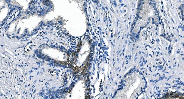
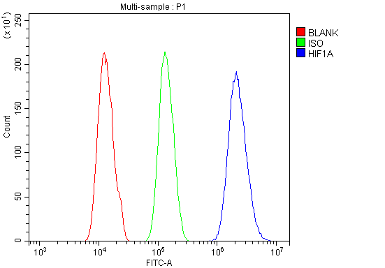
(Figure 6. Flow Cytometry analysis of A431 cells using anti-HIF-1 alpha/HIF1A antibody (AAA19213).Overlay histogram showing A431 cells stained with AAA19213 (Blue line). The cells were blocked with 10% normal goat serum. And then incubated with rabbit anti-HIF-1 alpha/HIF1A Antibody (AAA19213, 1μg/1x106 cells) for 30 min at 20 degree C. DyLight®488 conjugated goat anti-rabbit IgG (5-10μg/1x106 cells) was used as secondary antibody for 30 minutes at 20 degree C. Isotype control antibody (Green line) was rabbit IgG (1μg/1x106) used under the same conditions. Unlabelled sample (Red line) was also used as a control.)
FCM (Flow Cytometry)
(Figure 6. Flow Cytometry analysis of A431 cells using anti-HIF-1 alpha/HIF1A antibody (AAA19213).Overlay histogram showing A431 cells stained with AAA19213 (Blue line). The cells were blocked with 10% normal goat serum. And then incubated with rabbit anti-HIF-1 alpha/HIF1A Antibody (AAA19213, 1μg/1x106 cells) for 30 min at 20 degree C. DyLight®488 conjugated goat anti-rabbit IgG (5-10μg/1x106 cells) was used as secondary antibody for 30 minutes at 20 degree C. Isotype control antibody (Green line) was rabbit IgG (1μg/1x106) used under the same conditions. Unlabelled sample (Red line) was also used as a control.)
HIF-1 alpha/HIF1A, Polyclonal Antibody (Cat# AAA19213)
Full Name
Anti-HIF-1 alpha/HIF1A Antibody
Gene Names
HIF1A; HIF1; MOP1; PASD8; HIF-1A; bHLHe78; HIF-1alpha; HIF1-ALPHA
Reactivity
Human, Mouse, Rat
Applications
WB, IHC-P, FC/FACS/FCM, EIA
Purity
Immunogen affinity purified.


 FCM (Flow Cytometry)
(Figure 8. Flow Cytometry analysis of HepG2 cells using anti-MERTK antibody (AAA19219).Overlay histogram showing HepG2 cells stained with AAA19219 (Blue line). The cells were blocked with 10% normal goat serum. And then incubated with rabbit anti-MERTK Antibody (AAA19219, 1ug/1x106 cells) for 30 min at 20 degree C. DyLight488 conjugated goat anti-rabbit IgG (5-10ug/1x106 cells) was used as secondary antibody for 30 minutes at 20 degree C. Isotype control antibody (Green line) was rabbit IgG (1ug/1x106) used under the same conditions. Unlabelled sample (Red line) was also used as a control.)
FCM (Flow Cytometry)
(Figure 8. Flow Cytometry analysis of HepG2 cells using anti-MERTK antibody (AAA19219).Overlay histogram showing HepG2 cells stained with AAA19219 (Blue line). The cells were blocked with 10% normal goat serum. And then incubated with rabbit anti-MERTK Antibody (AAA19219, 1ug/1x106 cells) for 30 min at 20 degree C. DyLight488 conjugated goat anti-rabbit IgG (5-10ug/1x106 cells) was used as secondary antibody for 30 minutes at 20 degree C. Isotype control antibody (Green line) was rabbit IgG (1ug/1x106) used under the same conditions. Unlabelled sample (Red line) was also used as a control.)
MERTK, Polyclonal Antibody (Cat# AAA19219)
Full Name
Anti-MERTK Antibody
Gene Names
MERTK; MER; RP38; c-mer
Reactivity
Human, Mouse, Rat
Applications
WB, IHC-P, FC/FACS/FCM, EIA
Purity
Immunogen affinity purified.


 FCM (Flow Cytometry)
(Figure 7. Flow Cytometry analysis of U20S cells using anti- ADA antibody (AAA19184). Overlay histogram showing U20S cells stained with AAA19184 (Blue line).The cells were blocked with 10% normal goat serum. And then incubated with mouse anti- ADA Antibody (AAA19184, 1ug/1x106 cells) for 30 min at 20 degree C. DyLight488 conjugated goat anti-mouse IgG (BA1126, 5-10ug/1x106 cells) was used as secondary antibody for 30 minutes at 20 degree C. Isotype control antibody (Green line) was mouse IgG (1ug/1x106) used under the same conditions. Unlabelled sample (Red line) was also used as a control.)
FCM (Flow Cytometry)
(Figure 7. Flow Cytometry analysis of U20S cells using anti- ADA antibody (AAA19184). Overlay histogram showing U20S cells stained with AAA19184 (Blue line).The cells were blocked with 10% normal goat serum. And then incubated with mouse anti- ADA Antibody (AAA19184, 1ug/1x106 cells) for 30 min at 20 degree C. DyLight488 conjugated goat anti-mouse IgG (BA1126, 5-10ug/1x106 cells) was used as secondary antibody for 30 minutes at 20 degree C. Isotype control antibody (Green line) was mouse IgG (1ug/1x106) used under the same conditions. Unlabelled sample (Red line) was also used as a control.)
ADA, Monoclonal Antibody (Cat# AAA19184)
Full Name
Anti-ADA Antibody (monoclonal, 6D4)
Reactivity
Human
Applications
WB, IHC, FC/FACS
Purity
Immunogen Affinity Purified


 IF (Immunofluorescence)
(Figure 6. IF analysis of CD72 using anti- CD72 antibody (AAA19329).CD72 was detected in immunocytochemical section of HEPA1-6 cells. Enzyme antigen retrieval was performed using IHC enzyme antigen retrieval reagent for 15 mins. The cells were blocked with 10% goat serum. And then incubated with 5μg/mL rabbit anti- CD72 Antibody (AAA19329) overnight at 4 degree C. DyLight®488 Conjugated Goat Anti-Rabbit IgG was used as secondary antibody at 1:100 dilution and incubated for 30 minutes at 37 degree C. The section was counterstained with DAPI. Visualize using a fluorescence microscope and filter sets appropriate for the label used.)
IF (Immunofluorescence)
(Figure 6. IF analysis of CD72 using anti- CD72 antibody (AAA19329).CD72 was detected in immunocytochemical section of HEPA1-6 cells. Enzyme antigen retrieval was performed using IHC enzyme antigen retrieval reagent for 15 mins. The cells were blocked with 10% goat serum. And then incubated with 5μg/mL rabbit anti- CD72 Antibody (AAA19329) overnight at 4 degree C. DyLight®488 Conjugated Goat Anti-Rabbit IgG was used as secondary antibody at 1:100 dilution and incubated for 30 minutes at 37 degree C. The section was counterstained with DAPI. Visualize using a fluorescence microscope and filter sets appropriate for the label used.)
CD72, Polyclonal Antibody (Cat# AAA19329)
Full Name
Anti-CD72 Antibody
Gene Names
Cd72; CD72c; Ly-19; Ly-32; Lyb-2; Ly-m19
Reactivity
Mouse, Rat
Applications
WB, IHC-P, ICC, IF, FC/FACS/FCM, EIA
Purity
Immunogen affinity purified.


 IF (Immunofluorescence)
(Figure 7. IF analysis of METTL1 using anti- METTL1 antibody (AAA19336).METTL1 was detected in immunocytochemical section of A431 cells. Enzyme antigen retrieval was performed using IHC enzyme antigen retrieval reagent for 15 mins. The cells were blocked with 10% goat serum. And then incubated with 5μg/mL rabbit anti-METTL1 Antibody (AAA19336) overnight at 4 degree C. DyLight®488 Conjugated Goat Anti-Rabbit IgG was used as secondary antibody at 1:100 dilution and incubated for 30 minutes at 37 degree C. The section was counterstained with DAPI. Visualize using a fluorescence microscope and filter sets appropriate for the label used.)
IF (Immunofluorescence)
(Figure 7. IF analysis of METTL1 using anti- METTL1 antibody (AAA19336).METTL1 was detected in immunocytochemical section of A431 cells. Enzyme antigen retrieval was performed using IHC enzyme antigen retrieval reagent for 15 mins. The cells were blocked with 10% goat serum. And then incubated with 5μg/mL rabbit anti-METTL1 Antibody (AAA19336) overnight at 4 degree C. DyLight®488 Conjugated Goat Anti-Rabbit IgG was used as secondary antibody at 1:100 dilution and incubated for 30 minutes at 37 degree C. The section was counterstained with DAPI. Visualize using a fluorescence microscope and filter sets appropriate for the label used.)
METTL1, Polyclonal Antibody (Cat# AAA19336)
Full Name
Anti-METTL1 Antibody
Gene Names
METTL1; TRM8; TRMT8; C12orf1; YDL201w
Reactivity
Human, Rat
Applications
WB, IHC-P, ICC, IF, EIA
Purity
Immunogen affinity purified.


 FCM (Flow Cytometry)
(Figure 7. Flow Cytometry analysis of HEPA1-6 cells using anti-Rad9b antibody (AAA19340).Overlay histogram showing HEPA1-6 cells stained with AAA19340 (Blue line). The cells were blocked with 10% normal goat serum. And then incubated with rabbit anti-Rad9b Antibody (AAA19340, 1μg/1x106 cells) for 30 min at 20 degree C. DyLight®488 conjugated goat anti-rabbit IgG (5-10μg/1x106 cells) was used as secondary antibody for 30 minutes at 20 degree C. Isotype control antibody (Green line) was rabbit IgG (1μg/1x106) used under the same conditions. Unlabelled sample (Red line) was also used as a control.)
FCM (Flow Cytometry)
(Figure 7. Flow Cytometry analysis of HEPA1-6 cells using anti-Rad9b antibody (AAA19340).Overlay histogram showing HEPA1-6 cells stained with AAA19340 (Blue line). The cells were blocked with 10% normal goat serum. And then incubated with rabbit anti-Rad9b Antibody (AAA19340, 1μg/1x106 cells) for 30 min at 20 degree C. DyLight®488 conjugated goat anti-rabbit IgG (5-10μg/1x106 cells) was used as secondary antibody for 30 minutes at 20 degree C. Isotype control antibody (Green line) was rabbit IgG (1μg/1x106) used under the same conditions. Unlabelled sample (Red line) was also used as a control.)
Rad9b, Polyclonal Antibody (Cat# AAA19340)
Full Name
Anti-Rad9b Antibody
Gene Names
Rad9b; BC021784; A630082N15Rik
Reactivity
Mouse, Rat
Applications
WB, IHC-P, FC/FACS/FCM, EIA
Purity
Immunogen affinity purified.


 FCM (Flow Cytometry)
(Figure 6. Flow Cytometry analysis of U87 cells using anti-GRB10 antibody (AAA19244).Overlay histogram showing U87 cells stained with AAA19244 (Blue line). The cells were blocked with 10% normal goat serum. And then incubated with rabbit anti-GRB10 Antibody (AAA19244,1μg/1x106 cells) for 30 min at 20 degree C. DyLight®488 conjugated goat anti-rabbit IgG (5-10μg/1x106 cells) was used as secondary antibody for 30 minutes at 20 degree C. Isotype control antibody (Green line) was rabbit IgG (1μg/1x106) used under the same conditions. Unlabelled sample (Red line) was also used as a control.)
FCM (Flow Cytometry)
(Figure 6. Flow Cytometry analysis of U87 cells using anti-GRB10 antibody (AAA19244).Overlay histogram showing U87 cells stained with AAA19244 (Blue line). The cells were blocked with 10% normal goat serum. And then incubated with rabbit anti-GRB10 Antibody (AAA19244,1μg/1x106 cells) for 30 min at 20 degree C. DyLight®488 conjugated goat anti-rabbit IgG (5-10μg/1x106 cells) was used as secondary antibody for 30 minutes at 20 degree C. Isotype control antibody (Green line) was rabbit IgG (1μg/1x106) used under the same conditions. Unlabelled sample (Red line) was also used as a control.)
GRB10, Polyclonal Antibody (Cat# AAA19244)
Full Name
Anti-GRB10 Antibody
Gene Names
GRB10; RSS; IRBP; MEG1; GRB-IR; Grb-10
Reactivity
Human, Mouse, Rat
Applications
WB, IHC-P, FC/FACS/FCM, EIA
Purity
Immunogen affinity purified.


 FCM (Flow Cytometry)
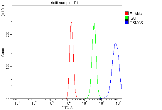
(Figure 7. Flow Cytometry analysis of 293T cells using anti-TBP-1/PSMC3 antibody (AAA19314).Overlay histogram showing 293T cells stained with AAA19314 (Blue line). The cells were blocked with 10% normal goat serum. And then incubated with rabbit anti-TBP-1/PSMC3 Antibody (AAA19314, 1μg/1x106 cells) for 30 min at 20 degree C. DyLight®488 conjugated goat anti-rabbit IgG (5-10μg/1x106 cells) was used as secondary antibody for 30 minutes at 20 degree C. Isotype control antibody (Green line) was rabbit IgG (1μg/1x106) used under the same conditions. Unlabelled sample (Red line) was also used as a control.)
FCM (Flow Cytometry)
(Figure 7. Flow Cytometry analysis of 293T cells using anti-TBP-1/PSMC3 antibody (AAA19314).Overlay histogram showing 293T cells stained with AAA19314 (Blue line). The cells were blocked with 10% normal goat serum. And then incubated with rabbit anti-TBP-1/PSMC3 Antibody (AAA19314, 1μg/1x106 cells) for 30 min at 20 degree C. DyLight®488 conjugated goat anti-rabbit IgG (5-10μg/1x106 cells) was used as secondary antibody for 30 minutes at 20 degree C. Isotype control antibody (Green line) was rabbit IgG (1μg/1x106) used under the same conditions. Unlabelled sample (Red line) was also used as a control.)
TBP-1/PSMC3, Polyclonal Antibody (Cat# AAA19314)
Full Name
Anti-TBP-1/PSMC3 Antibody
Gene Names
PSMC3; TBP1
Reactivity
Human, Mouse, Rat
Applications
WB, IHC-P, FC/FACS/FCM, EIA
Purity
Immunogen affinity purified.


 IF (Immunofluorescence)
(Figure 6. IF analysis of GRSF1 using anti- GRSF1 antibody (AAA19315).GRSF1 was detected in immunocytochemical section of MCF-7 cells. Enzyme antigen retrieval was performed using IHC enzyme antigen retrieval reagent for 15 mins. The cells were blocked with 10% goat serum. And then incubated with 5μg/mL rabbit anti- GRSF1 Antibody (AAA19315) overnight at 4 degree C. DyLight®488 Conjugated Goat Anti-Rabbit IgG was used as secondary antibody at 1:100 dilution and incubated for 30 minutes at 37 degree C. The section was counterstained with DAPI. Visualize using a fluorescence microscope and filter sets appropriate for the label used.)
IF (Immunofluorescence)
(Figure 6. IF analysis of GRSF1 using anti- GRSF1 antibody (AAA19315).GRSF1 was detected in immunocytochemical section of MCF-7 cells. Enzyme antigen retrieval was performed using IHC enzyme antigen retrieval reagent for 15 mins. The cells were blocked with 10% goat serum. And then incubated with 5μg/mL rabbit anti- GRSF1 Antibody (AAA19315) overnight at 4 degree C. DyLight®488 Conjugated Goat Anti-Rabbit IgG was used as secondary antibody at 1:100 dilution and incubated for 30 minutes at 37 degree C. The section was counterstained with DAPI. Visualize using a fluorescence microscope and filter sets appropriate for the label used.)
GRSF1, Polyclonal Antibody (Cat# AAA19315)
Full Name
Anti-GRSF1 Antibody
Reactivity
Human
Applications
WB, IHC-P, ICC, IF, FC/FACS/FCM, EIA
Purity
Immunogen affinity purified.


 FCM (Flow Cytometry)
(Figure 6. Flow Cytometry analysis of THP-1 cells using anti-PRMT8 antibody (AAA19321).Overlay histogram showing THP-1 cells stained with AAA19321 (Blue line). The cells were blocked with 10% normal goat serum. And then incubated with rabbit anti-PRMT8 Antibody (AAA19321,1μg/1x106 cells) for 30 min at 20 degree C. DyLight®488 conjugated goat anti-rabbit IgG (5-10μg/1x106 cells) was used as secondary antibody for 30 minutes at 20 degree C. Isotype control antibody (Green line) was rabbit IgG (1μg/1x106) used under the same conditions. Unlabelled sample (Red line) was also used as a control.)
FCM (Flow Cytometry)
(Figure 6. Flow Cytometry analysis of THP-1 cells using anti-PRMT8 antibody (AAA19321).Overlay histogram showing THP-1 cells stained with AAA19321 (Blue line). The cells were blocked with 10% normal goat serum. And then incubated with rabbit anti-PRMT8 Antibody (AAA19321,1μg/1x106 cells) for 30 min at 20 degree C. DyLight®488 conjugated goat anti-rabbit IgG (5-10μg/1x106 cells) was used as secondary antibody for 30 minutes at 20 degree C. Isotype control antibody (Green line) was rabbit IgG (1μg/1x106) used under the same conditions. Unlabelled sample (Red line) was also used as a control.)
PRMT8, Polyclonal Antibody (Cat# AAA19321)
Full Name
Anti-PRMT8 Antibody
Gene Names
PRMT8; HRMT1L3; HRMT1L4
Reactivity
Human, Rat
Applications
WB, IHC-P, FC/FACS/FCM, EIA
Purity
Immunogen affinity purified.


 FCM (Flow Cytometry)
(Figure 8. Flow Cytometry analysis of HepG2 cells using anti-REA/PHB2 antibody (AAA19374).Overlay histogram showing HepG2 cells stained with AAA19374 (Blue line). The cells were blocked with 10% normal goat serum. And then incubated with mouse anti- REA/PHB2 Antibody (AAA19374, 1μg/1x106 cells) for 30 min at 20 degree C. DyLight®488 conjugated goat anti-mouse IgG (BA1126, 5-10μg/1x106 cells) was used as secondary antibody for 30 minutes at 20 degree C. Isotype control antibody (Green line) was mouse IgG (1μg/1x106) used under the same conditions. Unlabelled sample (Red line) was also used as a control.)
FCM (Flow Cytometry)
(Figure 8. Flow Cytometry analysis of HepG2 cells using anti-REA/PHB2 antibody (AAA19374).Overlay histogram showing HepG2 cells stained with AAA19374 (Blue line). The cells were blocked with 10% normal goat serum. And then incubated with mouse anti- REA/PHB2 Antibody (AAA19374, 1μg/1x106 cells) for 30 min at 20 degree C. DyLight®488 conjugated goat anti-mouse IgG (BA1126, 5-10μg/1x106 cells) was used as secondary antibody for 30 minutes at 20 degree C. Isotype control antibody (Green line) was mouse IgG (1μg/1x106) used under the same conditions. Unlabelled sample (Red line) was also used as a control.)
REA/PHB2, Monoclonal Antibody (Cat# AAA19374)
Full Name
Anti-REA/PHB2 Antibody (monoclonal, 2B5)
Gene Names
PHB2; BAP; REA; p22; Bap37; BCAP37; PNAS-141
Reactivity
Human, Mouse, Rat
Applications
WB, IHC-P, FC/FACS/FCM
Purity
Immunogen affinity purified.


 FCM (Flow Cytometry)
(Figure 6. Flow Cytometry analysis of MCF-7 cells using anti-DDX1 antibody (AAA19379).Overlay histogram showing MCF-7 cells stained with AAA19379 (Blue line). The cells were blocked with 10% normal goat serum. And then incubated with mouse anti- DDX1 Antibody (AAA19379, 1μg/1x106 cells) for 30 min at 20 degree C. DyLight®488 conjugated goat anti-mouse IgG (BA1126, 5-10μg/1x106 cells) was used as secondary antibody for 30 minutes at 20 degree C. Isotype control antibody (Green line) was mouse IgG (1μg/1x106) used under the same conditions. Unlabelled sample (Red line) was also used as a control.)
FCM (Flow Cytometry)
(Figure 6. Flow Cytometry analysis of MCF-7 cells using anti-DDX1 antibody (AAA19379).Overlay histogram showing MCF-7 cells stained with AAA19379 (Blue line). The cells were blocked with 10% normal goat serum. And then incubated with mouse anti- DDX1 Antibody (AAA19379, 1μg/1x106 cells) for 30 min at 20 degree C. DyLight®488 conjugated goat anti-mouse IgG (BA1126, 5-10μg/1x106 cells) was used as secondary antibody for 30 minutes at 20 degree C. Isotype control antibody (Green line) was mouse IgG (1μg/1x106) used under the same conditions. Unlabelled sample (Red line) was also used as a control.)
DDX1, Monoclonal Antibody (Cat# AAA19379)
Full Name
Anti-DDX1 Antibody (monoclonal, 2D4)
Gene Names
DDX1; DBP-RB; UKVH5d
Reactivity
Human, Mouse, Rat
Applications
WB, IHC-P, FC/FACS/FCM
Purity
Immunogen affinity purified.


 IP (Immunoprecipitation)
(HSPA1A was immunoprecipitated using:Lane A:0.5 mg Hela Whole Cell Lysate2 uL anti-HSPA1A rabbit monoclonal antibody and 15 ul of 50 % Protein G agarose.Primary antibody:Anti-HSPA1A rabbit monoclonal antibody,at 1:200 dilution Secondary antibody:Dylight 800-labeled antibody to rabbit IgG (H+L), at 1:5000 dilution Developed using the odssey technique.Performed under reducing conditions.Predicted band size: 70 kDaObserved band size: 70 kDa)
IP (Immunoprecipitation)
(HSPA1A was immunoprecipitated using:Lane A:0.5 mg Hela Whole Cell Lysate2 uL anti-HSPA1A rabbit monoclonal antibody and 15 ul of 50 % Protein G agarose.Primary antibody:Anti-HSPA1A rabbit monoclonal antibody,at 1:200 dilution Secondary antibody:Dylight 800-labeled antibody to rabbit IgG (H+L), at 1:5000 dilution Developed using the odssey technique.Performed under reducing conditions.Predicted band size: 70 kDaObserved band size: 70 kDa)
HSP70, Monoclonal Antibody (Cat# AAA27747)
Full Name
Recombinant Anti-HSP70 Antibody, Rabbit Monoclonal
Gene Names
HSPA1B; HSP72; HSPA1; HSX70; HSP70-1; HSP70-2; HSP70.1; HSP70.2; HSP70-1B
Reactivity
Human
Applications
WB, EIA, IHC-P, FC/FACS/FCM, ICC, IF, IP
Purity
Protein A


 Application Data
(Figure 10 KD Validation in HeLa cells (Horinaka et al., 2005)HeLa cells were transfected with DR4siRNA or LacZ control siRNA. At 24 h after transfection, the cells were treated with or without 20 ?M luteolin for 24 h. Western blot analysis was carried out with anti-DR4 antibodies (1139). DR4 expression was markedly reduced after DR4 knockdown.)
Application Data
(Figure 10 KD Validation in HeLa cells (Horinaka et al., 2005)HeLa cells were transfected with DR4siRNA or LacZ control siRNA. At 24 h after transfection, the cells were treated with or without 20 ?M luteolin for 24 h. Western blot analysis was carried out with anti-DR4 antibodies (1139). DR4 expression was markedly reduced after DR4 knockdown.)
DR4, Polyclonal Antibody (Cat# AAA10952)
Full Name
DR4 Antibody
Gene Names
TNFRSF10A; DR4; APO2; CD261; TRAILR1; TRAILR-1
Reactivity
Human
Applications
EIA, WB, ICC
Purity
DR4 Antibody is Antibody is affinity chromatography purified via peptide column.


 WB (Western Blot)
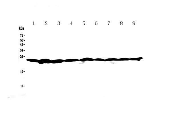
(Figure 1. Western blot analysis of Ran using anti-Ran antibody (AAA19131). Electrophoresis was performed on a 5-20% SDS-PAGE gel at 70V (Stacking gel) / 90V (Resolving gel) for 2-3 hours. The sample well of each lane was loaded with 50ug of sample under reducing conditions. Lane 1: rat brain tissue lysate,Lane 2: rat testis tissue lysate,Lane 3: rat thymus tissue lysate,Lane 4: mouse brain tissue lysate,Lane 5: mouse testis tissue lysate,Lane 6: mouse thymus tissue lysate,Lane 7: human A549 whole cell lysate,Lane 8: human 22RV1 whole cell lysate,Lane 9: human Hela whole cell lysate. After Electrophoresis, proteins were transferred to a Nitrocellulose membrane at 150mA for 50-90 minutes. Blocked the membrane with 5% Non-fat Milk/ TBS for 1.5 hour at RT. The membrane was incubated with rabbit anti-Ran antigen affinity purified polyclonal antibody at 0.5ug/mL overnight at 4 degree C, then washed with TBS-0.1%Tween 3 times with 5 minutes each and probed with a goat anti-rabbit IgG-HRP secondary antibody at a dilution of 1:10000 for 1.5 hour at RT. The signal is developed using an Enhanced Chemiluminescent detection (ECL) kit with Tanon 5200 system. A specific band was detected for Ran at approximately 24KD. The expected band size for Ran is at 24KD.)
WB (Western Blot)
(Figure 1. Western blot analysis of Ran using anti-Ran antibody (AAA19131). Electrophoresis was performed on a 5-20% SDS-PAGE gel at 70V (Stacking gel) / 90V (Resolving gel) for 2-3 hours. The sample well of each lane was loaded with 50ug of sample under reducing conditions. Lane 1: rat brain tissue lysate,Lane 2: rat testis tissue lysate,Lane 3: rat thymus tissue lysate,Lane 4: mouse brain tissue lysate,Lane 5: mouse testis tissue lysate,Lane 6: mouse thymus tissue lysate,Lane 7: human A549 whole cell lysate,Lane 8: human 22RV1 whole cell lysate,Lane 9: human Hela whole cell lysate. After Electrophoresis, proteins were transferred to a Nitrocellulose membrane at 150mA for 50-90 minutes. Blocked the membrane with 5% Non-fat Milk/ TBS for 1.5 hour at RT. The membrane was incubated with rabbit anti-Ran antigen affinity purified polyclonal antibody at 0.5ug/mL overnight at 4 degree C, then washed with TBS-0.1%Tween 3 times with 5 minutes each and probed with a goat anti-rabbit IgG-HRP secondary antibody at a dilution of 1:10000 for 1.5 hour at RT. The signal is developed using an Enhanced Chemiluminescent detection (ECL) kit with Tanon 5200 system. A specific band was detected for Ran at approximately 24KD. The expected band size for Ran is at 24KD.)
Ran, Polyclonal Antibody (Cat# AAA19131)
Full Name
Anti-Ran Picoband Antibody
Gene Names
RAN; TC4; Gsp1; ARA24
Reactivity
Human, Mouse, Rat
No cross reactivity with other proteins.
No cross reactivity with other proteins.
Applications
IHC, WB
Purity
Immunogen affinity purified


 FCM (Flow Cytometry)
(Figure 8. Flow Cytometry analysis of U87 cells using anti-Transketolase/TKT antibody (AAA19254).Overlay histogram showing U87 cells stained with AAA19254 (Blue line). The cells were blocked with 10% normal goat serum. And then incubated with rabbit anti-Transketolase/TKT Antibody (AAA19254,1μg/1x106 cells) for 30 min at 20 degree C. DyLight®488 conjugated goat anti-rabbit IgG (5-10μg/1x106 cells) was used as secondary antibody for 30 minutes at 20 degree C. Isotype control antibody (Green line) was rabbit IgG (1μg/1x106) used under the same conditions. Unlabelled sample (Red line) was also used as a control.)
FCM (Flow Cytometry)
(Figure 8. Flow Cytometry analysis of U87 cells using anti-Transketolase/TKT antibody (AAA19254).Overlay histogram showing U87 cells stained with AAA19254 (Blue line). The cells were blocked with 10% normal goat serum. And then incubated with rabbit anti-Transketolase/TKT Antibody (AAA19254,1μg/1x106 cells) for 30 min at 20 degree C. DyLight®488 conjugated goat anti-rabbit IgG (5-10μg/1x106 cells) was used as secondary antibody for 30 minutes at 20 degree C. Isotype control antibody (Green line) was rabbit IgG (1μg/1x106) used under the same conditions. Unlabelled sample (Red line) was also used as a control.)
Transketolase/TKT, Polyclonal Antibody (Cat# AAA19254)
Full Name
Anti-Transketolase/TKT Antibody
Gene Names
TKT; TK; TKT1; HEL107
Reactivity
Human, Mouse, Rat
Applications
WB, IHC-P, ICC, IF, FC/FACS/FCM, EIA
Purity
Immunogen affinity purified.


 FCM (Flow Cytometry)
(Figure 10. Flow Cytometry analysis of K562 cells using anti-RCC1 antibody (AAA19263).Overlay histogram showing K562 cells stained with AAA19263 (Blue line). The cells were blocked with 10% normal goat serum. And then incubated with rabbit anti-RCC1 Antibody (AAA19263,1μg/1x106 cells) for 30 min at 20 degree C. DyLight®488 conjugated goat anti-rabbit IgG (5-10μg/1x106 cells) was used as secondary antibody for 30 minutes at 20 degree C. Isotype control antibody (Green line) was rabbit IgG (1μg/1x106) used under the same conditions. Unlabelled sample (Red line) was also used as a control.)
FCM (Flow Cytometry)
(Figure 10. Flow Cytometry analysis of K562 cells using anti-RCC1 antibody (AAA19263).Overlay histogram showing K562 cells stained with AAA19263 (Blue line). The cells were blocked with 10% normal goat serum. And then incubated with rabbit anti-RCC1 Antibody (AAA19263,1μg/1x106 cells) for 30 min at 20 degree C. DyLight®488 conjugated goat anti-rabbit IgG (5-10μg/1x106 cells) was used as secondary antibody for 30 minutes at 20 degree C. Isotype control antibody (Green line) was rabbit IgG (1μg/1x106) used under the same conditions. Unlabelled sample (Red line) was also used as a control.)
RCC1, Polyclonal Antibody (Cat# AAA19263)
Full Name
Anti-RCC1 Antibody
Gene Names
RCC1; CHC1; RCC1-I; SNHG3-RCC1
Reactivity
Human, Mouse, Rat
Applications
WB, IHC-P, ICC, IF, FC/FACS/FCM, EIA
Purity
Immunogen affinity purified.


 FCM (Flow Cytometry)
(Figure 6. Flow Cytometry analysis of RAW264. 7 cells using anti-TRIM25/EFP antibody (AAA19272).Overlay histogram showing RAW264. 7 cells stained with AAA19272 (Blue line). The cells were blocked with 10% normal goat serum. And then incubated with rabbit anti-TRIM25/EFP Antibody (AAA19272, 1μg/1x106 cells) for 30 min at 20 degree C. DyLight®488 conjugated goat anti-rabbit IgG (5-10μg/1x106 cells) was used as secondary antibody for 30 minutes at 20 degree C. Isotype control antibody (Green line) was rabbit IgG (1μg/1x106) used under the same conditions. Unlabelled sample (Red line) was also used as a control.)
FCM (Flow Cytometry)
(Figure 6. Flow Cytometry analysis of RAW264. 7 cells using anti-TRIM25/EFP antibody (AAA19272).Overlay histogram showing RAW264. 7 cells stained with AAA19272 (Blue line). The cells were blocked with 10% normal goat serum. And then incubated with rabbit anti-TRIM25/EFP Antibody (AAA19272, 1μg/1x106 cells) for 30 min at 20 degree C. DyLight®488 conjugated goat anti-rabbit IgG (5-10μg/1x106 cells) was used as secondary antibody for 30 minutes at 20 degree C. Isotype control antibody (Green line) was rabbit IgG (1μg/1x106) used under the same conditions. Unlabelled sample (Red line) was also used as a control.)
TRIM25/EFP, Polyclonal Antibody (Cat# AAA19272)
Full Name
Anti-TRIM25/EFP Antibody
Gene Names
Trim25; EFP; Zfp147; AA960166; AL022677
Reactivity
Mouse, Rat
Applications
WB, IHC-P, ICC, IF, FC/FACS/FCM, EIA
Purity
Immunogen affinity purified.


 FCM (Flow Cytometry)
(Figure 8. Flow Cytometry analysis of HL-60 cells using anti-REA/PHB2 antibody (AAA19273).Overlay histogram showing HL-60 cells stained with AAA19273 (Blue line). The cells were blocked with 10% normal goat serum. And then incubated with rabbit anti-REA/PHB2 Antibody (AAA19273,1μg/1x106 cells) for 30 min at 20 degree C. DyLight®488 conjugated goat anti-rabbit IgG (5-10μg/1x106 cells) was used as secondary antibody for 30 minutes at 20 degree C. Isotype control antibody (Green line) was rabbit IgG (1μg/1x106) used under the same conditions. Unlabelled sample (Red line) was also used as a control.)
FCM (Flow Cytometry)
(Figure 8. Flow Cytometry analysis of HL-60 cells using anti-REA/PHB2 antibody (AAA19273).Overlay histogram showing HL-60 cells stained with AAA19273 (Blue line). The cells were blocked with 10% normal goat serum. And then incubated with rabbit anti-REA/PHB2 Antibody (AAA19273,1μg/1x106 cells) for 30 min at 20 degree C. DyLight®488 conjugated goat anti-rabbit IgG (5-10μg/1x106 cells) was used as secondary antibody for 30 minutes at 20 degree C. Isotype control antibody (Green line) was rabbit IgG (1μg/1x106) used under the same conditions. Unlabelled sample (Red line) was also used as a control.)
REA/PHB2, Polyclonal Antibody (Cat# AAA19273)
Full Name
Anti-REA/PHB2 Antibody
Gene Names
PHB2; BAP; REA; p22; Bap37; BCAP37; PNAS-141
Reactivity
Human, Mouse, Rat
Applications
WB, IHC-P, ICC, IF, FC/FACS/FCM, EIA
Purity
Immunogen affinity purified.


 FCM (Flow Cytometry)
(Figure 8. Flow Cytometry analysis of U87 cells using anti-PSMB5/MB1 antibody (AAA19274).Overlay histogram showing U87 cells stained with AAA19274 (Blue line). The cells were blocked with 10% normal goat serum. And then incubated with rabbit anti-PSMB5/MB1 Antibody (AAA19274, 1μg/1x106 cells) for 30 min at 20 degree C. DyLight®488 conjugated goat anti-rabbit IgG (5-10μg/1x106 cells) was used as secondary antibody for 30 minutes at 20 degree C. Isotype control antibody (Green line) was rabbit IgG (1μg/1x106) used under the same conditions. Unlabelled sample (Red line) was also used as a control.)
FCM (Flow Cytometry)
(Figure 8. Flow Cytometry analysis of U87 cells using anti-PSMB5/MB1 antibody (AAA19274).Overlay histogram showing U87 cells stained with AAA19274 (Blue line). The cells were blocked with 10% normal goat serum. And then incubated with rabbit anti-PSMB5/MB1 Antibody (AAA19274, 1μg/1x106 cells) for 30 min at 20 degree C. DyLight®488 conjugated goat anti-rabbit IgG (5-10μg/1x106 cells) was used as secondary antibody for 30 minutes at 20 degree C. Isotype control antibody (Green line) was rabbit IgG (1μg/1x106) used under the same conditions. Unlabelled sample (Red line) was also used as a control.)
PSMB5/MB1, Polyclonal Antibody (Cat# AAA19274)
Full Name
Anti-PSMB5/MB1 Antibody
Gene Names
PSMB5; X; MB1; LMPX
Reactivity
Human, Mouse, Rat
Applications
WB, IHC-P, ICC, IF, FC/FACS/FCM, EIA
Purity
Immunogen affinity purified.


 FCM (Flow Cytometry)
(Figure 8. Flow Cytometry analysis of K562 cells using anti-PDIA6 antibody (AAA19282).Overlay histogram showing K562 cells stained with AAA19282 (Blue line). The cells were blocked with 10% normal goat serum. And then incubated with rabbit anti-PDIA6 Antibody (AAA19282, 1μg/1x106 cells) for 30 min at 20 degree C. DyLight®488 conjugated goat anti-rabbit IgG (5-10μg/1x106 cells) was used as secondary antibody for 30 minutes at 20 degree C. Isotype control antibody (Green line) was rabbit IgG (1μg/1x106) used under the same conditions. Unlabelled sample (Red line) was also used as a control.)
FCM (Flow Cytometry)
(Figure 8. Flow Cytometry analysis of K562 cells using anti-PDIA6 antibody (AAA19282).Overlay histogram showing K562 cells stained with AAA19282 (Blue line). The cells were blocked with 10% normal goat serum. And then incubated with rabbit anti-PDIA6 Antibody (AAA19282, 1μg/1x106 cells) for 30 min at 20 degree C. DyLight®488 conjugated goat anti-rabbit IgG (5-10μg/1x106 cells) was used as secondary antibody for 30 minutes at 20 degree C. Isotype control antibody (Green line) was rabbit IgG (1μg/1x106) used under the same conditions. Unlabelled sample (Red line) was also used as a control.)
PDIA6, Polyclonal Antibody (Cat# AAA19282)
Full Name
Anti-PDIA6 Antibody
Gene Names
PDIA6; P5; ERP5; TXNDC7
Reactivity
Human, Mouse, Rat
Applications
WB, IHC-P, ICC, IF, FC/FACS/FCM, EIA
Purity
Immunogen affinity purified.


 FCM (Flow Cytometry)
(Figure 10. Flow Cytometry analysis of A431 cells using anti-CDC27 antibody (AAA19285).Overlay histogram showing A431 cells stained with AAA19285 (Blue line). The cells were blocked with 10% normal goat serum. And then incubated with rabbit anti-CDC27 Antibody (AAA19285, 1μg/1x106 cells) for 30 min at 20 degree C. DyLight®488 conjugated goat anti-rabbit IgG (5-10μg/1x106 cells) was used as secondary antibody for 30 minutes at 20 degree C. Isotype control antibody (Green line) was rabbit IgG (1μg/1x106) used under the same conditions. Unlabelled sample (Red line) was also used as a control.)
FCM (Flow Cytometry)
(Figure 10. Flow Cytometry analysis of A431 cells using anti-CDC27 antibody (AAA19285).Overlay histogram showing A431 cells stained with AAA19285 (Blue line). The cells were blocked with 10% normal goat serum. And then incubated with rabbit anti-CDC27 Antibody (AAA19285, 1μg/1x106 cells) for 30 min at 20 degree C. DyLight®488 conjugated goat anti-rabbit IgG (5-10μg/1x106 cells) was used as secondary antibody for 30 minutes at 20 degree C. Isotype control antibody (Green line) was rabbit IgG (1μg/1x106) used under the same conditions. Unlabelled sample (Red line) was also used as a control.)
CDC27, Polyclonal Antibody (Cat# AAA19285)
Full Name
Anti-CDC27 Antibody
Gene Names
CDC27; APC3; HNUC; NUC2; ANAPC3; CDC27Hs; D0S1430E; D17S978E
Reactivity
Human, Mouse, Rat
Applications
WB, IHC-P, ICC, IF, FC/FACS/FCM, EIA
Purity
Immunogen affinity purified.


 FCM (Flow Cytometry)
(Figure 9. Flow Cytometry analysis of MCF-7 cells using anti-Claudin 3/CLDN3 antibody (AAA19294).Overlay histogram showing MCF-7 cells stained with AAA19294 (Blue line). The cells were blocked with 10% normal goat serum. And then incubated with rabbit anti-Claudin 3/CLDN3 Antibody (AAA19294, 1μg/1x106 cells) for 30 min at 20 degree C. DyLight®488 conjugated goat anti-rabbit IgG (5-10μg/1x106 cells) was used as secondary antibody for 30 minutes at 20 degree C. Isotype control antibody (Green line) was rabbit IgG (1μg/1x106) used under the same conditions. Unlabelled sample (Red line) was also used as a control.)
FCM (Flow Cytometry)
(Figure 9. Flow Cytometry analysis of MCF-7 cells using anti-Claudin 3/CLDN3 antibody (AAA19294).Overlay histogram showing MCF-7 cells stained with AAA19294 (Blue line). The cells were blocked with 10% normal goat serum. And then incubated with rabbit anti-Claudin 3/CLDN3 Antibody (AAA19294, 1μg/1x106 cells) for 30 min at 20 degree C. DyLight®488 conjugated goat anti-rabbit IgG (5-10μg/1x106 cells) was used as secondary antibody for 30 minutes at 20 degree C. Isotype control antibody (Green line) was rabbit IgG (1μg/1x106) used under the same conditions. Unlabelled sample (Red line) was also used as a control.)
Claudin 3/CLDN3, Polyclonal Antibody (Cat# AAA19294)
Full Name
Anti-Claudin 3/CLDN3 Antibody
Gene Names
CLDN3; RVP1; HRVP1; C7orf1; CPE-R2; CPETR2
Reactivity
Human
Applications
WB, IHC-P, ICC, IF, FC/FACS/FCM, EIA
Purity
Immunogen affinity purified.


 FCM (Flow Cytometry)
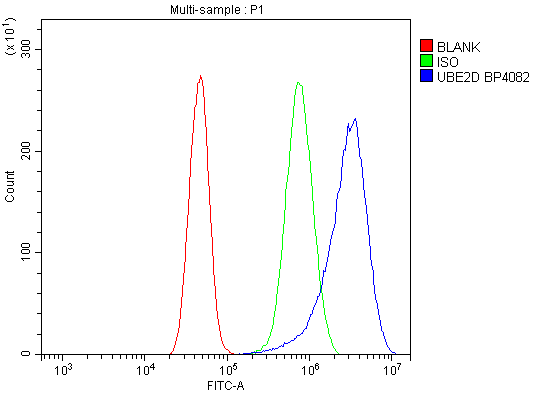
(Figure 7. Flow Cytometry analysis of A431 cells using anti-UBE2D1/2/3/4 antibody (AAA19298).Overlay histogram showing A431 cells stained with AAA19298 (Blue line). The cells were blocked with 10% normal goat serum. And then incubated with rabbit anti-UBE2D1/2/3/4 Antibody (AAA19298, 1μg/1x106 cells) for 30 min at 20 degree C. DyLight®488 conjugated goat anti-rabbit IgG (5-10μg/1x106 cells) was used as secondary antibody for 30 minutes at 20 degree C. Isotype control antibody (Green line) was rabbit IgG (1μg/1x106) used under the same conditions. Unlabelled sample (Red line) was also used as a control.)
FCM (Flow Cytometry)
(Figure 7. Flow Cytometry analysis of A431 cells using anti-UBE2D1/2/3/4 antibody (AAA19298).Overlay histogram showing A431 cells stained with AAA19298 (Blue line). The cells were blocked with 10% normal goat serum. And then incubated with rabbit anti-UBE2D1/2/3/4 Antibody (AAA19298, 1μg/1x106 cells) for 30 min at 20 degree C. DyLight®488 conjugated goat anti-rabbit IgG (5-10μg/1x106 cells) was used as secondary antibody for 30 minutes at 20 degree C. Isotype control antibody (Green line) was rabbit IgG (1μg/1x106) used under the same conditions. Unlabelled sample (Red line) was also used as a control.)
UBE2D1/2/3/4, Polyclonal Antibody (Cat# AAA19298)
Full Name
Anti-UBE2D1/2/3/4 Antibody
Gene Names
UBE2D1; SFT; UBCH5; UBC4/5; UBCH5A; E2(17)KB1
Reactivity
Human, Mouse, Rat
Applications
WB, IHC-P, ICC, IF, FC/FACS/FCM, EIA
Purity
Immunogen affinity purified.


 FCM (Flow Cytometry)
(Figure 9. Flow Cytometry analysis of THP-1 cells using anti-CHCHD10 antibody (AAA19310).Overlay histogram showing THP-1 cells stained with AAA19310 (Blue line). The cells were blocked with 10% normal goat serum. And then incubated with rabbit anti-CHCHD10 Antibody (AAA19310, 1μg/1x106 cells) for 30 min at 20 degree C. DyLight®488 conjugated goat anti-rabbit IgG (5-10μg/1x106 cells) was used as secondary antibody for 30 minutes at 20 degree C. Isotype control antibody (Green line) was rabbit IgG (1μg/1x106) used under the same conditions. Unlabelled sample (Red line) was also used as a control.)
FCM (Flow Cytometry)
(Figure 9. Flow Cytometry analysis of THP-1 cells using anti-CHCHD10 antibody (AAA19310).Overlay histogram showing THP-1 cells stained with AAA19310 (Blue line). The cells were blocked with 10% normal goat serum. And then incubated with rabbit anti-CHCHD10 Antibody (AAA19310, 1μg/1x106 cells) for 30 min at 20 degree C. DyLight®488 conjugated goat anti-rabbit IgG (5-10μg/1x106 cells) was used as secondary antibody for 30 minutes at 20 degree C. Isotype control antibody (Green line) was rabbit IgG (1μg/1x106) used under the same conditions. Unlabelled sample (Red line) was also used as a control.)
CHCHD10, Polyclonal Antibody (Cat# AAA19310)
Full Name
Anti-CHCHD10 Antibody
Gene Names
CHCHD10; FTDALS2; N27C7-4; C22orf16
Reactivity
Human, Mouse, Rat, Monkey
Applications
WB, IHC-P, ICC, IF, FC/FACS/FCM, EIA
Purity
Immunogen affinity purified.


 FCM (Flow Cytometry)
(Figure 7. Flow Cytometry analysis of 293T cells using anti-OTULIN antibody (AAA19319).Overlay histogram showing 293T cells stained with AAA19319 (Blue line). The cells were blocked with 10% normal goat serum. And then incubated with rabbit anti-OTULIN Antibody (AAA19319, 1μg/1x106 cells) for 30 min at 20 degree C. DyLight®488 conjugated goat anti-rabbit IgG (5-10μg/1x106 cells) was used as secondary antibody for 30 minutes at 20 degree C. Isotype control antibody (Green line) was rabbit IgG (1μg/1x106) used under the same conditions. Unlabelled sample (Red line) was also used as a control.)
FCM (Flow Cytometry)
(Figure 7. Flow Cytometry analysis of 293T cells using anti-OTULIN antibody (AAA19319).Overlay histogram showing 293T cells stained with AAA19319 (Blue line). The cells were blocked with 10% normal goat serum. And then incubated with rabbit anti-OTULIN Antibody (AAA19319, 1μg/1x106 cells) for 30 min at 20 degree C. DyLight®488 conjugated goat anti-rabbit IgG (5-10μg/1x106 cells) was used as secondary antibody for 30 minutes at 20 degree C. Isotype control antibody (Green line) was rabbit IgG (1μg/1x106) used under the same conditions. Unlabelled sample (Red line) was also used as a control.)
OTULIN, Polyclonal Antibody (Cat# AAA19319)
Full Name
Anti-OTULIN Antibody
Gene Names
OTULIN; GUM; FAM105B
Reactivity
Human, Mouse, Rat
Applications
WB, IHC-P, ICC, IF, FC/FACS/FCM, EIA
Purity
Immunogen affinity purified.


 FCM (Flow Cytometry)
(Figure 7. Flow Cytometry analysis of HELA cells using anti-DNAJC10 antibody (AAA19320).Overlay histogram showing HELA cells stained with AAA19320 (Blue line). The cells were blocked with 10% normal goat serum. And then incubated with rabbit anti-DNAJC10 Antibody (AAA19320, 1μg/1x106 cells) for 30 min at 20 degree C. DyLight®488 conjugated goat anti-rabbit IgG (5-10μg/1x106 cells) was used as secondary antibody for 30 minutes at 20 degree C. Isotype control antibody (Green line) was rabbit IgG (1μg/1x106) used under the same conditions. Unlabelled sample (Red line) was also used as a control.)
FCM (Flow Cytometry)
(Figure 7. Flow Cytometry analysis of HELA cells using anti-DNAJC10 antibody (AAA19320).Overlay histogram showing HELA cells stained with AAA19320 (Blue line). The cells were blocked with 10% normal goat serum. And then incubated with rabbit anti-DNAJC10 Antibody (AAA19320, 1μg/1x106 cells) for 30 min at 20 degree C. DyLight®488 conjugated goat anti-rabbit IgG (5-10μg/1x106 cells) was used as secondary antibody for 30 minutes at 20 degree C. Isotype control antibody (Green line) was rabbit IgG (1μg/1x106) used under the same conditions. Unlabelled sample (Red line) was also used as a control.)
DNAJC10, Polyclonal Antibody (Cat# AAA19320)
Full Name
Anti-DNAJC10 Antibody
Gene Names
DNAJC10; JPDI; MTHr; ERdj5; PDIA19
Reactivity
Human, Mouse, Rat
Applications
WB, IHC-P, ICC, IF, FC/FACS/FCM, EIA
Purity
Immunogen affinity purified.


 FCM (Flow Cytometry)
(Figure 11. Flow Cytometry analysis of THP-1 cells using anti-CDC123 antibody (AAA19323).Overlay histogram showing THP-1 cells stained with AAA19323 (Blue line). The cells were blocked with 10% normal goat serum. And then incubated with rabbit anti-CDC123 Antibody (AAA19323, 1μg/1x106 cells) for 30 min at 20 degree C. DyLight®488 conjugated goat anti-rabbit IgG (5-10μg/1x106 cells) was used as secondary antibody for 30 minutes at 20 degree C. Isotype control antibody (Green line) was rabbit IgG (1μg/1x106) used under the same conditions. Unlabelled sample (Red line) was also used as a control.)
FCM (Flow Cytometry)
(Figure 11. Flow Cytometry analysis of THP-1 cells using anti-CDC123 antibody (AAA19323).Overlay histogram showing THP-1 cells stained with AAA19323 (Blue line). The cells were blocked with 10% normal goat serum. And then incubated with rabbit anti-CDC123 Antibody (AAA19323, 1μg/1x106 cells) for 30 min at 20 degree C. DyLight®488 conjugated goat anti-rabbit IgG (5-10μg/1x106 cells) was used as secondary antibody for 30 minutes at 20 degree C. Isotype control antibody (Green line) was rabbit IgG (1μg/1x106) used under the same conditions. Unlabelled sample (Red line) was also used as a control.)
CDC123, Polyclonal Antibody (Cat# AAA19323)
Full Name
Anti-CDC123 Antibody
Gene Names
CDC123; D123; C10orf7
Reactivity
Human
Applications
WB, IHC-P, ICC, IF, FC/FACS/FCM, EIA
Purity
Immunogen affinity purified.


 IHC (Immunohistochemistry)
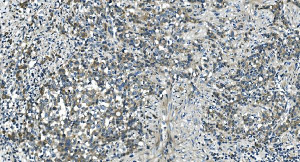
(Figure 8. IHC analysis of MEK2/MAP2K2 using anti-MEK2/MAP2K2 antibody (AAA19230).MEK2/MAP2K2 was detected in paraffin-embedded section of mouse brain tissue. Heat mediated antigen retrieval was performed in EDTA buffer (pH8. 0, epitope retrieval solution). The tissue section was blocked with 10% goat serum. The tissue section was then incubated with 2μg/ml rabbit anti-MEK2/MAP2K2 Antibody (AAA19230) overnight at 4 degree C. Biotinylated goat anti-rabbit IgG was used as secondary antibody and incubated for 30 minutes at 37 degree C. The tissue section was developed using Strepavidin-Biotin-Complex (SABC) (Catalog # with DAB as the chromogen.)
IHC (Immunohistochemistry)
(Figure 8. IHC analysis of MEK2/MAP2K2 using anti-MEK2/MAP2K2 antibody (AAA19230).MEK2/MAP2K2 was detected in paraffin-embedded section of mouse brain tissue. Heat mediated antigen retrieval was performed in EDTA buffer (pH8. 0, epitope retrieval solution). The tissue section was blocked with 10% goat serum. The tissue section was then incubated with 2μg/ml rabbit anti-MEK2/MAP2K2 Antibody (AAA19230) overnight at 4 degree C. Biotinylated goat anti-rabbit IgG was used as secondary antibody and incubated for 30 minutes at 37 degree C. The tissue section was developed using Strepavidin-Biotin-Complex (SABC) (Catalog # with DAB as the chromogen.)
MEK2/MAP2K2, Polyclonal Antibody (Cat# AAA19230)
Full Name
Anti-MEK2/MAP2K2 Antibody
Gene Names
MAP2K2; CFC4; MEK2; MKK2; MAPKK2; PRKMK2
Reactivity
Human, Mouse, Rat
Applications
WB, IHC-P, ICC, IF, FC/FACS/FCM, EIA
Purity
Immunogen affinity purified.


 FCM (Flow Cytometry)
(Figure 7. Flow Cytometry analysis of THP-1 cells using anti-S100A4 antibody (AAA19235).Overlay histogram showing THP-1 cells stained with AAA19235 (Blue line). The cells were blocked with 10% normal goat serum. And then incubated with rabbit anti-S100A4 Antibody (AAA19235, 1μg/1x106 cells) for 30 min at 20 degree C. DyLight®488 conjugated goat anti-rabbit IgG (5-10μg/1x106 cells) was used as secondary antibody for 30 minutes at 20 degree C. Isotype control antibody (Green line) was rabbit IgG (1μg/1x106) used under the same conditions. Unlabelled sample (Red line) was also used as a control.)
FCM (Flow Cytometry)
(Figure 7. Flow Cytometry analysis of THP-1 cells using anti-S100A4 antibody (AAA19235).Overlay histogram showing THP-1 cells stained with AAA19235 (Blue line). The cells were blocked with 10% normal goat serum. And then incubated with rabbit anti-S100A4 Antibody (AAA19235, 1μg/1x106 cells) for 30 min at 20 degree C. DyLight®488 conjugated goat anti-rabbit IgG (5-10μg/1x106 cells) was used as secondary antibody for 30 minutes at 20 degree C. Isotype control antibody (Green line) was rabbit IgG (1μg/1x106) used under the same conditions. Unlabelled sample (Red line) was also used as a control.)
S100A4, Polyclonal Antibody (Cat# AAA19235)
Full Name
Anti-S100A4 Antibody
Gene Names
S100A4; 42A; 18A2; CAPL; FSP1; MTS1; P9KA; PEL98
Reactivity
Human
Applications
WB, IHC-P, ICC, IF, FC/FACS/FCM, EIA
Purity
Immunogen affinity purified.


 FCM (Flow Cytometry)
(Figure 10. Flow Cytometry analysis of THP-1 cells using anti-HDAC5 antibody (AAA19236).Overlay histogram showing THP-1 cells stained with AAA19236 (Blue line). The cells were blocked with 10% normal goat serum. And then incubated with rabbit anti-HDAC5 Antibody (AAA19236, 1μg/1x106 cells) for 30 min at 20 degree C. DyLight®488 conjugated goat anti-rabbit IgG (5-10μg/1x106 cells) was used as secondary antibody for 30 minutes at 20 degree C. Isotype control antibody (Green line) was rabbit IgG (1μg/1x106) used under the same conditions. Unlabelled sample (Red line) was also used as a control.)
FCM (Flow Cytometry)
(Figure 10. Flow Cytometry analysis of THP-1 cells using anti-HDAC5 antibody (AAA19236).Overlay histogram showing THP-1 cells stained with AAA19236 (Blue line). The cells were blocked with 10% normal goat serum. And then incubated with rabbit anti-HDAC5 Antibody (AAA19236, 1μg/1x106 cells) for 30 min at 20 degree C. DyLight®488 conjugated goat anti-rabbit IgG (5-10μg/1x106 cells) was used as secondary antibody for 30 minutes at 20 degree C. Isotype control antibody (Green line) was rabbit IgG (1μg/1x106) used under the same conditions. Unlabelled sample (Red line) was also used as a control.)
HDAC5, Polyclonal Antibody (Cat# AAA19236)
Full Name
Anti-HDAC5 Antibody
Gene Names
HDAC5; HD5; NY-CO-9
Reactivity
Human, Mouse, Rat
Applications
WB, IHC-P, ICC, IF, FC/FACS/FCM, EIA
Purity
Immunogen affinity purified.


 FCM (Flow Cytometry)
(Figure 10. Flow Cytometry analysis of U20S cells using anti-PDIA5 antibody (AAA19331).Overlay histogram showing U20S cells stained with AAA19331 (Blue line). The cells were blocked with 10% normal goat serum. And then incubated with rabbit anti-PDIA5 Antibody (AAA19331, 1μg/1x106 cells) for 30 min at 20 degree C. DyLight®488 conjugated goat anti-rabbit IgG (5-10μg/1x106 cells) was used as secondary antibody for 30 minutes at 20 degree C. Isotype control antibody (Green line) was rabbit IgG (1μg/1x106) used under the same conditions. Unlabelled sample (Red line) was also used as a control.)
FCM (Flow Cytometry)
(Figure 10. Flow Cytometry analysis of U20S cells using anti-PDIA5 antibody (AAA19331).Overlay histogram showing U20S cells stained with AAA19331 (Blue line). The cells were blocked with 10% normal goat serum. And then incubated with rabbit anti-PDIA5 Antibody (AAA19331, 1μg/1x106 cells) for 30 min at 20 degree C. DyLight®488 conjugated goat anti-rabbit IgG (5-10μg/1x106 cells) was used as secondary antibody for 30 minutes at 20 degree C. Isotype control antibody (Green line) was rabbit IgG (1μg/1x106) used under the same conditions. Unlabelled sample (Red line) was also used as a control.)
PDIA5, Polyclonal Antibody (Cat# AAA19331)
Full Name
Anti-PDIA5 Antibody
Gene Names
PDIA5; PDIR
Reactivity
Human, Mouse, Rat
Applications
WB, IHC-P, ICC, IF, FC/FACS/FCM, EIA
Purity
Immunogen affinity purified.


 FCM (Flow Cytometry)
(Figure 12. Flow Cytometry analysis of HEPA1-6 cells using anti-LSM5 antibody (AAA19332).Overlay histogram showing HEPA1-6 cells stained with AAA19332 (Blue line). The cells were blocked with 10% normal goat serum. And then incubated with rabbit anti-LSM5 Antibody (AAA19332, 1μg/1x106 cells) for 30 min at 20 degree C. DyLight®488 conjugated goat anti-rabbit IgG (5-10μg/1x106 cells) was used as secondary antibody for 30 minutes at 20 degree C. Isotype control antibody (Green line) was rabbit IgG (1μg/1x106) used under the same conditions. Unlabelled sample (Red line) was also used as a control.)
FCM (Flow Cytometry)
(Figure 12. Flow Cytometry analysis of HEPA1-6 cells using anti-LSM5 antibody (AAA19332).Overlay histogram showing HEPA1-6 cells stained with AAA19332 (Blue line). The cells were blocked with 10% normal goat serum. And then incubated with rabbit anti-LSM5 Antibody (AAA19332, 1μg/1x106 cells) for 30 min at 20 degree C. DyLight®488 conjugated goat anti-rabbit IgG (5-10μg/1x106 cells) was used as secondary antibody for 30 minutes at 20 degree C. Isotype control antibody (Green line) was rabbit IgG (1μg/1x106) used under the same conditions. Unlabelled sample (Red line) was also used as a control.)
LSM5, Polyclonal Antibody (Cat# AAA19332)
Full Name
Anti-LSM5 Antibody
Gene Names
LSM5; YER146W
Reactivity
Human, Mouse, Rat
Applications
WB, IHC-P, ICC, IF, FC/FACS/FCM, EIA
Purity
Immunogen affinity purified.


 FCM (Flow Cytometry)
(Figure 9. Flow Cytometry analysis of U251 cells using anti-VPRBP/DCAF1 antibody (AAA19343).Overlay histogram showing U251 cells stained with AAA19343 (Blue line). The cells were blocked with 10% normal goat serum. And then incubated with rabbit anti-VPRBP/DCAF1 Antibody (AAA19343, 1μg/1x106 cells) for 30 min at 20 degree C. DyLight®488 conjugated goat anti-rabbit IgG (5-10μg/1x106 cells) was used as secondary antibody for 30 minutes at 20 degree C. Isotype control antibody (Green line) was rabbit IgG (1μg/1x106) used under the same conditions. Unlabelled sample (Red line) was also used as a control.)
FCM (Flow Cytometry)
(Figure 9. Flow Cytometry analysis of U251 cells using anti-VPRBP/DCAF1 antibody (AAA19343).Overlay histogram showing U251 cells stained with AAA19343 (Blue line). The cells were blocked with 10% normal goat serum. And then incubated with rabbit anti-VPRBP/DCAF1 Antibody (AAA19343, 1μg/1x106 cells) for 30 min at 20 degree C. DyLight®488 conjugated goat anti-rabbit IgG (5-10μg/1x106 cells) was used as secondary antibody for 30 minutes at 20 degree C. Isotype control antibody (Green line) was rabbit IgG (1μg/1x106) used under the same conditions. Unlabelled sample (Red line) was also used as a control.)
VPRBP/DCAF1, Polyclonal Antibody (Cat# AAA19343)
Full Name
Anti-VPRBP/DCAF1 Antibody
Gene Names
DCAF1; RIP; VPRBP
Reactivity
Human, Mouse, Rat
Applications
WB, IHC-P, ICC, IF, FC/FACS/FCM, EIA
Purity
Immunogen affinity purified.


 FCM (Flow Cytometry)
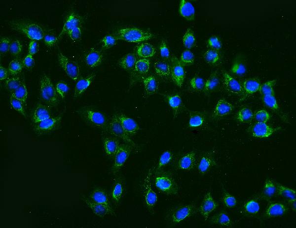
(Figure 6. Flow Cytometry analysis of HL-60 cells using anti- MCM2 antibody (AAA19351).Overlay histogram showing HL-60 cells stained with AAA19351 (Blue line). The cells were blocked with 10% normal goat serum. And then incubated with mouse anti-MCM2 Antibody (AAA19351, 1μg/1x106 cells) for 30 min at 20 degree C. DyLight®488 conjugated goat anti-mouse IgG (BA1126, 5-10μg/1x106 cells) was used as secondary antibody for 30 minutes at 20 degree C. Isotype control antibody (Green line) was mouse IgG (1μg/1x106) used under the same conditions. Unlabelled sample (Red line) was also used as a control.)
FCM (Flow Cytometry)
(Figure 6. Flow Cytometry analysis of HL-60 cells using anti- MCM2 antibody (AAA19351).Overlay histogram showing HL-60 cells stained with AAA19351 (Blue line). The cells were blocked with 10% normal goat serum. And then incubated with mouse anti-MCM2 Antibody (AAA19351, 1μg/1x106 cells) for 30 min at 20 degree C. DyLight®488 conjugated goat anti-mouse IgG (BA1126, 5-10μg/1x106 cells) was used as secondary antibody for 30 minutes at 20 degree C. Isotype control antibody (Green line) was mouse IgG (1μg/1x106) used under the same conditions. Unlabelled sample (Red line) was also used as a control.)
MCM2, Monoclonal Antibody (Cat# AAA19351)
Full Name
Anti-MCM2 Antibody (monoclonal, 11C4)
Gene Names
MCM2; BM28; CCNL1; CDCL1; cdc19; D3S3194; MITOTIN
Reactivity
Human, Monkey
Applications
WB, IHC-P, ICC, IF, FC/FACS/FCM
Purity
Immunogen affinity purified.


 FCM (Flow Cytometry)
(Figure 9. Flow Cytometry analysis of K562 cells using anti-MCM6 antibody (AAA19372).Overlay histogram showing K562 cells stained with AAA19372 (Blue line). The cells were blocked with 10% normal goat serum. And then incubated with mouse anti- MCM6 Antibody (AAA19372, 1μg/1x106 cells) for 30 min at 20 degree C. DyLight®488 conjugated goat anti-mouse IgG (BA1126, 5-10μg/1x106 cells) was used as secondary antibody for 30 minutes at 20 degree C. Isotype control antibody (Green line) was mouse IgG (1μg/1x106) used under the same conditions. Unlabelled sample (Red line) was also used as a control.)
FCM (Flow Cytometry)
(Figure 9. Flow Cytometry analysis of K562 cells using anti-MCM6 antibody (AAA19372).Overlay histogram showing K562 cells stained with AAA19372 (Blue line). The cells were blocked with 10% normal goat serum. And then incubated with mouse anti- MCM6 Antibody (AAA19372, 1μg/1x106 cells) for 30 min at 20 degree C. DyLight®488 conjugated goat anti-mouse IgG (BA1126, 5-10μg/1x106 cells) was used as secondary antibody for 30 minutes at 20 degree C. Isotype control antibody (Green line) was mouse IgG (1μg/1x106) used under the same conditions. Unlabelled sample (Red line) was also used as a control.)
MCM6, Monoclonal Antibody (Cat# AAA19372)
Full Name
Anti-MCM6 Antibody (monoclonal, 10I9)
Gene Names
MCM6; Mis5; P105MCM; MCG40308
Reactivity
Human
Applications
WB, IHC-P, ICC, IF, FC/FACS/FCM
Purity
Immunogen affinity purified.


 FCM (Flow Cytometry)
(Figure 10. Flow Cytometry analysis of A549 cells using anti- U2AF65/U2AF2 antibody (AAA19375).Overlay histogram showing A549 cells stained with AAA19375 (Blue line). The cells were blocked with 10% normal goat serum. And then incubated with mouse anti-U2AF65/U2AF2 Antibody (AAA19375, 1μg/1x106 cells) for 30 min at 20 degree C. DyLight®488 conjugated goat anti-mouse IgG (BA1126, 5-10μg/1x106 cells) was used as secondary antibody for 30 minutes at 20 degree C. Isotype control antibody (Green line) was mouse IgG (1μg/1x106) used under the same conditions. Unlabelled sample (Red line) was also used as a control.)
FCM (Flow Cytometry)
(Figure 10. Flow Cytometry analysis of A549 cells using anti- U2AF65/U2AF2 antibody (AAA19375).Overlay histogram showing A549 cells stained with AAA19375 (Blue line). The cells were blocked with 10% normal goat serum. And then incubated with mouse anti-U2AF65/U2AF2 Antibody (AAA19375, 1μg/1x106 cells) for 30 min at 20 degree C. DyLight®488 conjugated goat anti-mouse IgG (BA1126, 5-10μg/1x106 cells) was used as secondary antibody for 30 minutes at 20 degree C. Isotype control antibody (Green line) was mouse IgG (1μg/1x106) used under the same conditions. Unlabelled sample (Red line) was also used as a control.)
U2AF65/U2AF2, Monoclonal Antibody (Cat# AAA19375)
Full Name
Anti-U2AF65/U2AF2 Antibody (monoclonal, 10F4)
Gene Names
U2AF2; U2AF65
Reactivity
Human, Mouse, Rat
Applications
WB, IHC-P, ICC, IF, FC/FACS/FCM
Purity
Immunogen affinity purified.


 Application Data
(Published customer image: Increased accumulation of repair-associated macrophages surrounding collaterals in ischemic hind limbs is PAR2-dependent. (A) Stainings of CD206-positive macrophages (green) and SMA-positive vessels (red) in non-ischemic (control) and ischemic (ligated) hind limbs of WT, PAR1-/- and PAR2-/- mice are shown. Nuclei were visualized with DAPI (blue). Arrows indicate single macrophages in the non-ischemic adductor. Quantification of the average number of repair-associated macrophages per vessel is indicated on the right. (B) Correlation between the number of CD206-positive macrophages in the ischemic tissues and the expression of CD11b and (C) CD115 on monocytes. ** p)
Application Data
(Published customer image: Increased accumulation of repair-associated macrophages surrounding collaterals in ischemic hind limbs is PAR2-dependent. (A) Stainings of CD206-positive macrophages (green) and SMA-positive vessels (red) in non-ischemic (control) and ischemic (ligated) hind limbs of WT, PAR1-/- and PAR2-/- mice are shown. Nuclei were visualized with DAPI (blue). Arrows indicate single macrophages in the non-ischemic adductor. Quantification of the average number of repair-associated macrophages per vessel is indicated on the right. (B) Correlation between the number of CD206-positive macrophages in the ischemic tissues and the expression of CD11b and (C) CD115 on monocytes. ** p)
CD206, Monoclonal Antibody (Cat# AAA12123)
Full Name
RAT ANTI MOUSE CD206
Gene Names
Mrc1; MR; CD206; AW259686
Applications
FC/FACS, IF, IP


 FCM (Flow Cytometry)
(Figure 7. Flow Cytometry analysis of THP-1 cells using anti-PNPT1 antibody (AAA19306).Overlay histogram showing THP-1 cells stained with AAA19306 (Blue line). The cells were blocked with 10% normal goat serum. And then incubated with rabbit anti-PNPT1 Antibody (AAA19306, 1μg/1x106 cells) for 30 min at 20 degree C. DyLight®488 conjugated goat anti-rabbit IgG (5-10μg/1x106 cells) was used as secondary antibody for 30 minutes at 20 degree C. Isotype control antibody (Green line) was rabbit IgG (1μg/1x106) used under the same conditions. Unlabelled sample (Red line) was also used as a control.)
FCM (Flow Cytometry)
(Figure 7. Flow Cytometry analysis of THP-1 cells using anti-PNPT1 antibody (AAA19306).Overlay histogram showing THP-1 cells stained with AAA19306 (Blue line). The cells were blocked with 10% normal goat serum. And then incubated with rabbit anti-PNPT1 Antibody (AAA19306, 1μg/1x106 cells) for 30 min at 20 degree C. DyLight®488 conjugated goat anti-rabbit IgG (5-10μg/1x106 cells) was used as secondary antibody for 30 minutes at 20 degree C. Isotype control antibody (Green line) was rabbit IgG (1μg/1x106) used under the same conditions. Unlabelled sample (Red line) was also used as a control.)
PNPT1, Polyclonal Antibody (Cat# AAA19306)
Full Name
Anti-PNPT1 Antibody
Gene Names
PNPT1; OLD35; DFNB70; PNPASE; old-35; COXPD13
Reactivity
Human, Mouse, Rat
Applications
WB, IHC-P, ICC, IF, FC/FACS/FCM, EIA
Purity
Immunogen affinity purified.


 WB (Western Blot)
(Figure 1. Western blot analysis of CISD2 using anti-CISD2 antibody (AAA19311).Electrophoresis was performed on a 5-20% SDS-PAGE gel at 70V (Stacking gel) / 90V (Resolving gel) for 2-3 hours. The sample well of each lane was loaded with 50ug of sample under reducing conditions.Lane 1: human HEK293 whole cell lysatesLane 2: human HELA whole cell lysatesLane 3: human MCF-7 whole cell lysatesLane 4: monkey kidney tissue lysatesLane 5: human SW620 whole cell lysatesLane 6: human Raji whole cell lysatesLane 7: rat kidney tissue lysatesLane 8: mouse kidney tissue lysates.After Electrophoresis, proteins were transferred to a Nitrocellulose membrane at 150mA for 50-90 minutes. Blocked the membrane with 5% Non-fat Milk/ TBS for 1. 5 hour at RT. The membrane was incubated with rabbit anti-CISD2 antigen affinity purified polyclonal antibody (Catalog # AAA19311) at 0. 5 μg/mL overnight at 4 degree C, then washed with TBS-0. 1%Tween 3 times with 5 minutes each and probed with a goat anti-rabbit IgG-HRP secondary antibody at a dilution of 1:10000 for 1. 5 hour at RT. The signal is developed using an Enhanced Chemiluminescent detection (ECL) kit (Catalog # with Tanon 5200 system. A specific band was detected for CISD2 at approximately 15KD. The expected band size for CISD2 is at 15KD.)
WB (Western Blot)
(Figure 1. Western blot analysis of CISD2 using anti-CISD2 antibody (AAA19311).Electrophoresis was performed on a 5-20% SDS-PAGE gel at 70V (Stacking gel) / 90V (Resolving gel) for 2-3 hours. The sample well of each lane was loaded with 50ug of sample under reducing conditions.Lane 1: human HEK293 whole cell lysatesLane 2: human HELA whole cell lysatesLane 3: human MCF-7 whole cell lysatesLane 4: monkey kidney tissue lysatesLane 5: human SW620 whole cell lysatesLane 6: human Raji whole cell lysatesLane 7: rat kidney tissue lysatesLane 8: mouse kidney tissue lysates.After Electrophoresis, proteins were transferred to a Nitrocellulose membrane at 150mA for 50-90 minutes. Blocked the membrane with 5% Non-fat Milk/ TBS for 1. 5 hour at RT. The membrane was incubated with rabbit anti-CISD2 antigen affinity purified polyclonal antibody (Catalog # AAA19311) at 0. 5 μg/mL overnight at 4 degree C, then washed with TBS-0. 1%Tween 3 times with 5 minutes each and probed with a goat anti-rabbit IgG-HRP secondary antibody at a dilution of 1:10000 for 1. 5 hour at RT. The signal is developed using an Enhanced Chemiluminescent detection (ECL) kit (Catalog # with Tanon 5200 system. A specific band was detected for CISD2 at approximately 15KD. The expected band size for CISD2 is at 15KD.)
CISD2, Polyclonal Antibody (Cat# AAA19311)
Full Name
Anti-CISD2 Antibody
Gene Names
CISD2; ERIS; WFS2; ZCD2; NAF-1; Miner1
Reactivity
Human, Mouse, Rat, Monkey
Applications
WB, IHC-P, ICC, IF, FC/FACS/FCM, EIA
Purity
Immunogen affinity purified.


 FCM (Flow Cytometry)
(Figure 8. Flow Cytometry analysis of SiHa cells using anti-Astrin/Deepest/SPAG5 antibody (AAA19313).Overlay histogram showing SiHa cells stained with AAA19313 (Blue line). The cells were blocked with 10% normal goat serum. And then incubated with rabbit anti-Astrin/Deepest/SPAG5 Antibody (AAA19313, 1μg/1x106 cells) for 30 min at 20 degree C. DyLight®488 conjugated goat anti-rabbit IgG (5-10μg/1x106 cells) was used as secondary antibody for 30 minutes at 20 degree C. Isotype control antibody (Green line) was rabbit IgG (1μg/1x106) used under the same conditions. Unlabelled sample (Red line) was also used as a control.)
FCM (Flow Cytometry)
(Figure 8. Flow Cytometry analysis of SiHa cells using anti-Astrin/Deepest/SPAG5 antibody (AAA19313).Overlay histogram showing SiHa cells stained with AAA19313 (Blue line). The cells were blocked with 10% normal goat serum. And then incubated with rabbit anti-Astrin/Deepest/SPAG5 Antibody (AAA19313, 1μg/1x106 cells) for 30 min at 20 degree C. DyLight®488 conjugated goat anti-rabbit IgG (5-10μg/1x106 cells) was used as secondary antibody for 30 minutes at 20 degree C. Isotype control antibody (Green line) was rabbit IgG (1μg/1x106) used under the same conditions. Unlabelled sample (Red line) was also used as a control.)
Astrin/Deepest/SPAG5, Polyclonal Antibody (Cat# AAA19313)
Full Name
Anti-Astrin/Deepest/SPAG5 Antibody
Gene Names
SPAG5; MAP126; DEEPEST; hMAP126
Reactivity
Human
Applications
WB, IHC-P, ICC, IF, FC/FACS/FCM, EIA
Purity
Immunogen affinity purified.


 IHC (Immunohistochemistry)
(Figure 7. IHC analysis of GCHFR using anti-GCHFR antibody (AAA19322).GCHFR was detected in paraffin-embedded section of mouse liver tissue. Heat mediated antigen retrieval was performed in EDTA buffer (pH8. 0, epitope retrieval solution). The tissue section was blocked with 10% goat serum. The tissue section was then incubated with 2μg/ml rabbit anti-GCHFR Antibody (AAA19322) overnight at 4 degree C. Biotinylated goat anti-rabbit IgG was used as secondary antibody and incubated for 30 minutes at 37 degree C. The tissue section was developed using Strepavidin-Biotin-Complex (SABC) (Catalog # with DAB as the chromogen.)
IHC (Immunohistochemistry)
(Figure 7. IHC analysis of GCHFR using anti-GCHFR antibody (AAA19322).GCHFR was detected in paraffin-embedded section of mouse liver tissue. Heat mediated antigen retrieval was performed in EDTA buffer (pH8. 0, epitope retrieval solution). The tissue section was blocked with 10% goat serum. The tissue section was then incubated with 2μg/ml rabbit anti-GCHFR Antibody (AAA19322) overnight at 4 degree C. Biotinylated goat anti-rabbit IgG was used as secondary antibody and incubated for 30 minutes at 37 degree C. The tissue section was developed using Strepavidin-Biotin-Complex (SABC) (Catalog # with DAB as the chromogen.)
GCHFR, Polyclonal Antibody (Cat# AAA19322)
Full Name
Anti-GCHFR Antibody
Gene Names
GCHFR; P35; GFRP; HsT16933
Reactivity
Human, Mouse, Rat
Applications
WB, IHC-P, ICC, IF, FC/FACS/FCM, EIA
Purity
Immunogen affinity purified.