Filters
Clonality
Type
Reactivity
Gene Name
Isotype
Host
Application
Clone
1194 results for " E" - showing 1000-1050


 FCM (Flow Cytometry)
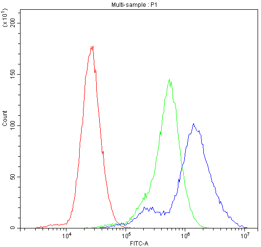
(Figure 6. Flow Cytometry analysis of HEPA1-6 cells using anti-Rad9/Rad9a antibody (AAA19288).Overlay histogram showing HEPA1-6 cells stained with AAA19288 (Blue line). The cells were blocked with 10% normal goat serum. And then incubated with rabbit anti-Rad9/Rad9a Antibody (AAA19288,1μg/1x106 cells) for 30 min at 20 degree C. DyLight®488 conjugated goat anti-rabbit IgG (5-10μg/1x106 cells) was used as secondary antibody for 30 minutes at 20 degree C. Isotype control antibody (Green line) was rabbit IgG (1μg/1x106) used under the same conditions. Unlabelled sample (Red line) was also used as a control.)
FCM (Flow Cytometry)
(Figure 6. Flow Cytometry analysis of HEPA1-6 cells using anti-Rad9/Rad9a antibody (AAA19288).Overlay histogram showing HEPA1-6 cells stained with AAA19288 (Blue line). The cells were blocked with 10% normal goat serum. And then incubated with rabbit anti-Rad9/Rad9a Antibody (AAA19288,1μg/1x106 cells) for 30 min at 20 degree C. DyLight®488 conjugated goat anti-rabbit IgG (5-10μg/1x106 cells) was used as secondary antibody for 30 minutes at 20 degree C. Isotype control antibody (Green line) was rabbit IgG (1μg/1x106) used under the same conditions. Unlabelled sample (Red line) was also used as a control.)
Rad9/Rad9a, Polyclonal Antibody (Cat# AAA19288)
Full Name
Anti-Rad9/Rad9a Antibody
Gene Names
Rad9a; Rad9
Reactivity
Mouse, Rat
Applications
WB, IHC-P, FC/FACS/FCM, EIA
Purity
Immunogen affinity purified.


 FCM (Flow Cytometry)
(Figure 7. Flow Cytometry analysis of A549 cells using anti-AGO3 antibody (AAA19290).Overlay histogram showing A549 cells stained with AAA19290 (Blue line). The cells were blocked with 10% normal goat serum. And then incubated with rabbit anti-AGO3 Antibody (AAA19290, 1μg/1x106 cells) for 30 min at 20 degree C. DyLight®488 conjugated goat anti-rabbit IgG (5-10μg/1x106 cells) was used as secondary antibody for 30 minutes at 20 degree C. Isotype control antibody (Green line) was rabbit IgG (1μg/1x106) used under the same conditions. Unlabelled sample (Red line) was also used as a control.)
FCM (Flow Cytometry)
(Figure 7. Flow Cytometry analysis of A549 cells using anti-AGO3 antibody (AAA19290).Overlay histogram showing A549 cells stained with AAA19290 (Blue line). The cells were blocked with 10% normal goat serum. And then incubated with rabbit anti-AGO3 Antibody (AAA19290, 1μg/1x106 cells) for 30 min at 20 degree C. DyLight®488 conjugated goat anti-rabbit IgG (5-10μg/1x106 cells) was used as secondary antibody for 30 minutes at 20 degree C. Isotype control antibody (Green line) was rabbit IgG (1μg/1x106) used under the same conditions. Unlabelled sample (Red line) was also used as a control.)
AGO3, Polyclonal Antibody (Cat# AAA19290)
Full Name
Anti-AGO3 Antibody
Gene Names
AGO3; EIF2C3
Reactivity
Human
Applications
WB, IHC-P, ICC, IF, FC/FACS/FCM, EIA
Purity
Immunogen affinity purified.


 FCM (Flow Cytometry)
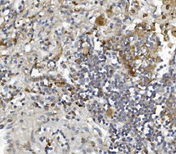
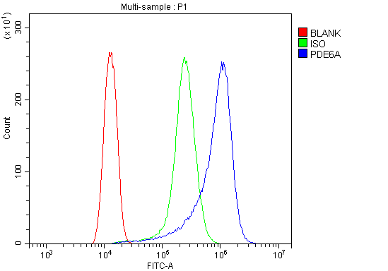
(Figure 6. Flow Cytometry analysis of HELA cells using anti-PDE6 alpha/PDE6A antibody (AAA19318).Overlay histogram showing HELA cells stained with AAA19318 (Blue line). The cells were blocked with 10% normal goat serum. And then incubated with rabbit anti-PDE6 alpha/PDE6A Antibody (AAA19318, 1μg/1x106 cells) for 30 min at 20 degree C. DyLight®488 conjugated goat anti-rabbit IgG (5-10μg/1x106 cells) was used as secondary antibody for 30 minutes at 20 degree C. Isotype control antibody (Green line) was rabbit IgG (1μg/1x106) used under the same conditions. Unlabelled sample (Red line) was also used as a control.)
FCM (Flow Cytometry)
(Figure 6. Flow Cytometry analysis of HELA cells using anti-PDE6 alpha/PDE6A antibody (AAA19318).Overlay histogram showing HELA cells stained with AAA19318 (Blue line). The cells were blocked with 10% normal goat serum. And then incubated with rabbit anti-PDE6 alpha/PDE6A Antibody (AAA19318, 1μg/1x106 cells) for 30 min at 20 degree C. DyLight®488 conjugated goat anti-rabbit IgG (5-10μg/1x106 cells) was used as secondary antibody for 30 minutes at 20 degree C. Isotype control antibody (Green line) was rabbit IgG (1μg/1x106) used under the same conditions. Unlabelled sample (Red line) was also used as a control.)
PDE6 alpha/PDE6A, Polyclonal Antibody (Cat# AAA19318)
Full Name
Anti-PDE6 alpha/PDE6A Antibody
Gene Names
PDE6A; PDEA; RP43; CGPR-A; PDE6A
Reactivity
Human, Mouse, Rat
Applications
WB, IHC-P, ICC, IF, FC/FACS/FCM, EIA
Purity
Immunogen affinity purified.


 FCM (Flow Cytometry)
(Figure 6. Flow Cytometry analysis of Hela cells using anti-Caspase-9/CASP9 antibody (AAA19215).Overlay histogram showing Hela cells stained with AAA19215 (Blue line). The cells were blocked with 10% normal goat serum. And then incubated with rabbit anti-Caspase-9/CASP9 Antibody (AAA19215, 1 μg/1x106 cells) for 30 min at 20 degree C. DyLight®488 conjugated goat anti-rabbit IgG (5-10 μg/1x106 cells) was used as secondary antibody for 30 minutes at 20 degree C. Isotype control antibody (Green line) was rabbit IgG (1 μg/1x106) used under the same conditions. Unlabelled sample (Red line) was also used as a control.)
FCM (Flow Cytometry)
(Figure 6. Flow Cytometry analysis of Hela cells using anti-Caspase-9/CASP9 antibody (AAA19215).Overlay histogram showing Hela cells stained with AAA19215 (Blue line). The cells were blocked with 10% normal goat serum. And then incubated with rabbit anti-Caspase-9/CASP9 Antibody (AAA19215, 1 μg/1x106 cells) for 30 min at 20 degree C. DyLight®488 conjugated goat anti-rabbit IgG (5-10 μg/1x106 cells) was used as secondary antibody for 30 minutes at 20 degree C. Isotype control antibody (Green line) was rabbit IgG (1 μg/1x106) used under the same conditions. Unlabelled sample (Red line) was also used as a control.)
Caspase-9/CASP9, Polyclonal Antibody (Cat# AAA19215)
Full Name
Anti-Caspase-9/CASP9 Antibody
Gene Names
CASP9; MCH6; APAF3; APAF-3; PPP1R56; ICE-LAP6
Reactivity
Human
Applications
WB, IHC-P, FC/FACS/FCM, EIA
Purity
Immunogen affinity purified.


 FCM (Flow Cytometry)
(Figure 7. Flow Cytometry analysis of U251 cells using anti-Serum Response Factor/SRF antibody (AAA19221).Overlay histogram showing U251 cells stained with AAA19221 (Blue line). The cells were blocked with 10% normal goat serum. And then incubated with rabbit anti-Serum Response Factor/SRF Antibody (AAA19221, 1μg/1x106 cells) for 30 min at 20 degree C. DyLight®488 conjugated goat anti-rabbit IgG (5-10μg/1x106 cells) was used as secondary antibody for 30 minutes at 20 degree C. Isotype control antibody (Green line) was rabbit IgG (1μg/1x106) used under the same conditions. Unlabelled sample (Red line) was also used as a control.)
FCM (Flow Cytometry)
(Figure 7. Flow Cytometry analysis of U251 cells using anti-Serum Response Factor/SRF antibody (AAA19221).Overlay histogram showing U251 cells stained with AAA19221 (Blue line). The cells were blocked with 10% normal goat serum. And then incubated with rabbit anti-Serum Response Factor/SRF Antibody (AAA19221, 1μg/1x106 cells) for 30 min at 20 degree C. DyLight®488 conjugated goat anti-rabbit IgG (5-10μg/1x106 cells) was used as secondary antibody for 30 minutes at 20 degree C. Isotype control antibody (Green line) was rabbit IgG (1μg/1x106) used under the same conditions. Unlabelled sample (Red line) was also used as a control.)
Serum Response Factor/SRF, Polyclonal Antibody (Cat# AAA19221)
Full Name
Anti-Serum Response Factor/SRF Antibody
Gene Names
SRF; MCM1
Reactivity
Human, Mouse, Rat
Applications
WB, IHC-P, ICC, IF, FC/FACS/FCM, EIA
Purity
Immunogen affinity purified.


 FCM (Flow Cytometry)
(Figure 6. Flow Cytometry analysis of MCF-7 cells using anti-Caspase-7/CASP7 antibody (AAA19232).Overlay histogram showing MCF-7 cells stained with AAA19232 (Blue line). The cells were blocked with 10% normal goat serum. And then incubated with rabbit anti-Caspase-7/CASP7 Antibody (AAA19232, 1μg/1x106 cells) for 30 min at 20 degree C. DyLight®488 conjugated goat anti-rabbit IgG (5-10μg/1x106 cells) was used as secondary antibody for 30 minutes at 20 degree C. Isotype control antibody (Green line) was rabbit IgG (1μg/1x106) used under the same conditions. Unlabelled sample (Red line) was also used as a control.)
FCM (Flow Cytometry)
(Figure 6. Flow Cytometry analysis of MCF-7 cells using anti-Caspase-7/CASP7 antibody (AAA19232).Overlay histogram showing MCF-7 cells stained with AAA19232 (Blue line). The cells were blocked with 10% normal goat serum. And then incubated with rabbit anti-Caspase-7/CASP7 Antibody (AAA19232, 1μg/1x106 cells) for 30 min at 20 degree C. DyLight®488 conjugated goat anti-rabbit IgG (5-10μg/1x106 cells) was used as secondary antibody for 30 minutes at 20 degree C. Isotype control antibody (Green line) was rabbit IgG (1μg/1x106) used under the same conditions. Unlabelled sample (Red line) was also used as a control.)
Caspase-7/CASP7, Polyclonal Antibody (Cat# AAA19232)
Full Name
Anti-Caspase-7/CASP7 Antibody
Gene Names
CASP7; MCH3; CMH-1; LICE2; CASP-7; ICE-LAP3
Reactivity
Human, Rat
Applications
WB, IHC-P, ICC, IF, FC/FACS/FCM, EIA
Purity
Immunogen affinity purified.


 FCM (Flow Cytometry)
(Figure 10. Flow Cytometry analysis of RH35 cells using anti-Prohibitin/PHB antibody (AAA19234).Overlay histogram showing RH35 cells stained with AAA19234 (Blue line). The cells were blocked with 10% normal goat serum. And then incubated with rabbit anti-Prohibitin/PHB Antibody (AAA19234, 1μg/1x106 cells) for 30 min at 20 degree C. DyLight®488 conjugated goat anti-rabbit IgG (5-10μg/1x106 cells) was used as secondary antibody for 30 minutes at 20 degree C. Isotype control antibody (Green line) was rabbit IgG (1μg/1x106) used under the same conditions. Unlabelled sample (Red line) was also used as a control.)
FCM (Flow Cytometry)
(Figure 10. Flow Cytometry analysis of RH35 cells using anti-Prohibitin/PHB antibody (AAA19234).Overlay histogram showing RH35 cells stained with AAA19234 (Blue line). The cells were blocked with 10% normal goat serum. And then incubated with rabbit anti-Prohibitin/PHB Antibody (AAA19234, 1μg/1x106 cells) for 30 min at 20 degree C. DyLight®488 conjugated goat anti-rabbit IgG (5-10μg/1x106 cells) was used as secondary antibody for 30 minutes at 20 degree C. Isotype control antibody (Green line) was rabbit IgG (1μg/1x106) used under the same conditions. Unlabelled sample (Red line) was also used as a control.)
Prohibitin/PHB, Polyclonal Antibody (Cat# AAA19234)
Full Name
Anti-Prohibitin/PHB Antibody
Gene Names
PHB; PHB1
Reactivity
Human, Mouse, Rat
Applications
WB, IHC-P, FC/FACS/FCM, EIA
Purity
Immunogen affinity purified.


 FCM (Flow Cytometry)
(Figure 9. Flow Cytometry analysis of U937 cells using anti-GNG7 antibody (AAA19335).Overlay histogram showing U937 cells stained with AAA19335 (Blue line). The cells were blocked with 10% normal goat serum. And then incubated with rabbit anti-GNG7 Antibody (AAA19335, 1μg/1x106 cells) for 30 min at 20 degree C. DyLight®488 conjugated goat anti-rabbit IgG (5-10μg/1x106 cells) was used as secondary antibody for 30 minutes at 20 degree C. Isotype control antibody (Green line) was rabbit IgG (1μg/1x106) used under the same conditions. Unlabelled sample (Red line) was also used as a control.)
FCM (Flow Cytometry)
(Figure 9. Flow Cytometry analysis of U937 cells using anti-GNG7 antibody (AAA19335).Overlay histogram showing U937 cells stained with AAA19335 (Blue line). The cells were blocked with 10% normal goat serum. And then incubated with rabbit anti-GNG7 Antibody (AAA19335, 1μg/1x106 cells) for 30 min at 20 degree C. DyLight®488 conjugated goat anti-rabbit IgG (5-10μg/1x106 cells) was used as secondary antibody for 30 minutes at 20 degree C. Isotype control antibody (Green line) was rabbit IgG (1μg/1x106) used under the same conditions. Unlabelled sample (Red line) was also used as a control.)
GNG7, Polyclonal Antibody (Cat# AAA19335)
Full Name
Anti-GNG7 Antibody
Reactivity
Human, Mouse, Rat
Applications
WB, IHC-P, FC/FACS/FCM, EIA
Purity
Immunogen affinity purified.


 FCM (Flow Cytometry)
(Figure 10. Flow Cytometry analysis of MCF-7 cells using anti-DDX1 antibody (AAA19378).Overlay histogram showing MCF-7 cells stained with AAA19378 (Blue line). The cells were blocked with 10% normal goat serum. And then incubated with mouse anti- DDX1 Antibody (AAA19378, 1μg/1x106 cells) for 30 min at 20 degree C. DyLight®488 conjugated goat anti-mouse IgG (BA1126, 5-10μg/1x106 cells) was used as secondary antibody for 30 minutes at 20 degree C. Isotype control antibody (Green line) was mouse IgG (1μg/1x106) used under the same conditions. Unlabelled sample (Red line) was also used as a control.)
FCM (Flow Cytometry)
(Figure 10. Flow Cytometry analysis of MCF-7 cells using anti-DDX1 antibody (AAA19378).Overlay histogram showing MCF-7 cells stained with AAA19378 (Blue line). The cells were blocked with 10% normal goat serum. And then incubated with mouse anti- DDX1 Antibody (AAA19378, 1μg/1x106 cells) for 30 min at 20 degree C. DyLight®488 conjugated goat anti-mouse IgG (BA1126, 5-10μg/1x106 cells) was used as secondary antibody for 30 minutes at 20 degree C. Isotype control antibody (Green line) was mouse IgG (1μg/1x106) used under the same conditions. Unlabelled sample (Red line) was also used as a control.)
DDX1, Monoclonal Antibody (Cat# AAA19378)
Full Name
Anti-DDX1 Antibody (monoclonal, 3I10)
Gene Names
DDX1; DBP-RB; UKVH5d
Reactivity
Human, Mouse, Rat
Applications
WB, IHC-P, FC/FACS/FCM
Purity
Immunogen affinity purified.


 FCM (Flow Cytometry)
(Figure 6. Flow Cytometry analysis of MCF-7 cells using anti- PCK2 antibody (AAA19384).Overlay histogram showing MCF-7 cells stained with AAA19384 (Blue line). The cells were blocked with 10% normal goat serum. And then incubated with mouse anti-PCK2 Antibody (AAA19384, 1μg/1x106 cells) for 30 min at 20 degree C. DyLight®488 conjugated goat anti-mouse IgG (BA1126, 5-10μg/1x106 cells) was used as secondary antibody for 30 minutes at 20 degree C. Isotype control antibody (Green line) was mouse IgG (1μg/1x106) used under the same conditions. Unlabelled sample (Red line) was also used as a control.)
FCM (Flow Cytometry)
(Figure 6. Flow Cytometry analysis of MCF-7 cells using anti- PCK2 antibody (AAA19384).Overlay histogram showing MCF-7 cells stained with AAA19384 (Blue line). The cells were blocked with 10% normal goat serum. And then incubated with mouse anti-PCK2 Antibody (AAA19384, 1μg/1x106 cells) for 30 min at 20 degree C. DyLight®488 conjugated goat anti-mouse IgG (BA1126, 5-10μg/1x106 cells) was used as secondary antibody for 30 minutes at 20 degree C. Isotype control antibody (Green line) was mouse IgG (1μg/1x106) used under the same conditions. Unlabelled sample (Red line) was also used as a control.)
PCK2, Monoclonal Antibody (Cat# AAA19384)
Full Name
Anti-PCK2 Antibody (monoclonal, 3F7)
Gene Names
PCK2; PEPCK; PEPCK2; PEPCK-M
Reactivity
Human, Mouse, Rat, Monkey
Applications
WB, IHC-P, ICC, IF, FC/FACS/FCM
Purity
Immunogen affinity purified.


 FCM (Flow Cytometry)
(Figure 6. Flow Cytometry analysis of SiHa cells using anti- Aldolase/ALDOA antibody (AAA19385).Overlay histogram showing SiHa cells stained with AAA19385 (Blue line). The cells were blocked with 10% normal goat serum. And then incubated with mouse anti-Aldolase/ALDOA Antibody (AAA19385, 1μg/1x106 cells) for 30 min at 20 degree C. DyLight®488 conjugated goat anti-mouse IgG (BA1126, 5-10μg/1x106 cells) was used as secondary antibody for 30 minutes at 20 degree C. Isotype control antibody (Green line) was mouse IgG (1μg/1x106) used under the same conditions. Unlabelled sample (Red line) was also used as a control.)
FCM (Flow Cytometry)
(Figure 6. Flow Cytometry analysis of SiHa cells using anti- Aldolase/ALDOA antibody (AAA19385).Overlay histogram showing SiHa cells stained with AAA19385 (Blue line). The cells were blocked with 10% normal goat serum. And then incubated with mouse anti-Aldolase/ALDOA Antibody (AAA19385, 1μg/1x106 cells) for 30 min at 20 degree C. DyLight®488 conjugated goat anti-mouse IgG (BA1126, 5-10μg/1x106 cells) was used as secondary antibody for 30 minutes at 20 degree C. Isotype control antibody (Green line) was mouse IgG (1μg/1x106) used under the same conditions. Unlabelled sample (Red line) was also used as a control.)
Aldolase/ALDOA, Monoclonal Antibody (Cat# AAA19385)
Full Name
Anti-Aldolase/ALDOA Antibody (monoclonal, 6H8)
Gene Names
ALDOA; ALDA; GSD12
Reactivity
Human
Applications
WB, IHC-P, ICC, IF, FC/FACS/FCM
Purity
Immunogen affinity purified.


 FCM (Flow Cytometry)
(Figure 6. Flow Cytometry analysis of A549 cells using anti-Ch TOG/CKAP5 antibody (AAA19386).Overlay histogram showing A549 cells stained with AAA19386 (Blue line). The cells were blocked with 10% normal goat serum. And then incubated with mouse anti- Ch TOG/CKAP5 Antibody (AAA19386, 1μg/1x106 cells) for 30 min at 20 degree C. DyLight®488 conjugated goat anti-mouse IgG (BA1126, 5-10μg/1x106 cells) was used as secondary antibody for 30 minutes at 20 degree C. Isotype control antibody (Green line) was mouse IgG (1μg/1x106) used under the same conditions. Unlabelled sample (Red line) was also used as a control.)
FCM (Flow Cytometry)
(Figure 6. Flow Cytometry analysis of A549 cells using anti-Ch TOG/CKAP5 antibody (AAA19386).Overlay histogram showing A549 cells stained with AAA19386 (Blue line). The cells were blocked with 10% normal goat serum. And then incubated with mouse anti- Ch TOG/CKAP5 Antibody (AAA19386, 1μg/1x106 cells) for 30 min at 20 degree C. DyLight®488 conjugated goat anti-mouse IgG (BA1126, 5-10μg/1x106 cells) was used as secondary antibody for 30 minutes at 20 degree C. Isotype control antibody (Green line) was mouse IgG (1μg/1x106) used under the same conditions. Unlabelled sample (Red line) was also used as a control.)
ch TOG/CKAP5, Monoclonal Antibody (Cat# AAA19386)
Full Name
Anti-ch TOG/CKAP5 Antibody (monoclonal, 3C13)
Gene Names
CKAP5; TOG; MSPS; TOGp; CHTOG; ch-TOG
Reactivity
Human, Mouse, Rat
Applications
WB, IHC-P, ICC, IF, FC/FACS/FCM
Purity
Immunogen affinity purified.


 IHC (Immunohistchemistry)
(AAA31057 at 1/200 staining human colon tissue sections by IHC-P. The tissue was formaldehyde fixed and a heat mediated antigen retrieval step in citrate buffer was performed. The tissue was then blocked and incubated with the antibody for 1.5 hours at 22 degree C. An HRP conjugated goat anti-rabbit antibody was used as the secondary.)
IHC (Immunohistchemistry)
(AAA31057 at 1/200 staining human colon tissue sections by IHC-P. The tissue was formaldehyde fixed and a heat mediated antigen retrieval step in citrate buffer was performed. The tissue was then blocked and incubated with the antibody for 1.5 hours at 22 degree C. An HRP conjugated goat anti-rabbit antibody was used as the secondary.)
Acetyl-Histone H3, Polyclonal Antibody (Cat# AAA31057)
Full Name
Acetyl-Histone H3 (Lys9) Antibody
Gene Names
HIST1H3A; H3/A; H3FA
Reactivity
Human, Mouse, Rat
Applications
WB, IHC, IF, ICC, EIA
Purity
Immunogen affinity purified


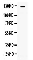
 WB (Western Blot)
(Western BlotSample: Recombinant GAL9, Human;Antibody: Rabbit Anti-Human GAL9 Ab)
WB (Western Blot)
(Western BlotSample: Recombinant GAL9, Human;Antibody: Rabbit Anti-Human GAL9 Ab)
Galectin 9 (GAL9), Active Protein (Cat# AAA21103)
Full Name
Active Galectin 9 (GAL9)
Gene Names
LGALS9; HUAT; LGALS9A
Reactivity
Homo sapiens (Human)
Applications
Cell culture; Activity Assays.
Purity
>90%


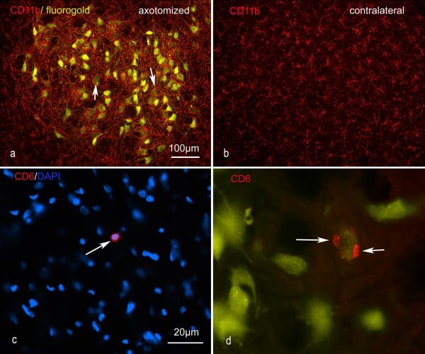 Application Data
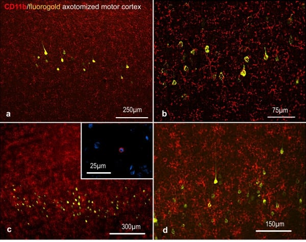
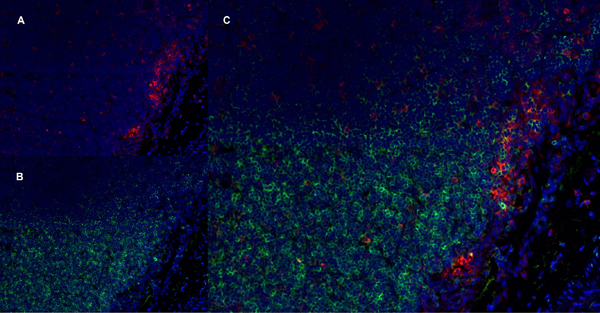
(Published customer image: Representative images of the inflammatory changes in the facial nucleus during axonal regeneration, one week following facial nerve transaction. a, b: CD11b immunoreactivity for microglia is increased in the axotomized facial nucleus, and microglia enwrap the facial motor neurons, e.g. at arrows. The regenerating neurons were retrogradely labelled with fluorogold. c, d: CD6- positive T-cells accumulated in the injured motor nucleus (arrows). They had little cytoplasm but dense nuclei (c) and were sometimes clustered around neurons retrogradely labelled with fluorogold (d). The scale bar in (a) also applies to (b) and that in (c) also applies to (d).From: Shokouhi et al. BMC Neuroscience 2010 11:13.)
Application Data
(Published customer image: Representative images of the inflammatory changes in the facial nucleus during axonal regeneration, one week following facial nerve transaction. a, b: CD11b immunoreactivity for microglia is increased in the axotomized facial nucleus, and microglia enwrap the facial motor neurons, e.g. at arrows. The regenerating neurons were retrogradely labelled with fluorogold. c, d: CD6- positive T-cells accumulated in the injured motor nucleus (arrows). They had little cytoplasm but dense nuclei (c) and were sometimes clustered around neurons retrogradely labelled with fluorogold (d). The scale bar in (a) also applies to (b) and that in (c) also applies to (d).From: Shokouhi et al. BMC Neuroscience 2010 11:13.)
CD11b, Monoclonal Antibody (Cat# AAA12146)
Full Name
MOUSE ANTI RAT CD11b:FITC
Gene Names
ITGAM; CD11B
Applications
FC/FACS


 WB (Western Blot)
(Western blot analysis of PAK1 expression in Etoposide treated 293 whole cell lysates, The lane on the left is treated with the antigen-specific peptide.)
WB (Western Blot)
(Western blot analysis of PAK1 expression in Etoposide treated 293 whole cell lysates, The lane on the left is treated with the antigen-specific peptide.)
PAK1, Polyclonal Antibody (Cat# AAA31096)
Full Name
PAK1 Antibody
Gene Names
PAK1; PAKalpha
Reactivity
Human, Mouse, Rat
Applications
WB, IHC, IF, ICC, EIA
Purity
The antiserum was purified by peptide affinity chromatography using SulfoLink Coupling Resin.


 FCM (Flow Cytometry)
(Figure 6. Flow Cytometry analysis of U-87 cells using anti-ATP citrate lyase antibody (AAA11689).Overlay histogram showing U-87 cells stained with AAA11689 (Blue line).The cells were blocked with 10% normal goat serum. And then incubated with rabbit anti-ATP citrate lyase Antibody (AAA11689,1ug/1x10^6 cells) for 30 min at 20 degree C. DyLight®488 conjugated goat anti-rabbit IgG (5-10ug/1x10^6 cells) was used as secondary antibody for 30 minutes at 20 degree C. Isotype control antibody (Green line) was rabbit IgG (1ug/1x106) used under the same conditions. Unlabelled sample (Red line) was also used as a control.)
FCM (Flow Cytometry)
(Figure 6. Flow Cytometry analysis of U-87 cells using anti-ATP citrate lyase antibody (AAA11689).Overlay histogram showing U-87 cells stained with AAA11689 (Blue line).The cells were blocked with 10% normal goat serum. And then incubated with rabbit anti-ATP citrate lyase Antibody (AAA11689,1ug/1x10^6 cells) for 30 min at 20 degree C. DyLight®488 conjugated goat anti-rabbit IgG (5-10ug/1x10^6 cells) was used as secondary antibody for 30 minutes at 20 degree C. Isotype control antibody (Green line) was rabbit IgG (1ug/1x106) used under the same conditions. Unlabelled sample (Red line) was also used as a control.)
ATP citrate lyase, Polyclonal Antibody (Cat# AAA11689)
Full Name
Anti-ATP citrate lyase Antibody
Gene Names
ACLY; ACL; ATPCL; CLATP
Reactivity
Human, Rat
Applications
WB, IHC
Purity
Immunogen affinity purified.


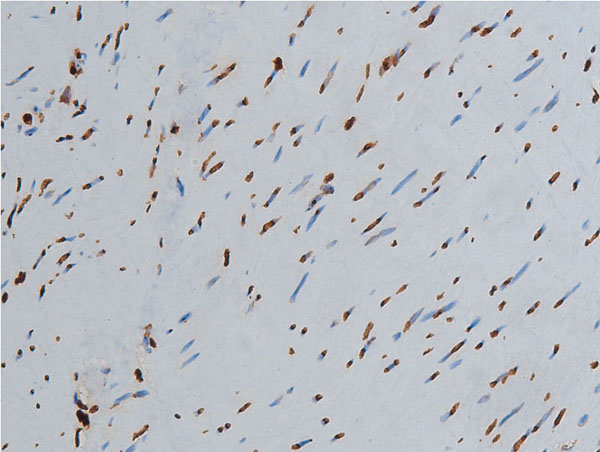
 Application Data
(Immunoperoxidase staining of rat lymph node cryosection with Mouse anti Rat CD25 followed by horseradish peroxidase conjugated Goat anti Mouse IgG1 as a detection reagent. High power)
Application Data
(Immunoperoxidase staining of rat lymph node cryosection with Mouse anti Rat CD25 followed by horseradish peroxidase conjugated Goat anti Mouse IgG1 as a detection reagent. High power)
CD25, Monoclonal Antibody (Cat# AAA11966)
Full Name
MOUSE ANTI RAT CD25
Gene Names
Il2ra; IL2RAC
Applications
EIA, FC/FACS, IP


 Application Data
(Published customer image: Representative images of the inflammatory changes in the facial nucleus during axonal regeneration, one week following facial nerve transaction. a, b: CD11b immunoreactivity for microglia is increased in the axotomized facial nucleus, and microglia enwrap the facial motor neurons, e.g. at arrows. The regenerating neurons were retrogradely labelled with fluorogold. c, d: CD6- positive T-cells accumulated in the injured motor nucleus (arrows). They had little cytoplasm but dense nuclei (c) and were sometimes clustered around neurons retrogradely labelled with fluorogold (d). The scale bar in (a) also applies to (b) and that in (c) also applies to (d).From: Shokouhi et al. BMC Neuroscience 2010 11:13.)
Application Data
(Published customer image: Representative images of the inflammatory changes in the facial nucleus during axonal regeneration, one week following facial nerve transaction. a, b: CD11b immunoreactivity for microglia is increased in the axotomized facial nucleus, and microglia enwrap the facial motor neurons, e.g. at arrows. The regenerating neurons were retrogradely labelled with fluorogold. c, d: CD6- positive T-cells accumulated in the injured motor nucleus (arrows). They had little cytoplasm but dense nuclei (c) and were sometimes clustered around neurons retrogradely labelled with fluorogold (d). The scale bar in (a) also applies to (b) and that in (c) also applies to (d).From: Shokouhi et al. BMC Neuroscience 2010 11:13.)
CD11b, Monoclonal Antibody (Cat# AAA11971)
Full Name
MOUSE ANTI RAT CD11b
Gene Names
ITGAM; CD11B
Applications
FC/FACS, IF, IP


 FCM (Flow Cytometry)
(Figure 8. Flow Cytometry analysis of U-87 cells using anti-MVP antibody (AAA19137).Overlay histogram showing U-87 cells stained with AAA19137 (Blue line).The cells were blocked with 10% normal goat serum. And then incubated with rabbit anti-MVP Antibody (AAA19137,1ug/1x10^6 cells) for 30 min at 20 degree C. DyLight®488 conjugated goat anti-rabbit IgG (5-10ug/1x10^6 cells) was used as secondary antibody for 30 minutes at 20 degree C. Isotype control antibody (Green line) was rabbit IgG (1ug/1x106) used under the same conditions. Unlabelled sample (Red line) was also used as a control.)
FCM (Flow Cytometry)
(Figure 8. Flow Cytometry analysis of U-87 cells using anti-MVP antibody (AAA19137).Overlay histogram showing U-87 cells stained with AAA19137 (Blue line).The cells were blocked with 10% normal goat serum. And then incubated with rabbit anti-MVP Antibody (AAA19137,1ug/1x10^6 cells) for 30 min at 20 degree C. DyLight®488 conjugated goat anti-rabbit IgG (5-10ug/1x10^6 cells) was used as secondary antibody for 30 minutes at 20 degree C. Isotype control antibody (Green line) was rabbit IgG (1ug/1x106) used under the same conditions. Unlabelled sample (Red line) was also used as a control.)
MVP, Polyclonal Antibody (Cat# AAA19137)
Full Name
Anti-MVP Picoband antibody
Gene Names
MVP; LRP; VAULT1
Reactivity
Human, Mouse, Rat
Applications
EIA, FC/FACS, IHC, ICC, WB


 Application Data
(At 25 degree C. The primary antibody was diluted at 1/200 and incubated with the sample for 1 hour at 37 degree C. An Alexa Fluor 594 conjugated goat anti-rabbit IgG (H+L) Ab, diluted at 1/600, was used as the secondary antibody.)
Application Data
(At 25 degree C. The primary antibody was diluted at 1/200 and incubated with the sample for 1 hour at 37 degree C. An Alexa Fluor 594 conjugated goat anti-rabbit IgG (H+L) Ab, diluted at 1/600, was used as the secondary antibody.)
Cyclin E1, Polyclonal Antibody (Cat# AAA31439)
Full Name
Phospho-Cyclin E1 (Ser399) Antibody
Gene Names
CCNE1; CCNE
Reactivity
Human, Mouse, Rat
Predicted Reactivity: Bovine (88%), Horse (88%), Sheep (88%), Chicken (88%), Xenopus (88%)
Predicted Reactivity: Bovine (88%), Horse (88%), Sheep (88%), Chicken (88%), Xenopus (88%)
Applications
WB, IHC, IF, ICC, EIA
Purity
The antibody is from purified rabbit serum by affinity purification via sequential chromatography on phospho-peptide and non-phospho-peptide affinity columns.


 IHC (Immunohistochemistry)
(AAA30929 at 1/200 staining human colon tissue sections by IHC-P. The tissue was formaldehyde fixed and a heat mediated antigen retrieval step in citrate buffer was performed. The tissue was then blocked and incubated with the antibody for 1.5 hours at 22 degree C. An HRP conjugated goat anti-rabbit antibody was used as the secondary.)
IHC (Immunohistochemistry)
(AAA30929 at 1/200 staining human colon tissue sections by IHC-P. The tissue was formaldehyde fixed and a heat mediated antigen retrieval step in citrate buffer was performed. The tissue was then blocked and incubated with the antibody for 1.5 hours at 22 degree C. An HRP conjugated goat anti-rabbit antibody was used as the secondary.)
Histone H3, Polyclonal Antibody (Cat# AAA30929)
Full Name
Histone H3 Antibody
Gene Names
HIST1H3A; H3/A; H3FA
Reactivity
Human, Mouse, Rat
Applications
WB, IHC, IF, ICC, EIA
Purity
The antiserum was purified by peptide affinity chromatography using SulfoLink Coupling Resin.


 FCM (Flow Cytometry)
(Figure 7. Flow Cytometry analysis of MCF-7 cells using anti-RGS6 antibody (AAA19308).Overlay histogram showing MCF-7 cells stained with AAA19308 (Blue line). The cells were blocked with 10% normal goat serum. And then incubated with rabbit anti-RGS6 Antibody (AAA19308, 1μg/1x106 cells) for 30 min at 20 degree C. DyLight®488 conjugated goat anti-rabbit IgG (5-10μg/1x106 cells) was used as secondary antibody for 30 minutes at 20 degree C. Isotype control antibody (Green line) was rabbit IgG (1μg/1x106) used under the same conditions. Unlabelled sample (Red line) was also used as a control.)
FCM (Flow Cytometry)
(Figure 7. Flow Cytometry analysis of MCF-7 cells using anti-RGS6 antibody (AAA19308).Overlay histogram showing MCF-7 cells stained with AAA19308 (Blue line). The cells were blocked with 10% normal goat serum. And then incubated with rabbit anti-RGS6 Antibody (AAA19308, 1μg/1x106 cells) for 30 min at 20 degree C. DyLight®488 conjugated goat anti-rabbit IgG (5-10μg/1x106 cells) was used as secondary antibody for 30 minutes at 20 degree C. Isotype control antibody (Green line) was rabbit IgG (1μg/1x106) used under the same conditions. Unlabelled sample (Red line) was also used as a control.)
RGS6, Polyclonal Antibody (Cat# AAA19308)
Full Name
Anti-RGS6 Antibody
Gene Names
RGS6; GAP
Reactivity
Human, Mouse, Rat
Applications
WB, IHC-P, FC/FACS/FCM, EIA
Purity
Immunogen affinity purified.


 FCM (Flow Cytometry)
(Figure 10. Flow Cytometry analysis of U87 cells using anti-GNG2 antibody (AAA19312).Overlay histogram showing U87 cells stained with AAA19312 (Blue line). The cells were blocked with 10% normal goat serum. And then incubated with rabbit anti-GNG2 Antibody (AAA19312, 1μg/1x106 cells) for 30 min at 20 degree C. DyLight®488 conjugated goat anti-rabbit IgG (5-10μg/1x106 cells) was used as secondary antibody for 30 minutes at 20 degree C. Isotype control antibody (Green line) was rabbit IgG (1μg/1x106) used under the same conditions. Unlabelled sample (Red line) was also used as a control.)
FCM (Flow Cytometry)
(Figure 10. Flow Cytometry analysis of U87 cells using anti-GNG2 antibody (AAA19312).Overlay histogram showing U87 cells stained with AAA19312 (Blue line). The cells were blocked with 10% normal goat serum. And then incubated with rabbit anti-GNG2 Antibody (AAA19312, 1μg/1x106 cells) for 30 min at 20 degree C. DyLight®488 conjugated goat anti-rabbit IgG (5-10μg/1x106 cells) was used as secondary antibody for 30 minutes at 20 degree C. Isotype control antibody (Green line) was rabbit IgG (1μg/1x106) used under the same conditions. Unlabelled sample (Red line) was also used as a control.)
GNG2, Polyclonal Antibody (Cat# AAA19312)
Full Name
Anti-GNG2 Antibody
Reactivity
Human, Mouse, Rat
Applications
WB, IHC-P, FC/FACS/FCM, EIA
Purity
Immunogen affinity purified.


 FCM (Flow Cytometry)
(Figure 7. Flow Cytometry analysis of Hela cells using anti- ETF/TEAD2 antibody (AAA19325).Overlay histogram showing Hela cells stained withAAA19325 (Blue line). The cells were blocked with 10% normal goat serum. And then incubated with rabbit anti- ETF/TEAD2 Antibody (AAA19325, 1μg/1x106 cells) for 30 min at 20 degree C. DyLight®488 conjugated goat anti-rabbit IgG (5-10μg/1x106 cells) was used as secondary antibody for 30 minutes at 20 degree C. Isotype control antibody (Green line) was rabbit IgG (1μg/1x106) used under the same conditions. Unlabelled sample (Red line) was also used as a control.)
FCM (Flow Cytometry)
(Figure 7. Flow Cytometry analysis of Hela cells using anti- ETF/TEAD2 antibody (AAA19325).Overlay histogram showing Hela cells stained withAAA19325 (Blue line). The cells were blocked with 10% normal goat serum. And then incubated with rabbit anti- ETF/TEAD2 Antibody (AAA19325, 1μg/1x106 cells) for 30 min at 20 degree C. DyLight®488 conjugated goat anti-rabbit IgG (5-10μg/1x106 cells) was used as secondary antibody for 30 minutes at 20 degree C. Isotype control antibody (Green line) was rabbit IgG (1μg/1x106) used under the same conditions. Unlabelled sample (Red line) was also used as a control.)
ETF/TEAD2, Polyclonal Antibody (Cat# AAA19325)
Full Name
Anti-ETF/TEAD2 Antibody
Gene Names
TEAD2; ETF; TEF4; TEF-4; TEAD-2
Reactivity
Human, Mouse, Rat
Applications
WB, IHC-P, FC/FACS/FCM, EIA
Purity
Immunogen affinity purified.


 FCM (Flow Cytometry)
(Figure 7. Flow Cytometry analysis of HELA cells using anti-MCU antibody (AAA19226).Overlay histogram showing HELA cells stained with AAA19226 (Blue line). The cells were blocked with 10% normal goat serum. And then incubated with rabbit anti-MCU Antibody (AAA19226, 1μg/1x106 cells) for 30 min at 20 degree C. DyLight®488 conjugated goat anti-rabbit IgG (5-10μg/1x106 cells) was used as secondary antibody for 30 minutes at 20 degree C. Isotype control antibody (Green line) was rabbit IgG (1μg/1x106) used under the same conditions. Unlabelled sample (Red line) was also used as a control.)
FCM (Flow Cytometry)
(Figure 7. Flow Cytometry analysis of HELA cells using anti-MCU antibody (AAA19226).Overlay histogram showing HELA cells stained with AAA19226 (Blue line). The cells were blocked with 10% normal goat serum. And then incubated with rabbit anti-MCU Antibody (AAA19226, 1μg/1x106 cells) for 30 min at 20 degree C. DyLight®488 conjugated goat anti-rabbit IgG (5-10μg/1x106 cells) was used as secondary antibody for 30 minutes at 20 degree C. Isotype control antibody (Green line) was rabbit IgG (1μg/1x106) used under the same conditions. Unlabelled sample (Red line) was also used as a control.)
MCU, Polyclonal Antibody (Cat# AAA19226)
Full Name
Anti-MCU Antibody
Gene Names
MCU; C10orf42; CCDC109A
Reactivity
Human, Mouse, Rat, Monkey
Applications
WB, IHC-P, FC/FACS/FCM, EIA
Purity
Immunogen affinity purified.


 FCM (Flow Cytometry)
(Figure 7. Flow Cytometry analysis of THP-1 cells using anti-PCBP2/hnRNP E2 antibody (AAA19259).Overlay histogram showing THP-1 cells stained with AAA19259 (Blue line). The cells were blocked with 10% normal goat serum. And then incubated with rabbit anti-PCBP2/hnRNP E2 Antibody (AAA19259, 1μg/1x106 cells) for 30 min at 20 degree C. DyLight®488 conjugated goat anti-rabbit IgG (5-10μg/1x106 cells) was used as secondary antibody for 30 minutes at 20 degree C. Isotype control antibody (Green line) was rabbit IgG (1μg/1x106) used under the same conditions. Unlabelled sample (Red line) was also used as a control.)
FCM (Flow Cytometry)
(Figure 7. Flow Cytometry analysis of THP-1 cells using anti-PCBP2/hnRNP E2 antibody (AAA19259).Overlay histogram showing THP-1 cells stained with AAA19259 (Blue line). The cells were blocked with 10% normal goat serum. And then incubated with rabbit anti-PCBP2/hnRNP E2 Antibody (AAA19259, 1μg/1x106 cells) for 30 min at 20 degree C. DyLight®488 conjugated goat anti-rabbit IgG (5-10μg/1x106 cells) was used as secondary antibody for 30 minutes at 20 degree C. Isotype control antibody (Green line) was rabbit IgG (1μg/1x106) used under the same conditions. Unlabelled sample (Red line) was also used as a control.)
PCBP2/hnRNP E2, Polyclonal Antibody (Cat# AAA19259)
Full Name
Anti-PCBP2/hnRNP E2 Antibody
Gene Names
PCBP2; HNRPE2; HNRNPE2; hnRNP-E2
Reactivity
Human, Mouse, Rat
Applications
WB, IHC-P, FC/FACS/FCM, EIA
Purity
Immunogen affinity purified.


 FCM (Flow Cytometry)
(Figure 7. Flow Cytometry analysis of Hela cells using anti-ATG9A antibody (AAA19280).Overlay histogram showing Hela cells stained with AAA19280 (Blue line). The cells were blocked with 10% normal goat serum. And then incubated with rabbit anti-ATG9A Antibody (AAA19280, 1μg/1x106 cells) for 30 min at 20 degree C. DyLight®488 conjugated goat anti-rabbit IgG (5-10μg/1x106 cells) was used as secondary antibody for 30 minutes at 20 degree C. Isotype control antibody (Green line) was rabbit IgG (1μg/1x106) used under the same conditions. Unlabelled sample (Red line) was also used as a control.)
FCM (Flow Cytometry)
(Figure 7. Flow Cytometry analysis of Hela cells using anti-ATG9A antibody (AAA19280).Overlay histogram showing Hela cells stained with AAA19280 (Blue line). The cells were blocked with 10% normal goat serum. And then incubated with rabbit anti-ATG9A Antibody (AAA19280, 1μg/1x106 cells) for 30 min at 20 degree C. DyLight®488 conjugated goat anti-rabbit IgG (5-10μg/1x106 cells) was used as secondary antibody for 30 minutes at 20 degree C. Isotype control antibody (Green line) was rabbit IgG (1μg/1x106) used under the same conditions. Unlabelled sample (Red line) was also used as a control.)
ATG9A, Polyclonal Antibody (Cat# AAA19280)
Full Name
Anti-ATG9A Antibody
Gene Names
ATG9A; mATG9; APG9L1; MGD3208
Reactivity
Human, Mouse, Rat
Applications
WB, IHC-P, FC/FACS/FCM, EIA
Purity
Immunogen affinity purified.


 FCM (Flow Cytometry)
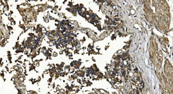
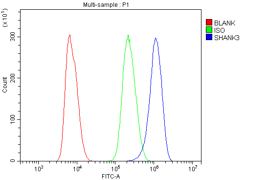
(Figure 6. Flow Cytometry analysis of NRK cells using anti-SHANK3 antibody (AAA19237).Overlay histogram showing NRK cells stained with AAA19237 (Blue line). The cells were blocked with 10% normal goat serum. And then incubated with rabbit anti-SHANK3 Antibody (AAA19237,1μg/1x106 cells) for 30 min at 20 degree C. DyLight®488 conjugated goat anti-rabbit IgG (5-10μg/1x106 cells) was used as secondary antibody for 30 minutes at 20 degree C. Isotype control antibody (Green line) was rabbit IgG (1μg/1x106) used under the same conditions. Unlabelled sample (Red line) was also used as a control.)
FCM (Flow Cytometry)
(Figure 6. Flow Cytometry analysis of NRK cells using anti-SHANK3 antibody (AAA19237).Overlay histogram showing NRK cells stained with AAA19237 (Blue line). The cells were blocked with 10% normal goat serum. And then incubated with rabbit anti-SHANK3 Antibody (AAA19237,1μg/1x106 cells) for 30 min at 20 degree C. DyLight®488 conjugated goat anti-rabbit IgG (5-10μg/1x106 cells) was used as secondary antibody for 30 minutes at 20 degree C. Isotype control antibody (Green line) was rabbit IgG (1μg/1x106) used under the same conditions. Unlabelled sample (Red line) was also used as a control.)
SHANK3, Polyclonal Antibody (Cat# AAA19237)
Full Name
Anti-SHANK3 Antibody
Gene Names
SHANK3; PSAP2; SCZD15; PROSAP2; SPANK-2; DEL22q13.3
Reactivity
Human, Mouse, Rat
Applications
WB, IHC-P, FC/FACS/FCM, EIA
Purity
Immunogen affinity purified.


 FCM (Flow Cytometry)
(Figure 6. Flow Cytometry analysis of MCF-7 cells using anti-ALK-1/ACVRL1 antibody (AAA19242).Overlay histogram showing MCF-7 cells stained with AAA19242 (Blue line). The cells were blocked with 10% normal goat serum. And then incubated with rabbit anti-ALK-1/ACVRL1 Antibody (AAA19242, 1μg/1x106 cells) for 30 min at 20 degree C. DyLight®488 conjugated goat anti-rabbit IgG (5-10μg/1x106 cells) was used as secondary antibody for 30 minutes at 20 degree C. Isotype control antibody (Green line) was rabbit IgG (1μg/1x106) used under the same conditions. Unlabelled sample (Red line) was also used as a control.)
FCM (Flow Cytometry)
(Figure 6. Flow Cytometry analysis of MCF-7 cells using anti-ALK-1/ACVRL1 antibody (AAA19242).Overlay histogram showing MCF-7 cells stained with AAA19242 (Blue line). The cells were blocked with 10% normal goat serum. And then incubated with rabbit anti-ALK-1/ACVRL1 Antibody (AAA19242, 1μg/1x106 cells) for 30 min at 20 degree C. DyLight®488 conjugated goat anti-rabbit IgG (5-10μg/1x106 cells) was used as secondary antibody for 30 minutes at 20 degree C. Isotype control antibody (Green line) was rabbit IgG (1μg/1x106) used under the same conditions. Unlabelled sample (Red line) was also used as a control.)
ALK-1/ACVRL1, Polyclonal Antibody (Cat# AAA19242)
Full Name
Anti-ALK-1/ACVRL1 Antibody
Gene Names
ACVRL1; HHT; ALK1; HHT2; ORW2; SKR3; ALK-1; TSR-I; ACVRLK1
Reactivity
Human, Mouse, Rat
Applications
WB, IHC-P, FC/FACS/FCM, EIA
Purity
Immunogen affinity purified.


 FCM (Flow Cytometry)
(Figure 6. Flow Cytometry analysis of A431 cells using anti-TMPRSS3 antibody (AAA19289).Overlay histogram showing A431 cells stained with AAA19289 (Blue line). The cells were blocked with 10% normal goat serum. And then incubated with rabbit anti-TMPRSS3 Antibody (AAA19289, 1μg/1x106 cells) for 30 min at 20 degree C. DyLight®488 conjugated goat anti-rabbit IgG (5-10μg/1x106 cells) was used as secondary antibody for 30 minutes at 20 degree C. Isotype control antibody (Green line) was rabbit IgG (1μg/1x106) used under the same conditions. Unlabelled sample (Red line) was also used as a control.)
FCM (Flow Cytometry)
(Figure 6. Flow Cytometry analysis of A431 cells using anti-TMPRSS3 antibody (AAA19289).Overlay histogram showing A431 cells stained with AAA19289 (Blue line). The cells were blocked with 10% normal goat serum. And then incubated with rabbit anti-TMPRSS3 Antibody (AAA19289, 1μg/1x106 cells) for 30 min at 20 degree C. DyLight®488 conjugated goat anti-rabbit IgG (5-10μg/1x106 cells) was used as secondary antibody for 30 minutes at 20 degree C. Isotype control antibody (Green line) was rabbit IgG (1μg/1x106) used under the same conditions. Unlabelled sample (Red line) was also used as a control.)
TMPRSS3, Polyclonal Antibody (Cat# AAA19289)
Full Name
Anti-TMPRSS3 Antibody
Gene Names
TMPRSS3; DFNB8; DFNB10; ECHOS1; TADG12
Reactivity
Human, Mouse, Rat
Applications
WB, IHC-P, FC/FACS/FCM, EIA
Purity
Immunogen affinity purified.


 FCM (Flow Cytometry)
(Figure 6. Flow Cytometry analysis of PC-3 cells using anti-MitoNEET/CISD1 antibody (AAA19293).Overlay histogram showing PC-3 cells stained with AAA19293 (Blue line). The cells were blocked with 10% normal goat serum. And then incubated with rabbit anti-MitoNEET/CISD1 Antibody (AAA19293,1μg/1x106 cells) for 30 min at 20 degree C. DyLight®488 conjugated goat anti-rabbit IgG (5-10μg/1x106 cells) was used as secondary antibody for 30 minutes at 20 degree C. Isotype control antibody (Green line) was rabbit IgG (1μg/1x106) used under the same conditions. Unlabelled sample (Red line) was also used as a control.)
FCM (Flow Cytometry)
(Figure 6. Flow Cytometry analysis of PC-3 cells using anti-MitoNEET/CISD1 antibody (AAA19293).Overlay histogram showing PC-3 cells stained with AAA19293 (Blue line). The cells were blocked with 10% normal goat serum. And then incubated with rabbit anti-MitoNEET/CISD1 Antibody (AAA19293,1μg/1x106 cells) for 30 min at 20 degree C. DyLight®488 conjugated goat anti-rabbit IgG (5-10μg/1x106 cells) was used as secondary antibody for 30 minutes at 20 degree C. Isotype control antibody (Green line) was rabbit IgG (1μg/1x106) used under the same conditions. Unlabelled sample (Red line) was also used as a control.)
MitoNEET/CISD1, Polyclonal Antibody (Cat# AAA19293)
Full Name
Anti-MitoNEET/CISD1 Antibody
Gene Names
CISD1; ZCD1; MDS029; C10orf70; mitoNEET
Reactivity
Human, Mouse, Rat, Monkey
Applications
WB, IHC-P, FC/FACS/FCM, EIA
Purity
Immunogen affinity purified.


 FCM (Flow Cytometry)
(Figure 7. Flow Cytometry analysis of HEPG2 cells using anti-ALDH1L1 antibody (AAA19297).Overlay histogram showing HEPG2 cells stained with AAA19297 (Blue line). The cells were blocked with 10% normal goat serum. And then incubated with rabbit anti-ALDH1L1 Antibody (AAA19297 1μg/1x106 cells) for 30 min at 20 degree C. DyLight®488 conjugated goat anti-rabbit IgG (5-10μg/1x106 cells) was used as secondary antibody for 30 minutes at 20 degree C. Isotype control antibody (Green line) was rabbit IgG (1μg/1x106) used under the same conditions. Unlabelled sample (Red line) was also used as a control.)
FCM (Flow Cytometry)
(Figure 7. Flow Cytometry analysis of HEPG2 cells using anti-ALDH1L1 antibody (AAA19297).Overlay histogram showing HEPG2 cells stained with AAA19297 (Blue line). The cells were blocked with 10% normal goat serum. And then incubated with rabbit anti-ALDH1L1 Antibody (AAA19297 1μg/1x106 cells) for 30 min at 20 degree C. DyLight®488 conjugated goat anti-rabbit IgG (5-10μg/1x106 cells) was used as secondary antibody for 30 minutes at 20 degree C. Isotype control antibody (Green line) was rabbit IgG (1μg/1x106) used under the same conditions. Unlabelled sample (Red line) was also used as a control.)
ALDH1L1, Polyclonal Antibody (Cat# AAA19297)
Full Name
Anti-ALDH1L1 Antibody
Gene Names
ALDH1L1; FDH; FTHFD; 10-fTHF; 10-FTHFDH
Reactivity
Human, Mouse, Rat, Monkey
Applications
WB, IHC-P, ICC, IF, FC/FACS/FCM, EIA
Purity
Immunogen affinity purified.


 FCM (Flow Cytometry)
(Figure 8. Flow Cytometry analysis of A549 cells using anti-Transketolase/TKT antibody (AAA19368).Overlay histogram showing A549 cells stained with AAA19368 (Blue line). The cells were blocked with 10% normal goat serum. And then incubated with mouse anti- Transketolase/TKT Antibody (AAA19368, 1μg/1x106 cells) for 30 min at 20 degree C. DyLight®488 conjugated goat anti-mouse IgG (BA1126, 5-10μg/1x106 cells) was used as secondary antibody for 30 minutes at 20 degree C. Isotype control antibody (Green line) was mouse IgG (1μg/1x106) used under the same conditions. Unlabelled sample (Red line) was also used as a control.)
FCM (Flow Cytometry)
(Figure 8. Flow Cytometry analysis of A549 cells using anti-Transketolase/TKT antibody (AAA19368).Overlay histogram showing A549 cells stained with AAA19368 (Blue line). The cells were blocked with 10% normal goat serum. And then incubated with mouse anti- Transketolase/TKT Antibody (AAA19368, 1μg/1x106 cells) for 30 min at 20 degree C. DyLight®488 conjugated goat anti-mouse IgG (BA1126, 5-10μg/1x106 cells) was used as secondary antibody for 30 minutes at 20 degree C. Isotype control antibody (Green line) was mouse IgG (1μg/1x106) used under the same conditions. Unlabelled sample (Red line) was also used as a control.)
Transketolase/TKT, Monoclonal Antibody (Cat# AAA19368)
Full Name
Anti-Transketolase/TKT Antibody (monoclonal, 2I3)
Gene Names
TKT; TK; TKT1; HEL107
Reactivity
Human, Mouse, Rat
Applications
WB, IHC-P, ICC, IF, FC/FACS/FCM
Purity
Immunogen affinity purified.


 FCM (Flow Cytometry)
(Figure 6. Flow Cytometry analysis of THP-1 cells using anti-TLR1 antibody (AAA19134).Overlay histogram showing THP-1 cells stained with AAA19134 (Blue line).The cells were blocked with 10% normal goat serum. And then incubated with rabbit anti-TLR1 Antibody (AAA19134,1ug/1x10^6 cells) for 30 min at 20 degree C. DyLight®488 conjugated goat anti-rabbit IgG (5-10ug/1x10^6 cells) was used as secondary antibody for 30 minutes at 20 degree C. Isotype control antibody (Green line) was rabbit IgG (1ug/1x106) used under the same conditions. Unlabelled sample (Red line) was also used as a control.)
FCM (Flow Cytometry)
(Figure 6. Flow Cytometry analysis of THP-1 cells using anti-TLR1 antibody (AAA19134).Overlay histogram showing THP-1 cells stained with AAA19134 (Blue line).The cells were blocked with 10% normal goat serum. And then incubated with rabbit anti-TLR1 Antibody (AAA19134,1ug/1x10^6 cells) for 30 min at 20 degree C. DyLight®488 conjugated goat anti-rabbit IgG (5-10ug/1x10^6 cells) was used as secondary antibody for 30 minutes at 20 degree C. Isotype control antibody (Green line) was rabbit IgG (1ug/1x106) used under the same conditions. Unlabelled sample (Red line) was also used as a control.)
TLR1, Polyclonal Antibody (Cat# AAA19134)
Full Name
Anti-TLR1 Picoband antibody
Gene Names
TLR1; TIL; CD281; rsc786; TIL. LPRS5
Reactivity
Human, Mouse, Rat
No cross reactivity with other proteins.
No cross reactivity with other proteins.
Applications
EIA, FC/FACS, IHC, ICC, WB


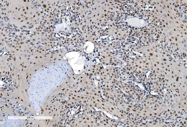
 IHC (Immunohistchemistry)
(Figure 6. IHC analysis of Flt3 / CD135 using anti-Flt3 / CD135 antibody (AAA19130).Flt3 / CD135 was detected in paraffin-embedded section of mouse kidney tissue. Heat mediated antigen retrieval was performed in citrate buffer (pH6, epitope retrieval solution) for 20 mins. The tissue section was blocked with 10% goat serum. The tissue section was then incubated with 1ug/ml rabbit anti-Flt3 / CD135 Antibody (AAA19130) overnight at 4 degree C. Biotinylated goat anti-rabbit IgG was used as secondary antibody and incubated for 30 minutes at 37 degree C. The tissue section was developed using Strepavidin-Biotin-Complex (SABC) with DAB as the chromogen.)
IHC (Immunohistchemistry)
(Figure 6. IHC analysis of Flt3 / CD135 using anti-Flt3 / CD135 antibody (AAA19130).Flt3 / CD135 was detected in paraffin-embedded section of mouse kidney tissue. Heat mediated antigen retrieval was performed in citrate buffer (pH6, epitope retrieval solution) for 20 mins. The tissue section was blocked with 10% goat serum. The tissue section was then incubated with 1ug/ml rabbit anti-Flt3 / CD135 Antibody (AAA19130) overnight at 4 degree C. Biotinylated goat anti-rabbit IgG was used as secondary antibody and incubated for 30 minutes at 37 degree C. The tissue section was developed using Strepavidin-Biotin-Complex (SABC) with DAB as the chromogen.)
Flt3/CD135, Polyclonal Antibody (Cat# AAA19130)
Full Name
Anti-Flt3/CD135 Picoband Antibody
Reactivity
Mouse, Rat
No cross reactivity with other proteins.
No cross reactivity with other proteins.
Applications
EIA, IHC, WB
Purity
Immunogen affinity purified


 IHC (Immunohistochemistry)
(Figure 8. IHC analysis of beta Catenin using anti-beta Catenin antibody (AAA19129).beta Catenin was detected in paraffin-embedded section of rat small intestine tissue. Heat mediated antigen retrieval was performed in citrate buffer (pH6, epitope retrieval solution) for 20 mins. The tissue section was blocked with 10% goat serum. The tissue section was then incubated with 1ug/ml rabbit anti-beta Catenin Antibody (AAA19129) overnight at 4 degree C. Biotinylated goat anti-rabbit IgG was used as secondary antibody and incubated for 30 minutes at 37 degree C. The tissue section was developed using Strepavidin-Biotin-Complex (SABC) with DAB as the chromogen.)
IHC (Immunohistochemistry)
(Figure 8. IHC analysis of beta Catenin using anti-beta Catenin antibody (AAA19129).beta Catenin was detected in paraffin-embedded section of rat small intestine tissue. Heat mediated antigen retrieval was performed in citrate buffer (pH6, epitope retrieval solution) for 20 mins. The tissue section was blocked with 10% goat serum. The tissue section was then incubated with 1ug/ml rabbit anti-beta Catenin Antibody (AAA19129) overnight at 4 degree C. Biotinylated goat anti-rabbit IgG was used as secondary antibody and incubated for 30 minutes at 37 degree C. The tissue section was developed using Strepavidin-Biotin-Complex (SABC) with DAB as the chromogen.)
beta Catenin, Polyclonal Antibody (Cat# AAA19129)
Full Name
Anti-beta Catenin Picoband Antibody
Gene Names
CTNNB1; EVR7; CTNNB; MRD19; armadillo
Reactivity
Human, Mouse, Rat
No cross reactivity with other proteins.
No cross reactivity with other proteins.
Applications
EIA, IHC, WB
Purity
Immunogen affinity purified


 IHC (Immunohistchemistry)
(Figure 6. IHC analysis of HP1 gamma using anti-HP1 gamma antibody (AAA19145).HP1 gamma was detected in paraffin-embedded section of rat small intestine tissue. Heat mediated antigen retrieval was performed in citrate buffer (pH6, epitope retrieval solution) for 20 mins. The tissue section was blocked with 10% goat serum. The tissue section was then incubated with 1ug/ml rabbit anti-HP1 gamma Antibody (AAA19145) overnight at 4 degree C. Biotinylated goat anti-rabbit IgG was used as secondary antibody and incubated for 30 minutes at 37 degree C. The tissue section was developed using Strepavidin-Biotin-Complex (SABC) with DAB as the chromogen.)
IHC (Immunohistchemistry)
(Figure 6. IHC analysis of HP1 gamma using anti-HP1 gamma antibody (AAA19145).HP1 gamma was detected in paraffin-embedded section of rat small intestine tissue. Heat mediated antigen retrieval was performed in citrate buffer (pH6, epitope retrieval solution) for 20 mins. The tissue section was blocked with 10% goat serum. The tissue section was then incubated with 1ug/ml rabbit anti-HP1 gamma Antibody (AAA19145) overnight at 4 degree C. Biotinylated goat anti-rabbit IgG was used as secondary antibody and incubated for 30 minutes at 37 degree C. The tissue section was developed using Strepavidin-Biotin-Complex (SABC) with DAB as the chromogen.)
HP1 gamma, Polyclonal Antibody (Cat# AAA19145)
Full Name
Anti-HP1 gamma Picoband antibody
Gene Names
CBX3; HECH; HP1-GAMMA; HP1Hs-gamma
Reactivity
Human, Mouse, Rat
No cross reactivity with other proteins.
No cross reactivity with other proteins.
Applications
EIA, IHC, WB


 IHC (Immunohistchemistry)
(Figure 6. IHC analysis of Cyclophilin B using anti-Cyclophilin B antibody (AAA19167).Cyclophilin B was detected in paraffin-embedded section of rat thyroid gland tissue. Heat mediated antigen retrieval was performed in citrate buffer (pH6, epitope retrieval solution) for 20 mins. The tissue section was blocked with 10% goat serum. The tissue section was then incubated with 1ug/ml rabbit anti-Cyclophilin B Antibody (AAA19167) overnight at 4 degree C. Biotinylated goat anti-rabbit IgG was used as secondary antibody and incubated for 30 minutes at 37 degree C. The tissue section was developed using Strepavidin-Biotin-Complex (SABC) with DAB as the chromogen.)
IHC (Immunohistchemistry)
(Figure 6. IHC analysis of Cyclophilin B using anti-Cyclophilin B antibody (AAA19167).Cyclophilin B was detected in paraffin-embedded section of rat thyroid gland tissue. Heat mediated antigen retrieval was performed in citrate buffer (pH6, epitope retrieval solution) for 20 mins. The tissue section was blocked with 10% goat serum. The tissue section was then incubated with 1ug/ml rabbit anti-Cyclophilin B Antibody (AAA19167) overnight at 4 degree C. Biotinylated goat anti-rabbit IgG was used as secondary antibody and incubated for 30 minutes at 37 degree C. The tissue section was developed using Strepavidin-Biotin-Complex (SABC) with DAB as the chromogen.)
Cyclophilin B, Polyclonal Antibody (Cat# AAA19167)
Full Name
Anti-Cyclophilin B Picoband Antibody
Gene Names
PPIB; OI9; CYPB; SCYLP; CYP-S1; HEL-S-39
Reactivity
Human, Mouse, Rat
No cross reactivity with other proteins.
No cross reactivity with other proteins.
Applications
EIA, IHC, WB
Purity
Immunogen affinity purified


 IHC (Immunohistchemistry)
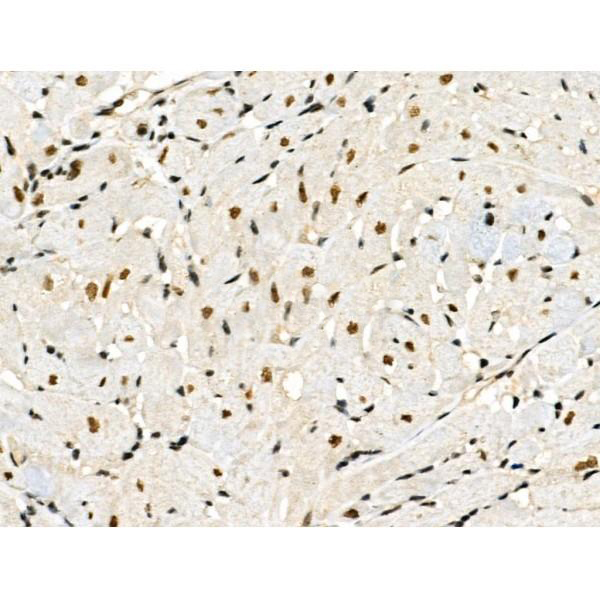
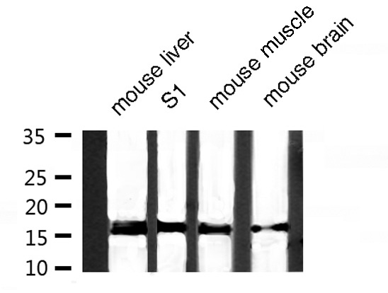
(At 1/100 staining Mouse liver tissue by IHC-P. The sample was formaldehyde fixed and a heat mediated antigen retrieval step in citrate buffer was performed. The sample was then blocked and incubated with the primary antibody at 4 degree C overnight. An HRP conjugated anti-Rabbit antibody was used as the secondary antibody.)
IHC (Immunohistchemistry)
(At 1/100 staining Mouse liver tissue by IHC-P. The sample was formaldehyde fixed and a heat mediated antigen retrieval step in citrate buffer was performed. The sample was then blocked and incubated with the primary antibody at 4 degree C overnight. An HRP conjugated anti-Rabbit antibody was used as the secondary antibody.)
NDEL1, Polyclonal Antibody (Cat# AAA31462)
Full Name
Phospho-NDEL1 (Thr219) Antibody
Gene Names
NDEL1; EOPA; NDE2; NUDEL; MITAP1; NDE1L1
Reactivity
Human, Mouse, Rat
Predicted Reactivity: Pig (100%), Bovine (100%), Horse (100%), Sheep (100%), Rabbit (100%), Dog (100%), Chicken (100%), Xenopus (100%)
Predicted Reactivity: Pig (100%), Bovine (100%), Horse (100%), Sheep (100%), Rabbit (100%), Dog (100%), Chicken (100%), Xenopus (100%)
Applications
WB, IHC, EIA
Purity
The antibody is from purified rabbit serum by affinity purification via sequential chromatography on phospho-peptide and non-phospho-peptide affinity columns.


 Application Data
(Published customer image: Representative images of the inflammatory changes in the facial nucleus during axonal regeneration, one week following facial nerve transaction. a, b: CD11b immunoreactivity for microglia is increased in the axotomized facial nucleus, and microglia enwrap the facial motor neurons, e.g. at arrows. The regenerating neurons were retrogradely labelled with fluorogold. c, d: CD6- positive T-cells accumulated in the injured motor nucleus (arrows). They had little cytoplasm but dense nuclei (c) and were sometimes clustered around neurons retrogradely labelled with fluorogold (d). The scale bar in (a) also applies to (b) and that in (c) also applies to (d).From: Shokouhi et al. BMC Neuroscience 2010 11:13.)
Application Data
(Published customer image: Representative images of the inflammatory changes in the facial nucleus during axonal regeneration, one week following facial nerve transaction. a, b: CD11b immunoreactivity for microglia is increased in the axotomized facial nucleus, and microglia enwrap the facial motor neurons, e.g. at arrows. The regenerating neurons were retrogradely labelled with fluorogold. c, d: CD6- positive T-cells accumulated in the injured motor nucleus (arrows). They had little cytoplasm but dense nuclei (c) and were sometimes clustered around neurons retrogradely labelled with fluorogold (d). The scale bar in (a) also applies to (b) and that in (c) also applies to (d).From: Shokouhi et al. BMC Neuroscience 2010 11:13.)
CD11b, Monoclonal Antibody (Cat# AAA11876)
Full Name
MOUSE ANTI RAT CD11b:FITC
Gene Names
ITGAM; CD11B
Applications
FC/FACS


 IHC (Immunohistchemistry)
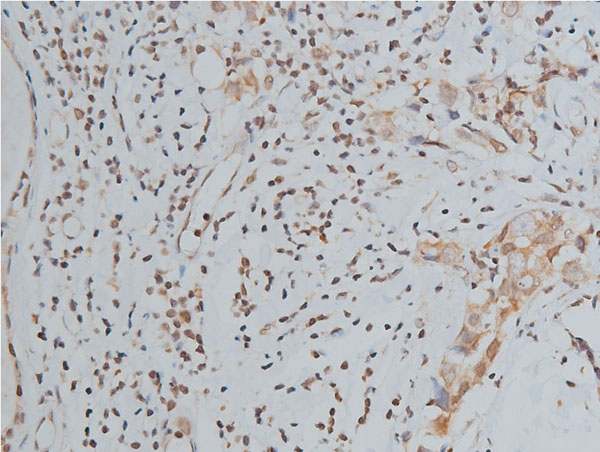
(AAA31095 at 1/100 staining Human kidney tissue by IHC-P. The sample was formaldehyde fixed and a heat mediated antigen retrieval step in citrate buffer was performed. The sample was then blocked and incubated with the antibody for 1.5 hours at 22 degree C. An HRP conjugated goat anti-rabbit antibody was used as the secondary.)
IHC (Immunohistchemistry)
(AAA31095 at 1/100 staining Human kidney tissue by IHC-P. The sample was formaldehyde fixed and a heat mediated antigen retrieval step in citrate buffer was performed. The sample was then blocked and incubated with the antibody for 1.5 hours at 22 degree C. An HRP conjugated goat anti-rabbit antibody was used as the secondary.)
Histone H3, Polyclonal Antibody (Cat# AAA31095)
Full Name
Histone H3 Antibody
Gene Names
HIST1H3A; H3/A; H3FA
Reactivity
Human, Mouse, Rat
Applications
WB, IHC, IF, ICC, EIA
Purity
The antiserum was purified by peptide affinity chromatography using SulfoLink Coupling Resin.


 ICC (Immunocytochemistry)
(Figure 7. IHC analysis of ERAB using anti-ERAB antibody (AAA11668).ERAB was detected in immunocytochemical section of SMMC-7721 cell. Heat mediated antigen retrieval was performed in citrate buffer (pH6, epitope retrieval solution) for 20 mins. The tissue section was blocked with 10% goat serum. The tissue section was then incubated with 1ug/ml rabbit anti-ERAB Antibody (AAA11668) overnight at 4 degree C. Biotinylated goat anti-rabbit IgG was used as secondary antibody and incubated for 30 minutes at 37 degree C. The tissue section was developed using Strepavidin-Biotin-Complex (SABC) with DAB as the chromogen.)
ICC (Immunocytochemistry)
(Figure 7. IHC analysis of ERAB using anti-ERAB antibody (AAA11668).ERAB was detected in immunocytochemical section of SMMC-7721 cell. Heat mediated antigen retrieval was performed in citrate buffer (pH6, epitope retrieval solution) for 20 mins. The tissue section was blocked with 10% goat serum. The tissue section was then incubated with 1ug/ml rabbit anti-ERAB Antibody (AAA11668) overnight at 4 degree C. Biotinylated goat anti-rabbit IgG was used as secondary antibody and incubated for 30 minutes at 37 degree C. The tissue section was developed using Strepavidin-Biotin-Complex (SABC) with DAB as the chromogen.)
ERAB, Polyclonal Antibody (Cat# AAA11668)
Full Name
Anti-ERAB Antibody
Gene Names
HSD17B10; ABAD; CAMR; ERAB; HCD2; MHBD; HADH2; MRPP2; MRX17; MRX31; SCHAD; MRXS10; SDR5C1; 17b-HSD10; DUPXp11.22
Reactivity
Human, Mouse
Applications
WB, IHC
Purity
Immunogen Affinity Purified


 WB (Western Blot)
(Sample: Recombinant GAL3, Human; Antibody: Rabbit Anti-Human GAL3 Ab)
WB (Western Blot)
(Sample: Recombinant GAL3, Human; Antibody: Rabbit Anti-Human GAL3 Ab)
Galectin 3 (GAL3), Active Protein (Cat# AAA21140)
Full Name
Active Galectin 3 (GAL3)
Gene Names
LGALS3; L31; GAL3; MAC2; CBP35; GALBP; GALIG; LGALS2
Reactivity
Homo sapiens (Human)
Applications
Cell culture; Activity Assays.
Purity
>98%


 Application Data
TCP1 delta, Polyclonal Antibody (Cat# AAA11672)
Full Name
Anti-TCP1 delta Antibody
Gene Names
CCT4; SRB; Cctd; CCT-DELTA
Reactivity
Human, Mouse, Rat
No cross reactivity with other proteins.
No cross reactivity with other proteins.
Applications
WB, IHC
Purity
Immunogen affinity purified.


 Application Data
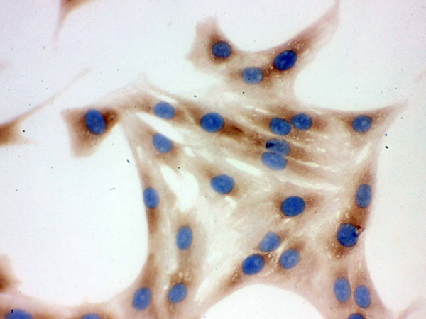
(At 25 degree C. The primary antibody was diluted at 1/200 and incubated with the sample for 1 hour at 37 degree C. An Alexa Fluor 594 conjugated goat anti-rabbit IgG (H+L) Ab, diluted at 1/600, was used as the secondary antibody.)
Application Data
(At 25 degree C. The primary antibody was diluted at 1/200 and incubated with the sample for 1 hour at 37 degree C. An Alexa Fluor 594 conjugated goat anti-rabbit IgG (H+L) Ab, diluted at 1/600, was used as the secondary antibody.)
p70 S6 Kinase, Polyclonal Antibody (Cat# AAA31408)
Full Name
Phospho-p70 S6 Kinase (Ser427) Antibody
Gene Names
RPS6KB1; S6K; PS6K; S6K1; STK14A; p70-S6K; p70 S6KA; p70-alpha; S6K-beta-1; p70(S6K)-alpha
Reactivity
Human, Mouse, Rat
Predicted Reactivity: Pig (100%), Bovine (100%), Horse (100%), Sheep (100%), Rabbit (100%), Dog (100%), Chicken (100%), Xenopus (100%)
Predicted Reactivity: Pig (100%), Bovine (100%), Horse (100%), Sheep (100%), Rabbit (100%), Dog (100%), Chicken (100%), Xenopus (100%)
Applications
WB, IHC, IF, ICC, EIA
Purity
The antibody is from purified rabbit serum by affinity purification via sequential chromatography on phospho-peptide and non-phospho-peptide affinity columns.


 Application Data
(Immunoperoxidase staining of rat lymph node cryosection with Mouse anti Rat CD25 followed by horseradish peroxidase conjugated Goat anti Mouse IgG1 as a detection reagent. High power)
Application Data
(Immunoperoxidase staining of rat lymph node cryosection with Mouse anti Rat CD25 followed by horseradish peroxidase conjugated Goat anti Mouse IgG1 as a detection reagent. High power)
CD25, Monoclonal Antibody (Cat# AAA11965)
Full Name
MOUSE ANTI RAT CD25
Gene Names
Il2ra; IL2RAC
Applications
EIA, FC/FACS, IP


 FCM (Flow Cytometry)
(Figure 6. Flow Cytometry analysis of Hela cells using anti-PDE4D antibody (AAA19143).Overlay histogram showing Hela cells stained with AAA19143 (Blue line).The cells were blocked with 10% normal goat serum. And then incubated with rabbit anti-PDE4D Antibody (AAA19143,1ug/1x10^6 cells) for 30 min at 20 degree C. DyLight®488 conjugated goat anti-rabbit IgG (5-10ug/1x10^6 cells) was used as secondary antibody for 30 minutes at 20 degree C. Isotype control antibody (Green line) was rabbit IgG (1ug/1x106) used under the same conditions. Unlabelled sample (Red line) was also used as a control.)
FCM (Flow Cytometry)
(Figure 6. Flow Cytometry analysis of Hela cells using anti-PDE4D antibody (AAA19143).Overlay histogram showing Hela cells stained with AAA19143 (Blue line).The cells were blocked with 10% normal goat serum. And then incubated with rabbit anti-PDE4D Antibody (AAA19143,1ug/1x10^6 cells) for 30 min at 20 degree C. DyLight®488 conjugated goat anti-rabbit IgG (5-10ug/1x10^6 cells) was used as secondary antibody for 30 minutes at 20 degree C. Isotype control antibody (Green line) was rabbit IgG (1ug/1x106) used under the same conditions. Unlabelled sample (Red line) was also used as a control.)
PDE4D, Polyclonal Antibody (Cat# AAA19143)
Full Name
Anti-PDE4D Picoband antibody
Gene Names
PDE4D; DPDE3; PDE43; STRK1; ACRDYS2; HSPDE4D; PDE4DN2
Reactivity
Human, Mouse, Rat
No cross reactivity with other proteins.
No cross reactivity with other proteins.
Applications
EIA, FC/FACS, IHC, ICC, WB


 Application Data
(Published customer image: Representative images of the inflammatory changes in the facial nucleus during axonal regeneration, one week following facial nerve transaction. a, b: CD11b immunoreactivity for microglia is increased in the axotomized facial nucleus, and microglia enwrap the facial motor neurons, e.g. at arrows. The regenerating neurons were retrogradely labelled with fluorogold. c, d: CD6- positive T-cells accumulated in the injured motor nucleus (arrows). They had little cytoplasm but dense nuclei (c) and were sometimes clustered around neurons retrogradely labelled with fluorogold (d). The scale bar in (a) also applies to (b) and that in (c) also applies to (d).From: Shokouhi et al. BMC Neuroscience 2010 11:13.)
Application Data
(Published customer image: Representative images of the inflammatory changes in the facial nucleus during axonal regeneration, one week following facial nerve transaction. a, b: CD11b immunoreactivity for microglia is increased in the axotomized facial nucleus, and microglia enwrap the facial motor neurons, e.g. at arrows. The regenerating neurons were retrogradely labelled with fluorogold. c, d: CD6- positive T-cells accumulated in the injured motor nucleus (arrows). They had little cytoplasm but dense nuclei (c) and were sometimes clustered around neurons retrogradely labelled with fluorogold (d). The scale bar in (a) also applies to (b) and that in (c) also applies to (d).From: Shokouhi et al. BMC Neuroscience 2010 11:13.)
CD11b, Monoclonal Antibody (Cat# AAA11970)
Full Name
MOUSE ANTI RAT CD11b
Gene Names
ITGAM; CD11B
Applications
FC/FACS, IF, IP


 IHC (Immunohistochemistry)
(Figure 7. IHC analysis of MYLK using anti-MYLK antibody (AAA19154).MYLK was detected in paraffin-embedded section of rat lung tissue. Heat mediated antigen retrieval was performed in citrate buffer (pH6, epitope retrieval solution) for 20 mins. The tissue section was blocked with 10% goat serum. The tissue section was then incubated with 1ug/ml rabbit anti-MYLK Antibody (AAA19154) overnight at 4 degree C. Biotinylated goat anti-rabbit IgG was used as secondary antibody and incubated for 30 minutes at 37 degree C. The tissue section was developed using Strepavidin-Biotin-Complex (SABC) with DAB as the chromogen. )
IHC (Immunohistochemistry)
(Figure 7. IHC analysis of MYLK using anti-MYLK antibody (AAA19154).MYLK was detected in paraffin-embedded section of rat lung tissue. Heat mediated antigen retrieval was performed in citrate buffer (pH6, epitope retrieval solution) for 20 mins. The tissue section was blocked with 10% goat serum. The tissue section was then incubated with 1ug/ml rabbit anti-MYLK Antibody (AAA19154) overnight at 4 degree C. Biotinylated goat anti-rabbit IgG was used as secondary antibody and incubated for 30 minutes at 37 degree C. The tissue section was developed using Strepavidin-Biotin-Complex (SABC) with DAB as the chromogen. )
MYLK, Polyclonal Antibody (Cat# AAA19154)
Full Name
Anti-MYLK Picoband antibody
Gene Names
MYLK; KRP; AAT7; MLCK; MLCK1; MYLK1; smMLCK; MLCK108; MLCK210; MSTP083
Reactivity
Human, Mouse, Rat
No cross reactivity with other proteins.
No cross reactivity with other proteins.
Applications
EIA, IHC, WB