Filters
Clonality
Type
Reactivity
Gene Name
Isotype
Host
Application
Clone
1194 results for " E" - showing 900-950


 IHC (Immunohistochemistry)
(DAB staining on IHC-P; Samples: Human Kidney Tissue.)
IHC (Immunohistochemistry)
(DAB staining on IHC-P; Samples: Human Kidney Tissue.)
Extracellular Matrix Metalloproteinase Inducer, Polyclonal Antibody (Cat# AAA20867)
Full Name
Polyclonal Antibody to Extracellular Matrix Metalloproteinase Inducer (EMMPRIN)
Gene Names
BSG; OK; 5F7; TCSF; CD147; EMMPRIN
Reactivity
Human
Applications
WB, ICC, IHC, EIA
Purity
Affinity Chromatography


 IHC (Immunohistochemistry)
(DAB staining on IHC-P; Samples: Human Breast Cancer Tissue.)
IHC (Immunohistochemistry)
(DAB staining on IHC-P; Samples: Human Breast Cancer Tissue.)
Pyruvate Dehydrogenase Complex Component X, Polyclonal Antibody (Cat# AAA20946)
Full Name
Polyclonal Antibody to Pyruvate Dehydrogenase Complex Component X (PDHX)
Gene Names
PDHX; E3BP; OPDX; PDX1; proX; DLDBP; PDHXD
Reactivity
Human
Applications
WB, ICC, IHC, EIA
Purity
Affinity Chromatography


 FCM (Flow Cytometry)
(Flow cytometric analysis of Hela cells with Histone H4 (acetyl K16) antibody at 1/100 dilution (red) compared with an unlabelled control (cells without incubation with primary antibody; black). Alexa Fluor 488-conjugated goat anti-rabbit IgG was used as the secondary antibody.)
FCM (Flow Cytometry)
(Flow cytometric analysis of Hela cells with Histone H4 (acetyl K16) antibody at 1/100 dilution (red) compared with an unlabelled control (cells without incubation with primary antibody; black). Alexa Fluor 488-conjugated goat anti-rabbit IgG was used as the secondary antibody.)
Histone H4, Monoclonal Antibody (Cat# AAA30539)
Full Name
Histone H4 (Acetyl K16) Antibody
Gene Names
HIST2H4B; H4/o
Reactivity
Human, Mouse, Rat
Applications
WB, ICC, IF, IHC, FC/FACS
Purity
ProA affinity purified


 Application Data
(Published clone specific image Alloimmunity-associated cytotoxicity is mediated through the NKG2D receptor. (A) Liver expression of nkg2d on day ten after liver transplantation. (B) Representative NKG2D expression levels in blood NK cells (left) and monocytes (right) of allogeneic (black) and syngeneic (grey) recipients. Isotype was used as control (dashed lines). (C) Sorted blood NK cell cytotoxicity inhibition with anti-NKG2D antibody or with anti-NKp30 antibody. (D) Levels of NKG2D ligand (rae1l, rrlt and irp94) expression in the liver on day ten after transplantation. (E) Levels of NKG2D ligand (rae1l, rrlt and irp94) expression in rat HCC cell lines. (F) Representative level of recombinant NKG2D-Fc binding to rat HCC cells lines. *p)
Application Data
(Published clone specific image Alloimmunity-associated cytotoxicity is mediated through the NKG2D receptor. (A) Liver expression of nkg2d on day ten after liver transplantation. (B) Representative NKG2D expression levels in blood NK cells (left) and monocytes (right) of allogeneic (black) and syngeneic (grey) recipients. Isotype was used as control (dashed lines). (C) Sorted blood NK cell cytotoxicity inhibition with anti-NKG2D antibody or with anti-NKp30 antibody. (D) Levels of NKG2D ligand (rae1l, rrlt and irp94) expression in the liver on day ten after transplantation. (E) Levels of NKG2D ligand (rae1l, rrlt and irp94) expression in rat HCC cell lines. (F) Representative level of recombinant NKG2D-Fc binding to rat HCC cells lines. *p)
CD172a, Monoclonal Antibody (Cat# AAA11852)
Full Name
MOUSE ANTI RAT CD172a:Biotin
Gene Names
Sirpa; Bit; Ptpns1; SHPS-1
Applications
FC/FACS


 WB (Western Blot)
(Western Blot (WB) Sample: Recombinant ADIPOR1, Human; Antibody: Rabbit Anti-Human ADIPOR1 Ab)
WB (Western Blot)
(Western Blot (WB) Sample: Recombinant ADIPOR1, Human; Antibody: Rabbit Anti-Human ADIPOR1 Ab)
Adiponectin Receptor 1 (ADIPOR1), Active Protein (Cat# AAA21107)
Full Name
Active Adiponectin Receptor 1 (ADIPOR1)
Gene Names
ADIPOR1; CGI45; PAQR1; ACDCR1; CGI-45; TESBP1A
Reactivity
Homo sapiens (Human)
Applications
Cell culture; Activity Assays.
Purity
>98%


 WB (Western Blot)
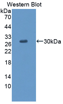
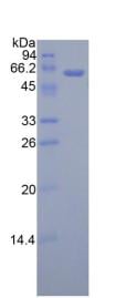
(Sample: Recombinant MASP2, Human;Antibody: Rabbit Anti-Human MASP2 Ab)
WB (Western Blot)
(Sample: Recombinant MASP2, Human;Antibody: Rabbit Anti-Human MASP2 Ab)
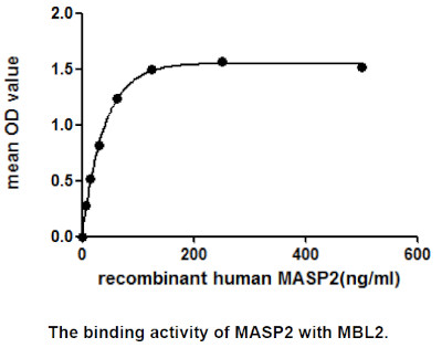
Mannose Associated Serine Protease 2 (MASP2), Active Protein (Cat# AAA21058)
Full Name
Active Mannose Associated Serine Protease 2 (MASP2)
Gene Names
MASP2; sMAP; MAP19; MASP-2; MASP1P1
Reactivity
Homo sapiens (Human)
Applications
Cell culture; Activity Assays; In vivo assays.
Purity
>98%


 WB (Western Blot)
(Anti-HA Tag mouse monoclonal antibody at 1:1000 dilutionLane A: HA-GST (Recombinant protein)(30ng)Lane B: GST-HA (Recombinant protein)(10ng)Lane C: HA-ARG1-myc transfected 293 cell lysate (2ug)Lane D: HA-mFABP4-myc transfected 293 cell lysate (2ug)Lane E: myc-mFABP4-HA transfected 293 cell lysate (0.5ug)Lane F: myc-ARG1-HA transfected 293 cell lysate (2ug)SecondaryGoat Anti-Mouse IgG H&L (Dylight800) at 1/15000 dilution.Developed using the Odyssey technique. Performed under reducing conditions.)
WB (Western Blot)
(Anti-HA Tag mouse monoclonal antibody at 1:1000 dilutionLane A: HA-GST (Recombinant protein)(30ng)Lane B: GST-HA (Recombinant protein)(10ng)Lane C: HA-ARG1-myc transfected 293 cell lysate (2ug)Lane D: HA-mFABP4-myc transfected 293 cell lysate (2ug)Lane E: myc-mFABP4-HA transfected 293 cell lysate (0.5ug)Lane F: myc-ARG1-HA transfected 293 cell lysate (2ug)SecondaryGoat Anti-Mouse IgG H&L (Dylight800) at 1/15000 dilution.Developed using the Odyssey technique. Performed under reducing conditions.)
HA, Monoclonal Antibody (Cat# AAA27712)
Full Name
Anti-HA tag Antibody, Mouse Monoclonal
Reactivity
HA tag
Applications
WB, EIA, FC/FACS/FCM, ICC, IF, IP
Purity
Protein A


 IHC (Immunohistochemistry)
(Anti-Desmin Picoband antibody, AAA11607-7.JPGIHC(P): Rat Cardiac Muscle Tissue)
IHC (Immunohistochemistry)
(Anti-Desmin Picoband antibody, AAA11607-7.JPGIHC(P): Rat Cardiac Muscle Tissue)
Desmin, Polyclonal Antibody (Cat# AAA11607)
Full Name
Anti-Desmin Antibody
Gene Names
DES; CSM1; CSM2; LGMD2R
Reactivity
Human, Mouse, Rat
Applications
WB, IHC, IHC
Purity
Immunogen affinity purified.


 Application Data
(Formalin fixed, paraffin embedded human breast cancer biopsy stained with Mouse anti Human CD44 antibody followed by HRP-polymer detection and DAB substrate development (high power) following antigen retrieval using citrate buffer at pH6.2)
Application Data
(Formalin fixed, paraffin embedded human breast cancer biopsy stained with Mouse anti Human CD44 antibody followed by HRP-polymer detection and DAB substrate development (high power) following antigen retrieval using citrate buffer at pH6.2)
CD44, Monoclonal Antibody (Cat# AAA12016)
Full Name
MOUSE ANTI HUMAN CD44
Gene Names
CD44; IN; LHR; MC56; MDU2; MDU3; MIC4; Pgp1; CDW44; CSPG8; HCELL; HUTCH-I; ECMR-III
Reactivity
Cynomolgus monkey
Applications
FC/FACS, IF


 SDS-PAGE
Islet Cell Autoantigen 1 (ICA1), Recombinant Protein (Cat# AAA20268)
Full Name
Recombinant Islet Cell Autoantigen 1 (ICA1)
Gene Names
ICA1; ICA69; ICAp69
Reactivity
Homo sapiens (Human)
Applications
WB
Purity
> 95%


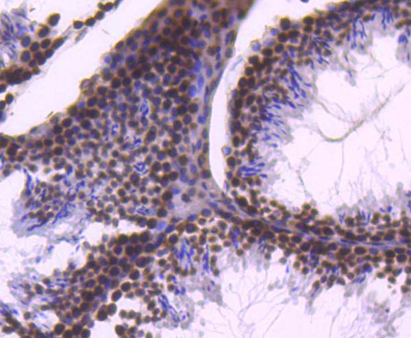
 IHC (Immunohistchemistry)
(Immunohistochemical analysis of (AAA28653) on paraffin-embedded Human breastcarcinoma tissue. Tissue was fixed withformaldehyde at room temperature. Heatinduced epitope retrieval was performed byEDTA buffer (pH9. 0). Samples wereincubated with primary antibody(1:100) for 1hour at room temperature. Undiluted CRFAnti-Polyvalent HRP Polymer antibody wasused as the secondary antibody.)
IHC (Immunohistchemistry)
(Immunohistochemical analysis of (AAA28653) on paraffin-embedded Human breastcarcinoma tissue. Tissue was fixed withformaldehyde at room temperature. Heatinduced epitope retrieval was performed byEDTA buffer (pH9. 0). Samples wereincubated with primary antibody(1:100) for 1hour at room temperature. Undiluted CRFAnti-Polyvalent HRP Polymer antibody wasused as the secondary antibody.)
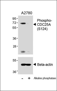
Phospho-CDC25A (S124), Polyclonal Antibody (Cat# AAA28653)
Full Name
Phospho-CDC25A (S124) Antibody
Gene Names
CDC25A; CDC25A2
Reactivity
Human (Predicted Reactivity: Rat)
Applications
WB, EIA, IHC
Purity
Peptide Affinity Purified Rabbit Polyclonal Antibody (Pab)


 WB (Western Blot)
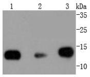
(Western blot of anti-CD44 mAb against HeLa (1) and HUVE-12(2) cell lysate.)
WB (Western Blot)
(Western blot of anti-CD44 mAb against HeLa (1) and HUVE-12(2) cell lysate.)
CD44, Monoclonal Antibody (Cat# AAA12358)
Full Name
Mouse Monoclonal [clone 8E2F3] (IgG1) to Human CD44
Gene Names
CD44; IN; LHR; MC56; MDU2; MDU3; MIC4; Pgp1; CDW44; CSPG8; HCELL; HUTCH-I; ECMR-III
Reactivity
Human, Mouse
Applications
IHC - Paraffin, IF, WB, FC/FACS, EIA
Purity
Ascites


 Application Data
(Published clone specific image Alloimmunity-associated cytotoxicity is mediated through the NKG2D receptor. (A) Liver expression of nkg2d on day ten after liver transplantation. (B) Representative NKG2D expression levels in blood NK cells (left) and monocytes (right) of allogeneic (black) and syngeneic (grey) recipients. Isotype was used as control (dashed lines). (C) Sorted blood NK cell cytotoxicity inhibition with anti-NKG2D antibody or with anti-NKp30 antibody. (D) Levels of NKG2D ligand (rae1l, rrlt and irp94) expression in the liver on day ten after transplantation. (E) Levels of NKG2D ligand (rae1l, rrlt and irp94) expression in rat HCC cell lines. (F) Representative level of recombinant NKG2D-Fc binding to rat HCC cells lines. *p)
Application Data
(Published clone specific image Alloimmunity-associated cytotoxicity is mediated through the NKG2D receptor. (A) Liver expression of nkg2d on day ten after liver transplantation. (B) Representative NKG2D expression levels in blood NK cells (left) and monocytes (right) of allogeneic (black) and syngeneic (grey) recipients. Isotype was used as control (dashed lines). (C) Sorted blood NK cell cytotoxicity inhibition with anti-NKG2D antibody or with anti-NKp30 antibody. (D) Levels of NKG2D ligand (rae1l, rrlt and irp94) expression in the liver on day ten after transplantation. (E) Levels of NKG2D ligand (rae1l, rrlt and irp94) expression in rat HCC cell lines. (F) Representative level of recombinant NKG2D-Fc binding to rat HCC cells lines. *p)
CD172a, Monoclonal Antibody (Cat# AAA11875)
Full Name
MOUSE ANTI RAT CD172a:FITC
Gene Names
Sirpa; Bit; Ptpns1; SHPS-1
Applications
FC/FACS


 WB (Western Blot)
(Sample: Recombinant TRAIL, Human;Antibody: Rabbit Anti-Human TRAIL Ab)
WB (Western Blot)
(Sample: Recombinant TRAIL, Human;Antibody: Rabbit Anti-Human TRAIL Ab)
Tumor Necrosis Factor Related Apoptosis Inducing Ligand (TRAIL), Active Protein (Cat# AAA21106)
Full Name
Active Tumor Necrosis Factor Related Apoptosis Inducing Ligand (TRAIL)
Gene Names
TNFSF10; TL2; APO2L; CD253; TRAIL; Apo-2L; TNLG6A
Reactivity
Homo sapiens (Human)
Applications
Cell culture; Activity Assays; In vivo assays
Purity
>95%


 WB (Western Blot)
(Western Blot Sample: Recombinant LCAT, Mouse;Antibody: Rabbit Anti-Mouse LCAT Ab)
WB (Western Blot)
(Western Blot Sample: Recombinant LCAT, Mouse;Antibody: Rabbit Anti-Mouse LCAT Ab)
Lecithin Cholesterol Acyltransferase (LCAT), Active Protein (Cat# AAA21104)
Full Name
Active Lecithin Cholesterol Acyltransferase (LCAT)
Gene Names
Lcat; AI046659; D8Wsu61e
Reactivity
Mus musculus (Mouse)
Applications
Cell culture; Activity Assays; In vivo assays.
Purity
>98%


 IHC (Immunohistochemistry)
(Figure 7. IHC analysis of CRM1 using anti-CRM1 antibody (AAA11673).CRM1 was detected in frozen section of rat testis tissue. Heat mediated antigen retrieval was performed in citrate buffer (pH6, epitope retrieval solution) for 20 mins. The tissue section was blocked with 10% goat serum. The tissue section was then incubated with 1ug/ml rabbit anti-CRM1 Antibody (AAA11673) overnight at 4 degree C. Biotinylated goat anti-rabbit IgG was used as secondary antibody and incubated for 30 minutes at 37 degree C. The tissue section was developed using Strepavidin-Biotin-Complex (SABC) with DAB as the chromogen.)
IHC (Immunohistochemistry)
(Figure 7. IHC analysis of CRM1 using anti-CRM1 antibody (AAA11673).CRM1 was detected in frozen section of rat testis tissue. Heat mediated antigen retrieval was performed in citrate buffer (pH6, epitope retrieval solution) for 20 mins. The tissue section was blocked with 10% goat serum. The tissue section was then incubated with 1ug/ml rabbit anti-CRM1 Antibody (AAA11673) overnight at 4 degree C. Biotinylated goat anti-rabbit IgG was used as secondary antibody and incubated for 30 minutes at 37 degree C. The tissue section was developed using Strepavidin-Biotin-Complex (SABC) with DAB as the chromogen.)
CRM1, Polyclonal Antibody (Cat# AAA11673)
Full Name
Anti-CRM1 Antibody
Gene Names
XPO1; emb; CRM1; exp1
Reactivity
Human, Mouse, Rat
No cross reactivity with other proteins.
No cross reactivity with other proteins.
Applications
WB, IHC
Purity
Immunogen affinity purified.


 Application Data
(Formalin fixed, paraffin embedded human breast cancer biopsy stained with Mouse anti Human CD44 antibody followed by HRP-polymer detection and DAB substrate development (high power) following antigen retrieval using citrate buffer at pH6.2)
Application Data
(Formalin fixed, paraffin embedded human breast cancer biopsy stained with Mouse anti Human CD44 antibody followed by HRP-polymer detection and DAB substrate development (high power) following antigen retrieval using citrate buffer at pH6.2)
CD44, Monoclonal Antibody (Cat# AAA12208)
Full Name
MOUSE ANTI HUMAN CD44
Gene Names
CD44; IN; LHR; MC56; MDU2; MDU3; MIC4; Pgp1; CDW44; CSPG8; HCELL; HUTCH-I; ECMR-III
Applications
FC/FACS, IF


 FCM (Flow Cytometry)
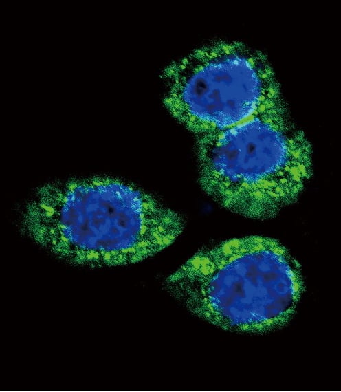
(Flow cytometry analysis of Hsp105alpha in Hep3B cell line, staining at 2-5ug for 1x10⁶cells (red line). The secondary antibody used goat anti-mouse IgG Alexa fluor 488 conjugate. Isotype control antibody was mouse IgG (black line).)
FCM (Flow Cytometry)
(Flow cytometry analysis of Hsp105alpha in Hep3B cell line, staining at 2-5ug for 1x10⁶cells (red line). The secondary antibody used goat anti-mouse IgG Alexa fluor 488 conjugate. Isotype control antibody was mouse IgG (black line).)
HSP105alpha, Monoclonal Antibody (Cat# AAA11727)
Full Name
HSP105alpha antibody
Gene Names
HSPH1; HSP105; HSP105A; HSP105B; NY-CO-25
Reactivity
Human
Applications
ELISA, WB, Flow cytometry, ICC/IF
Purity
By protein-G affinity chromatography


 WB (Western Blot)
(Applications: Western BlotSample: Rat PFCDetection: Protein Simple, Simple Western size-based assay"We selected this product as a polyclonal ab for our Tg Rat colony. We are interested in adding enzymatic catabolism/clearance of amyloid beta isoforms into our body of research. We purchased this product because it was cheap and we are looking at some 'proof-of-concept' and didn't need remarkable specificity. We still have much to investigate.")
WB (Western Blot)
(Applications: Western BlotSample: Rat PFCDetection: Protein Simple, Simple Western size-based assay"We selected this product as a polyclonal ab for our Tg Rat colony. We are interested in adding enzymatic catabolism/clearance of amyloid beta isoforms into our body of research. We purchased this product because it was cheap and we are looking at some 'proof-of-concept' and didn't need remarkable specificity. We still have much to investigate.")
CD10, Polyclonal Antibody (Cat# AAA11606)
Full Name
Anti-CD10 Antibody
Gene Names
MME; NEP; SFE; CD10; CALLA
Reactivity
Human, Mouse, Rat
Applications
WB, IHC
Purity
Immunogen affinity purified.


 WB (Western Blot)
(Western blot analysis of Catenin 1 Antibody expression in MCF-7 cells lysates.The lane on the left is treated with the antigen-specific peptide..The lane on the left is treated with the antigen-specific peptide.)
WB (Western Blot)
(Western blot analysis of Catenin 1 Antibody expression in MCF-7 cells lysates.The lane on the left is treated with the antigen-specific peptide..The lane on the left is treated with the antigen-specific peptide.)
Catenin alpha1, Polyclonal Antibody (Cat# AAA31080)
Full Name
Catenin alpha1 Antibody
Gene Names
CTNNA1; MDPT2; CAP102
Reactivity
Human, Mouse, Rat
Applications
WB, EIA
Purity
The antiserum was purified by peptide affinity chromatography using SulfoLink Coupling Resin.


 Application Data
(Published clone specific image Alloimmunity-associated cytotoxicity is mediated through the NKG2D receptor. (A) Liver expression of nkg2d on day ten after liver transplantation. (B) Representative NKG2D expression levels in blood NK cells (left) and monocytes (right) of allogeneic (black) and syngeneic (grey) recipients. Isotype was used as control (dashed lines). (C) Sorted blood NK cell cytotoxicity inhibition with anti-NKG2D antibody or with anti-NKp30 antibody. (D) Levels of NKG2D ligand (rae1l, rrlt and irp94) expression in the liver on day ten after transplantation. (E) Levels of NKG2D ligand (rae1l, rrlt and irp94) expression in rat HCC cell lines. (F) Representative level of recombinant NKG2D-Fc binding to rat HCC cells lines. *p)
Application Data
(Published clone specific image Alloimmunity-associated cytotoxicity is mediated through the NKG2D receptor. (A) Liver expression of nkg2d on day ten after liver transplantation. (B) Representative NKG2D expression levels in blood NK cells (left) and monocytes (right) of allogeneic (black) and syngeneic (grey) recipients. Isotype was used as control (dashed lines). (C) Sorted blood NK cell cytotoxicity inhibition with anti-NKG2D antibody or with anti-NKp30 antibody. (D) Levels of NKG2D ligand (rae1l, rrlt and irp94) expression in the liver on day ten after transplantation. (E) Levels of NKG2D ligand (rae1l, rrlt and irp94) expression in rat HCC cell lines. (F) Representative level of recombinant NKG2D-Fc binding to rat HCC cells lines. *p)
CD172a, Monoclonal Antibody (Cat# AAA11968)
Full Name
MOUSE ANTI RAT CD172a
Gene Names
Sirpa; Bit; Ptpns1; SHPS-1
Applications
FC/FACS, WB


 Application Data
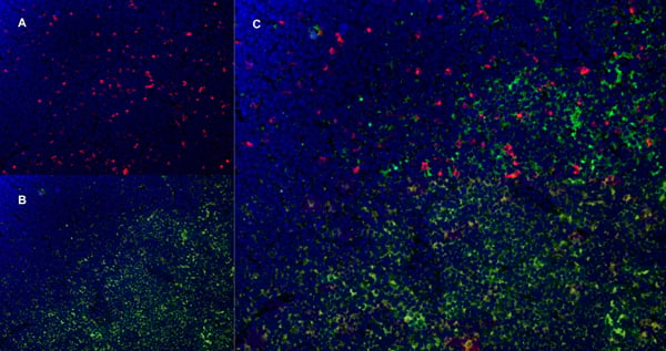
(C:FGFR2/isolectinB4 (C) and FGFR1/isolectinB4 (D) staining of apparent mesenchymal cells and the subpopulation of endothelial cells. Virtually all other dispersed apparent mesenchymal cells express FGFR1 and FGFR2 (merged image in E). F: FGFR2 (F) and FGFR1 (G) staining in clustered cells of epithelial origin (inferred by morphology here) demonstrating that epithelial cells express both FGFR1 and FGFR2 (merged image with DAPI staining in H).)
Application Data
(C:FGFR2/isolectinB4 (C) and FGFR1/isolectinB4 (D) staining of apparent mesenchymal cells and the subpopulation of endothelial cells. Virtually all other dispersed apparent mesenchymal cells express FGFR1 and FGFR2 (merged image in E). F: FGFR2 (F) and FGFR1 (G) staining in clustered cells of epithelial origin (inferred by morphology here) demonstrating that epithelial cells express both FGFR1 and FGFR2 (merged image with DAPI staining in H).)
FGFR2, Polyclonal Antibody (Cat# AAA26854)
Full Name
FGFR2, NT (FGFR2, BEK, KGFR, KSAM, Fibroblast growth factor receptor 2, K-sam, Keratinocyte growth factor receptor, CD332) (FITC)
Gene Names
FGFR2; BEK; JWS; BBDS; CEK3; CFD1; ECT1; KGFR; TK14; TK25; BFR-1; CD332; K-SAM
Reactivity
Human, Monkey, Mouse, Rat
Applications
WB, IHC, IF, FC/FACS
Purity
Purified by Protein G Affinity Chromatography.


 Application Data
(Published clone specific image Alloimmunity-associated cytotoxicity is mediated through the NKG2D receptor. (A) Liver expression of nkg2d on day ten after liver transplantation. (B) Representative NKG2D expression levels in blood NK cells (left) and monocytes (right) of allogeneic (black) and syngeneic (grey) recipients. Isotype was used as control (dashed lines). (C) Sorted blood NK cell cytotoxicity inhibition with anti-NKG2D antibody or with anti-NKp30 antibody. (D) Levels of NKG2D ligand (rae1l, rrlt and irp94) expression in the liver on day ten after transplantation. (E) Levels of NKG2D ligand (rae1l, rrlt and irp94) expression in rat HCC cell lines. (F) Representative level of recombinant NKG2D-Fc binding to rat HCC cells lines. *p)
Application Data
(Published clone specific image Alloimmunity-associated cytotoxicity is mediated through the NKG2D receptor. (A) Liver expression of nkg2d on day ten after liver transplantation. (B) Representative NKG2D expression levels in blood NK cells (left) and monocytes (right) of allogeneic (black) and syngeneic (grey) recipients. Isotype was used as control (dashed lines). (C) Sorted blood NK cell cytotoxicity inhibition with anti-NKG2D antibody or with anti-NKp30 antibody. (D) Levels of NKG2D ligand (rae1l, rrlt and irp94) expression in the liver on day ten after transplantation. (E) Levels of NKG2D ligand (rae1l, rrlt and irp94) expression in rat HCC cell lines. (F) Representative level of recombinant NKG2D-Fc binding to rat HCC cells lines. *p)
CD172a, Monoclonal Antibody (Cat# AAA11967)
Full Name
MOUSE ANTI RAT CD172a
Gene Names
Sirpa; Bit; Ptpns1; SHPS-1
Applications
FC/FACS, WB

 SDS-PAGE
(SDS-PAGE of ~14 kDa Active Human Recombinant Alpha Synuclein Protein Monomer (SPR-321). Lane 1: Molecular Weight Ladder (MW). Lane 2: BSA (5 ug). Lane 3: BSA (2.5 ug). Lane 4: Active Alpha Synuclein Protein Monomer (5 ug) (SPR-321). Lane 5: Active Alpha Synuclein Protein Monomer (2.5 ug) (SPR-321).)
SDS-PAGE
(SDS-PAGE of ~14 kDa Active Human Recombinant Alpha Synuclein Protein Monomer (SPR-321). Lane 1: Molecular Weight Ladder (MW). Lane 2: BSA (5 ug). Lane 3: BSA (2.5 ug). Lane 4: Active Alpha Synuclein Protein Monomer (5 ug) (SPR-321). Lane 5: Active Alpha Synuclein Protein Monomer (2.5 ug) (SPR-321).)
Alpha Synuclein, Active Protein (Cat# AAA27658)
Full Name
Alpha Synuclein Protein
Gene Names
SNCA; PD1; NACP; PARK1; PARK4
Applications
WB
Purity
Purity: >95%
Purification: Ion-exchange Purified
Purification: Ion-exchange Purified


 SDS-PAGE
Granulin (GRN), Recombinant Protein (Cat# AAA20248)
Full Name
Recombinant Granulin (GRN)
Gene Names
GRN; GEP; GP88; PEPI; PGRN; CLN11; PCDGF
Reactivity
Homo sapiens (Human)
Applications
WB
Purity
> 95%


 FCM (Flow Cytometry)
(Flow cytometric analysis of Hela cells with Histone H2B antibody at 1/50 dilution (red) compared with an unlabelled control (cells without incubation with primary antibody; black). Alexa Fluor 488-conjugated goat anti rabbit IgG was used as the secondary antibody.)
FCM (Flow Cytometry)
(Flow cytometric analysis of Hela cells with Histone H2B antibody at 1/50 dilution (red) compared with an unlabelled control (cells without incubation with primary antibody; black). Alexa Fluor 488-conjugated goat anti rabbit IgG was used as the secondary antibody.)
Histone H2B, Monoclonal Antibody (Cat# AAA30192)
Full Name
Histone H2B Antibody
Gene Names
HIST1H2BK; H2BK; H2B/S; H2BFT; H2BFAiii
Reactivity
Human, Mouse, Rat
Applications
WB, ICC, IF, IHC, IP, FC/FACS
Purity
ProA affinity purified


 WB (Western Blot)
(Anti-Beta-Tubulin mouse monoclonal antibody at 1:2000 dilution Lane A: HepG2 Whole Cell Lysate Lane B: Daudi Whole Cell Lysate Lane C: MOLT-4 Whole Cell Lysate Lane D: A549 Whole Cell Lysate Lane E: 293T Whole Cell Lysate Lane F: HelaS3 Whole Cell Lysate Lysates/proteins at 30 ug per lane. Secondary Goat Anti-Mouse IgG H&L (Dylight800) at 1/15000 dilution. Developed using the Odyssey technique. Performed under reducing conditions. Predicted band size:50 kDa Observed band size:54 kDa)
WB (Western Blot)
(Anti-Beta-Tubulin mouse monoclonal antibody at 1:2000 dilution Lane A: HepG2 Whole Cell Lysate Lane B: Daudi Whole Cell Lysate Lane C: MOLT-4 Whole Cell Lysate Lane D: A549 Whole Cell Lysate Lane E: 293T Whole Cell Lysate Lane F: HelaS3 Whole Cell Lysate Lysates/proteins at 30 ug per lane. Secondary Goat Anti-Mouse IgG H&L (Dylight800) at 1/15000 dilution. Developed using the Odyssey technique. Performed under reducing conditions. Predicted band size:50 kDa Observed band size:54 kDa)
Beta-Tubulin, Monoclonal Antibody (Cat# AAA27713)
Full Name
Beta-Tubulin Loading Control Antibody, Mouse MAb
Reactivity
Human
Applications
WB, IHC-P, ICC, IF, IP
Purity
Protein A


 Application Data
(C:FGFR2/isolectinB4 (C) and FGFR1/isolectinB4 (D) staining of apparent mesenchymal cells and the subpopulation of endothelial cells. Virtually all other dispersed apparent mesenchymal cells express FGFR1 and FGFR2 (merged image in E). F: FGFR2 (F) and FGFR1 (G) staining in clustered cells of epithelial origin (inferred by morphology here) demonstrating that epithelial cells express both FGFR1 and FGFR2 (merged image with DAPI staining in H).)
Application Data
(C:FGFR2/isolectinB4 (C) and FGFR1/isolectinB4 (D) staining of apparent mesenchymal cells and the subpopulation of endothelial cells. Virtually all other dispersed apparent mesenchymal cells express FGFR1 and FGFR2 (merged image in E). F: FGFR2 (F) and FGFR1 (G) staining in clustered cells of epithelial origin (inferred by morphology here) demonstrating that epithelial cells express both FGFR1 and FGFR2 (merged image with DAPI staining in H).)
FGFR2, Polyclonal Antibody (Cat# AAA14790)
Full Name
FGFR2, NT (FGFR2, BEK, KGFR, KSAM, Fibroblast growth factor receptor 2, K-sam, Keratinocyte growth factor receptor, CD332)
Gene Names
FGFR2; BEK; JWS; BBDS; CEK3; CFD1; ECT1; KGFR; TK14; TK25; BFR-1; CD332; K-SAM
Reactivity
Human, Monkey, Mouse, Rat
Applications
EL/EIA, WB, IHC, FC/FACS, IF
Purity
Affinity Purified
Purified by Protein A affinity chromatography.
Purified by Protein A affinity chromatography.


 IHC (Immunohistochemistry)
(Figure 7. IHC analysis of Periostin using anti-Periostin antibody (AAA19149).Periostin was detected in paraffin-embedded section of rat small intestine tissue. Heat mediated antigen retrieval was performed in citrate buffer (pH6, epitope retrieval solution) for 20 mins. The tissue section was blocked with 10% goat serum. The tissue section was then incubated with 1ug/ml rabbit anti-Periostin Antibody (AAA19149) overnight at 4 degree C. Biotinylated goat anti-rabbit IgG was used as secondary antibody and incubated for 30 minutes at 37 degree C. The tissue section was developed using Strepavidin-Biotin-Complex (SABC) with DAB as the chromogen.)
IHC (Immunohistochemistry)
(Figure 7. IHC analysis of Periostin using anti-Periostin antibody (AAA19149).Periostin was detected in paraffin-embedded section of rat small intestine tissue. Heat mediated antigen retrieval was performed in citrate buffer (pH6, epitope retrieval solution) for 20 mins. The tissue section was blocked with 10% goat serum. The tissue section was then incubated with 1ug/ml rabbit anti-Periostin Antibody (AAA19149) overnight at 4 degree C. Biotinylated goat anti-rabbit IgG was used as secondary antibody and incubated for 30 minutes at 37 degree C. The tissue section was developed using Strepavidin-Biotin-Complex (SABC) with DAB as the chromogen.)
Periostin, Polyclonal Antibody (Cat# AAA19149)
Full Name
Anti-Periostin Picoband Antibody
Gene Names
Postn; Plf
Reactivity
Mouse, Rat
No cross reactivity with other proteins.
No cross reactivity with other proteins.
Applications
EIA, IHC, WB
Purity
Immunogen affinity purified


 IHC (Immunohistchemistry)
(Immunohistochemical analysis of paraffin-embedded rectum cancer tissues using EIF2AK2 mouse mAb with DAB staining.)
IHC (Immunohistchemistry)
(Immunohistochemical analysis of paraffin-embedded rectum cancer tissues using EIF2AK2 mouse mAb with DAB staining.)
PKR, Monoclonal Antibody (Cat# AAA14157)
Full Name
Anti-PKR Mouse mAb
Gene Names
EIF2AK2; PKR; PRKR; EIF2AK1; PPP1R83
Reactivity
Human
Applications
WB
CD169, Monoclonal Antibody (Cat# AAA11973)
Full Name
MOUSE ANTI RAT CD169
Applications
FC/FACS, IF, IP


 Application Data
(C:FGFR2/isolectinB4 (C) and FGFR1/isolectinB4 (D) staining of apparent mesenchymal cells and the subpopulation of endothelial cells. Virtually all other dispersed apparent mesenchymal cells express FGFR1 and FGFR2 (merged image in E). F: FGFR2 (F) and FGFR1 (G) staining in clustered cells of epithelial origin (inferred by morphology here) demonstrating that epithelial cells express both FGFR1 and FGFR2 (merged image with DAPI staining in H).)
Application Data
(C:FGFR2/isolectinB4 (C) and FGFR1/isolectinB4 (D) staining of apparent mesenchymal cells and the subpopulation of endothelial cells. Virtually all other dispersed apparent mesenchymal cells express FGFR1 and FGFR2 (merged image in E). F: FGFR2 (F) and FGFR1 (G) staining in clustered cells of epithelial origin (inferred by morphology here) demonstrating that epithelial cells express both FGFR1 and FGFR2 (merged image with DAPI staining in H).)
FGFR2, Polyclonal Antibody (Cat# AAA26855)
Full Name
FGFR2, NT (FGFR2, BEK, KGFR, KSAM, Fibroblast growth factor receptor 2, K-sam, Keratinocyte growth factor receptor, CD332) (Azide free) (HRP)
Gene Names
FGFR2; BEK; JWS; BBDS; CEK3; CFD1; ECT1; KGFR; TK14; TK25; BFR-1; CD332; K-SAM
Reactivity
Human, Monkey, Mouse, Rat
Applications
IHC, EIA, WB
Purity
Purified by Protein G Affinity Chromatography.


 Application Data
(C:FGFR2/isolectinB4 (C) and FGFR1/isolectinB4 (D) staining of apparent mesenchymal cells and the subpopulation of endothelial cells. Virtually all other dispersed apparent mesenchymal cells express FGFR1 and FGFR2 (merged image in E). F: FGFR2 (F) and FGFR1 (G) staining in clustered cells of epithelial origin (inferred by morphology here) demonstrating that epithelial cells express both FGFR1 and FGFR2 (merged image with DAPI staining in H).)
Application Data
(C:FGFR2/isolectinB4 (C) and FGFR1/isolectinB4 (D) staining of apparent mesenchymal cells and the subpopulation of endothelial cells. Virtually all other dispersed apparent mesenchymal cells express FGFR1 and FGFR2 (merged image in E). F: FGFR2 (F) and FGFR1 (G) staining in clustered cells of epithelial origin (inferred by morphology here) demonstrating that epithelial cells express both FGFR1 and FGFR2 (merged image with DAPI staining in H).)
FGFR2, Polyclonal Antibody (Cat# AAA26853)
Full Name
FGFR2, NT (FGFR2, BEK, KGFR, KSAM, Fibroblast growth factor receptor 2, K-sam, Keratinocyte growth factor receptor, CD332) (Biotin)
Gene Names
FGFR2; BEK; JWS; BBDS; CEK3; CFD1; ECT1; KGFR; TK14; TK25; BFR-1; CD332; K-SAM
Reactivity
Human, Monkey, Mouse, Rat
Applications
FC/FACS, EIA, IF, IHC, WB
Purity
Purified by Protein G Affinity Chromatography.


 Application Data
(C:FGFR2/isolectinB4 (C) and FGFR1/isolectinB4 (D) staining of apparent mesenchymal cells and the subpopulation of endothelial cells. Virtually all other dispersed apparent mesenchymal cells express FGFR1 and FGFR2 (merged image in E). F: FGFR2 (F) and FGFR1 (G) staining in clustered cells of epithelial origin (inferred by morphology here) demonstrating that epithelial cells express both FGFR1 and FGFR2 (merged image with DAPI staining in H).)
Application Data
(C:FGFR2/isolectinB4 (C) and FGFR1/isolectinB4 (D) staining of apparent mesenchymal cells and the subpopulation of endothelial cells. Virtually all other dispersed apparent mesenchymal cells express FGFR1 and FGFR2 (merged image in E). F: FGFR2 (F) and FGFR1 (G) staining in clustered cells of epithelial origin (inferred by morphology here) demonstrating that epithelial cells express both FGFR1 and FGFR2 (merged image with DAPI staining in H).)
FGFR2, Polyclonal Antibody (Cat# AAA26851)
Full Name
FGFR2, NT (FGFR2, BEK, KGFR, KSAM, Fibroblast growth factor receptor 2, K-sam, Keratinocyte growth factor receptor, CD332) (AP)
Gene Names
FGFR2; BEK; JWS; BBDS; CEK3; CFD1; ECT1; KGFR; TK14; TK25; BFR-1; CD332; K-SAM
Reactivity
Human, Monkey, Mouse, Rat
Applications
IF, EIA, IHC, WB
Purity
Purified by Protein G Affinity Chromatography.


 IHC (Immunohistchemistry)
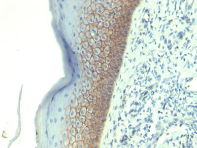
(Formalin-paraffin human Placenta stained with E-Cadherin MAb (CDH1/1525).)
IHC (Immunohistchemistry)
(Formalin-paraffin human Placenta stained with E-Cadherin MAb (CDH1/1525).)
E-Cadherin/CD324, Monoclonal Antibody (Cat# AAA23896)
Full Name
E-Cadherin/CD324 (Intercellular Junction Marker)
Gene Names
CDH1; UVO; CDHE; ECAD; LCAM; Arc-1; BCDS1; CD324
Reactivity
Human.
Does not react with Mouse and Rat. Others not known.
Does not react with Mouse and Rat. Others not known.
Applications
FC/FACS, IF, WB, IHC


 DB (Dot Blot)
(Dot blot analysis using Rabbit Anti-Amyloid Fibrils (OC) Polyclonal Antibody (SPC-507). Tissue: Cell lysates. Species: Human. Primary Antibody: Rabbit Anti-Amyloid Fibrils (OC) Polyclonal Antibody (SPC-507) at 1:500, 1:5000. Beta Amyloid HEPES-NaCl aggregation, showing 1:500 (L) and 1:5000 (R) time lapse dot blot.)
DB (Dot Blot)
(Dot blot analysis using Rabbit Anti-Amyloid Fibrils (OC) Polyclonal Antibody (SPC-507). Tissue: Cell lysates. Species: Human. Primary Antibody: Rabbit Anti-Amyloid Fibrils (OC) Polyclonal Antibody (SPC-507) at 1:500, 1:5000. Beta Amyloid HEPES-NaCl aggregation, showing 1:500 (L) and 1:5000 (R) time lapse dot blot.)
Amyloid Fibrils (OC), Polyclonal Antibody (Cat# AAA17803)
Full Name
Amyloid Fibrils (OC) Antibody: ATTO 488
Reactivity
Human. Potentially mouse and rat based on species homology.
Applications
IP, ICC, IF, IHC, EIA, WB, DB
Purity
Protein A Purified
CD169, Monoclonal Antibody (Cat# AAA11972)
Full Name
MOUSE ANTI RAT CD169
Applications
FC/FACS, IF, IP


 IHC (Immunohistchemistry)
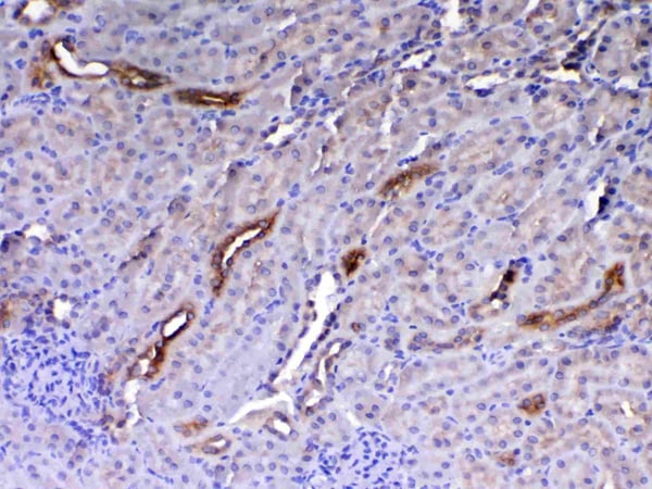
(Figure 6. IHC analysis of GALE using anti-GALE antibody (AAA19135).GALE was detected in paraffin-embedded section of rat kidney tissue. Heat mediated antigen retrieval was performed in citrate buffer (pH6, epitope retrieval solution) for 20 mins. The tissue section was blocked with 10% goat serum. The tissue section was then incubated with 1ug/ml rabbit anti-GALE Antibody (AAA19135) overnight at 4 degree C. Biotinylated goat anti-rabbit IgG was used as secondary antibody and incubated for 30 minutes at 37 degree C. The tissue section was developed using Strepavidin-Biotin-Complex (SABC) with DAB as the chromogen.)
IHC (Immunohistchemistry)
(Figure 6. IHC analysis of GALE using anti-GALE antibody (AAA19135).GALE was detected in paraffin-embedded section of rat kidney tissue. Heat mediated antigen retrieval was performed in citrate buffer (pH6, epitope retrieval solution) for 20 mins. The tissue section was blocked with 10% goat serum. The tissue section was then incubated with 1ug/ml rabbit anti-GALE Antibody (AAA19135) overnight at 4 degree C. Biotinylated goat anti-rabbit IgG was used as secondary antibody and incubated for 30 minutes at 37 degree C. The tissue section was developed using Strepavidin-Biotin-Complex (SABC) with DAB as the chromogen.)
GALE, Polyclonal Antibody (Cat# AAA19135)
Full Name
Anti-GALE Picoband antibody
Gene Names
GALE; SDR1E1
Reactivity
Human, Mouse, Rat
No cross reactivity with other proteins.
No cross reactivity with other proteins.
Applications
EIA, IHC, WB


 IHC (Immunohistchemistry)
(Figure 6. IHC analysis of Integrin alpha 5 using anti-Integrin alpha 5 antibody (AAA19156).Integrin alpha 5 was detected in paraffin-embedded section of human placenta tissue. Heat mediated antigen retrieval was performed in citrate buffer (pH6, epitope retrieval solution) for 20 mins. The tissue section was blocked with 10% goat serum. The tissue section was then incubated with 1ug/ml rabbit anti-Integrin alpha 5 Antibody (AAA19156) overnight at 4 degree C. Biotinylated goat anti-rabbit IgG was used as secondary antibody and incubated for 30 minutes at 37 degree C. The tissue section was developed using Strepavidin-Biotin-Complex (SABC) with DAB as the chromogen.)
IHC (Immunohistchemistry)
(Figure 6. IHC analysis of Integrin alpha 5 using anti-Integrin alpha 5 antibody (AAA19156).Integrin alpha 5 was detected in paraffin-embedded section of human placenta tissue. Heat mediated antigen retrieval was performed in citrate buffer (pH6, epitope retrieval solution) for 20 mins. The tissue section was blocked with 10% goat serum. The tissue section was then incubated with 1ug/ml rabbit anti-Integrin alpha 5 Antibody (AAA19156) overnight at 4 degree C. Biotinylated goat anti-rabbit IgG was used as secondary antibody and incubated for 30 minutes at 37 degree C. The tissue section was developed using Strepavidin-Biotin-Complex (SABC) with DAB as the chromogen.)
Integrin alpha 5, Polyclonal Antibody (Cat# AAA19156)
Full Name
Anti-Integrin alpha 5 Picoband antibody
Gene Names
ITGA5; FNRA; CD49e; VLA-5; VLA5A
Reactivity
Human, Mouse, Rat
No cross reactivity with other proteins.
No cross reactivity with other proteins.
Applications
EIA, IHC, WB


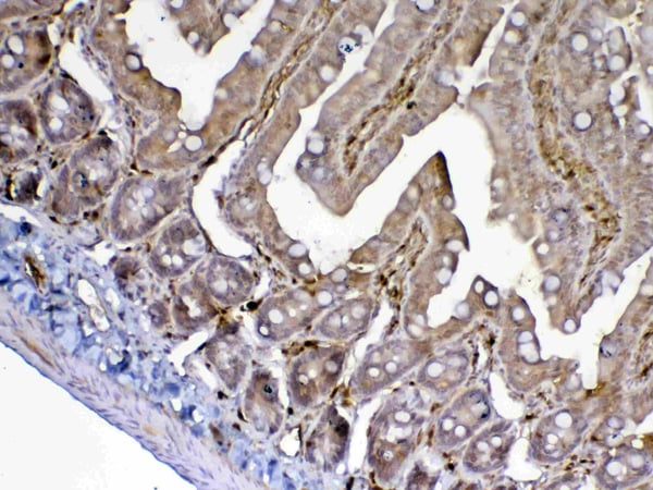
 IHC (Immunohistchemistry)
(Figure 6. IHC analysis of HE4 using anti-HE4 antibody (AAA19161).HE4 was detected in paraffin-embedded section of rat small intestine tissue. Heat mediated antigen retrieval was performed in citrate buffer (pH6, epitope retrieval solution) for 20 mins. The tissue section was blocked with 10% goat serum. The tissue section was then incubated with 1ug/ml rabbit anti-HE4 Antibody (AAA19161) overnight at 4 degree C. Biotinylated goat anti-rabbit IgG was used as secondary antibody and incubated for 30 minutes at 37 degree C. The tissue section was developed using Strepavidin-Biotin-Complex (SABC) with DAB as the chromogen.)
IHC (Immunohistchemistry)
(Figure 6. IHC analysis of HE4 using anti-HE4 antibody (AAA19161).HE4 was detected in paraffin-embedded section of rat small intestine tissue. Heat mediated antigen retrieval was performed in citrate buffer (pH6, epitope retrieval solution) for 20 mins. The tissue section was blocked with 10% goat serum. The tissue section was then incubated with 1ug/ml rabbit anti-HE4 Antibody (AAA19161) overnight at 4 degree C. Biotinylated goat anti-rabbit IgG was used as secondary antibody and incubated for 30 minutes at 37 degree C. The tissue section was developed using Strepavidin-Biotin-Complex (SABC) with DAB as the chromogen.)
HE4, Polyclonal Antibody (Cat# AAA19161)
Full Name
Anti-HE4 Picoband Antibody
Gene Names
Wfdc2; re4
Reactivity
Mouse, Rat
No cross reactivity with other proteins.
No cross reactivity with other proteins.
Applications
IHC, WB
Purity
Immunogen affinity purified

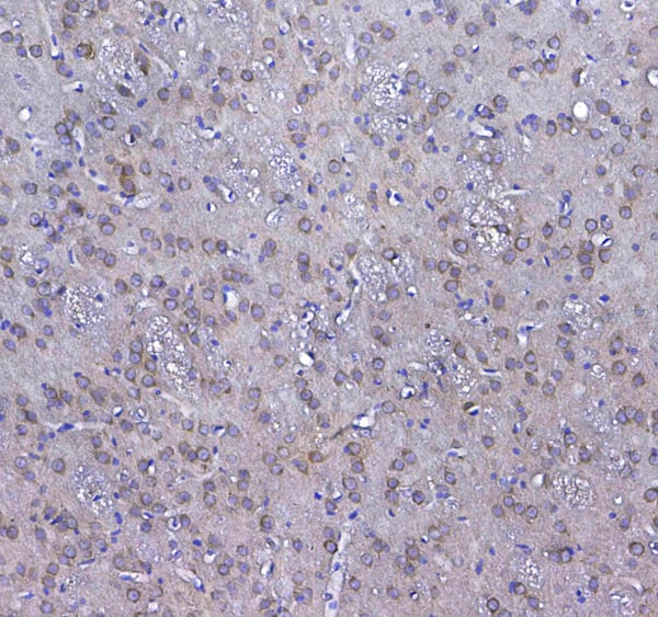


 IHC (Immunohistochemistry)
(Figure 8. IHC analysis of GSTM3 using anti-GSTM3 antibody (AAA19168).GSTM3 was detected in paraffin-embedded section of rat testis tissue. Heat mediated antigen retrieval was performed in citrate buffer (pH6, epitope retrieval solution) for 20 mins. The tissue section was blocked with 10% goat serum. The tissue section was then incubated with 1ug/ml rabbit anti-GSTM3 Antibody (AAA19168) overnight at 4 degree C. Biotinylated goat anti-rabbit IgG was used as secondary antibody and incubated for 30 minutes at 37 degree C. The tissue section was developed using Strepavidin-Biotin-Complex (SABC) with DAB as the chromogen.)
IHC (Immunohistochemistry)
(Figure 8. IHC analysis of GSTM3 using anti-GSTM3 antibody (AAA19168).GSTM3 was detected in paraffin-embedded section of rat testis tissue. Heat mediated antigen retrieval was performed in citrate buffer (pH6, epitope retrieval solution) for 20 mins. The tissue section was blocked with 10% goat serum. The tissue section was then incubated with 1ug/ml rabbit anti-GSTM3 Antibody (AAA19168) overnight at 4 degree C. Biotinylated goat anti-rabbit IgG was used as secondary antibody and incubated for 30 minutes at 37 degree C. The tissue section was developed using Strepavidin-Biotin-Complex (SABC) with DAB as the chromogen.)
GSTM3, Polyclonal Antibody (Cat# AAA19168)
Full Name
Anti-GSTM3 Picoband antibody
Gene Names
GSTM3; GST5; GSTB; GTM3; GSTM3-3
Reactivity
Human, Mouse, Rat
No cross reactivity with other proteins.
No cross reactivity with other proteins.
Applications
EIA, IHC, WB


 FCM (Flow Cytometry)
(Figure 8. Flow Cytometry analysis of Jurkat cells using anti-Ki67 antibody (AAA19350).Overlay histogram showing Jurkat cells stained with AAA19350 (Blue line). The cells were blocked with 10% normal goat serum. And then incubated with mouse anti-Ki67 Antibody (AAA19350, 1μg/1x106 cells) for 30 min at 20 degree C. DyLight®488 conjugated goat anti-mouse IgG (BA1126, 5-10μg/1x106 cells) was used as secondary antibody for 30 minutes at 20 degree C. Isotype control antibody (Green line) was mouse IgG (1μg/1x106) used under the same conditions. Unlabelled sample (Red line) was also used as a control.)
FCM (Flow Cytometry)
(Figure 8. Flow Cytometry analysis of Jurkat cells using anti-Ki67 antibody (AAA19350).Overlay histogram showing Jurkat cells stained with AAA19350 (Blue line). The cells were blocked with 10% normal goat serum. And then incubated with mouse anti-Ki67 Antibody (AAA19350, 1μg/1x106 cells) for 30 min at 20 degree C. DyLight®488 conjugated goat anti-mouse IgG (BA1126, 5-10μg/1x106 cells) was used as secondary antibody for 30 minutes at 20 degree C. Isotype control antibody (Green line) was mouse IgG (1μg/1x106) used under the same conditions. Unlabelled sample (Red line) was also used as a control.)
Ki67, Monoclonal Antibody (Cat# AAA19350)
Full Name
Anti-Ki67 Antibody (monoclonal, 5E12)
Gene Names
MKI67; KIA
Reactivity
Human
Applications
IHC-P, ICC, IF, FC/FACS/FCM
Purity
Immunogen affinity purified.

 IF (Immunofluorescence)
(Immunostaining of mixed neuron/glial cultures stained with anti-UCHL1 antibody (AAA14227), green, 1:500) and rabbit anti-GFAP antibody , red, 1:1000. The blue stains nuclear DNA. The anti-UCHL1 stains strongly the cell body and dendrites of neurons, while anti-GFAP specifically labels astrocytes.)
IF (Immunofluorescence)
(Immunostaining of mixed neuron/glial cultures stained with anti-UCHL1 antibody (AAA14227), green, 1:500) and rabbit anti-GFAP antibody , red, 1:1000. The blue stains nuclear DNA. The anti-UCHL1 stains strongly the cell body and dendrites of neurons, while anti-GFAP specifically labels astrocytes.)
Ubiquitin C-terminal Hydrolase 1 (UCHL1) ck, Polyclonal Antibody (Cat# AAA14227)
Full Name
Anti-Ubiquitin C Terminal Hydrolase 1 (UCHL1)
Gene Names
UCHL1; NDGOA; PARK5; PGP95; PGP9.5; Uch-L1; HEL-117; PGP 9.5
Reactivity
Bovine, Human, Mouse, Pig, Rat
Expected Reactivity: Canine, Feline, Goat, Guinea Pig, Hamster, Horse, Non-Human Primate, Rabbit, Sheep, Vole
Expected Reactivity: Canine, Feline, Goat, Guinea Pig, Hamster, Horse, Non-Human Primate, Rabbit, Sheep, Vole
Applications
WB, IF
Purity
Total IgY fraction


 Application Data
(C:FGFR2/isolectinB4 (C) and FGFR1/isolectinB4 (D) staining of apparent mesenchymal cells and the subpopulation of endothelial cells. Virtually all other dispersed apparent mesenchymal cells express FGFR1 and FGFR2 (merged image in E). F: FGFR2 (F) and FGFR1 (G) staining in clustered cells of epithelial origin (inferred by morphology here) demonstrating that epithelial cells express both FGFR1 and FGFR2 (merged image with DAPI staining in H).)
Application Data
(C:FGFR2/isolectinB4 (C) and FGFR1/isolectinB4 (D) staining of apparent mesenchymal cells and the subpopulation of endothelial cells. Virtually all other dispersed apparent mesenchymal cells express FGFR1 and FGFR2 (merged image in E). F: FGFR2 (F) and FGFR1 (G) staining in clustered cells of epithelial origin (inferred by morphology here) demonstrating that epithelial cells express both FGFR1 and FGFR2 (merged image with DAPI staining in H).)
FGFR2, Polyclonal Antibody (Cat# AAA26856)
Full Name
FGFR2, NT (FGFR2, BEK, KGFR, KSAM, Fibroblast growth factor receptor 2, K-sam, Keratinocyte growth factor receptor, CD332) (PE)
Gene Names
FGFR2; BEK; JWS; BBDS; CEK3; CFD1; ECT1; KGFR; TK14; TK25; BFR-1; CD332; K-SAM
Reactivity
Human, Monkey, Mouse, Rat
Applications
WB, IHC, IF, FC/FACS
Purity
Purified by Protein G Affinity Chromatography.


 IHC (Immunohistchemistry)
(Figure 6. IHC analysis of Cytoglobin using anti- Cytoglobin antibody (AAA11681).Cytoglobin was detected in paraffin-embedded section of human lung cancer tissues. Heat mediated antigen retrieval was performed in citrate buffer (pH6, epitope retrieval solution) for 20 mins. The tissue section was blocked with 10% goat serum. The tissue section was then incubated with 1ug/ml rabbit anti- Cytoglobin Antibody (AAA11681) overnight at 4 degree C. Biotinylated goat anti-rabbit IgG was used as secondary antibody and incubated for 30 minutes at 37 degree C. The tissue section was developed using Strepavidin-Biotin-Complex (SABC) with DAB as the chromogen.)
IHC (Immunohistchemistry)
(Figure 6. IHC analysis of Cytoglobin using anti- Cytoglobin antibody (AAA11681).Cytoglobin was detected in paraffin-embedded section of human lung cancer tissues. Heat mediated antigen retrieval was performed in citrate buffer (pH6, epitope retrieval solution) for 20 mins. The tissue section was blocked with 10% goat serum. The tissue section was then incubated with 1ug/ml rabbit anti- Cytoglobin Antibody (AAA11681) overnight at 4 degree C. Biotinylated goat anti-rabbit IgG was used as secondary antibody and incubated for 30 minutes at 37 degree C. The tissue section was developed using Strepavidin-Biotin-Complex (SABC) with DAB as the chromogen.)
Cytoglobin, Polyclonal Antibody (Cat# AAA11681)
Full Name
Anti-Cytoglobin Antibody
Gene Names
CYGB; HGB; STAP
Reactivity
Human, Mouse, Rat
Applications
WB, IHC
Purity
Immunogen affinity purified.


 IHC (Immunohistchemistry)
(Figure 6. IHC analysis of ADA using anti-ADA antibody (AAA19140).ADA was detected in paraffin-embedded section of rat spleen tissue. Heat mediated antigen retrieval was performed in citrate buffer (pH6, epitope retrieval solution) for 20 mins. The tissue section was blocked with 10% goat serum. The tissue section was then incubated with 1ug/ml rabbit anti-ADA Antibody (AAA19140) overnight at 4 degree C. Biotinylated goat anti-rabbit IgG was used as secondary antibody and incubated for 30 minutes at 37 degree C. The tissue section was developed using Strepavidin-Biotin-Complex (SABC) with DAB as the chromogen.)
IHC (Immunohistchemistry)
(Figure 6. IHC analysis of ADA using anti-ADA antibody (AAA19140).ADA was detected in paraffin-embedded section of rat spleen tissue. Heat mediated antigen retrieval was performed in citrate buffer (pH6, epitope retrieval solution) for 20 mins. The tissue section was blocked with 10% goat serum. The tissue section was then incubated with 1ug/ml rabbit anti-ADA Antibody (AAA19140) overnight at 4 degree C. Biotinylated goat anti-rabbit IgG was used as secondary antibody and incubated for 30 minutes at 37 degree C. The tissue section was developed using Strepavidin-Biotin-Complex (SABC) with DAB as the chromogen.)
ADA/Adenosine Deaminase, Polyclonal Antibody (Cat# AAA19140)
Full Name
Anti-ADA/Adenosine Deaminase Picoband antibody
Reactivity
Mouse, Rat
No cross reactivity with other proteins.
No cross reactivity with other proteins.
Applications
EIA, IHC, WB


 IHC (Immunohistchemistry)
(Figure 6. IHC analysis of CPI17 alpha using anti- CPI17 alpha antibody (AAA19176).CPI17 alpha was detected in paraffin-embedded section of human placenta tissues. Heat mediated antigen retrieval was performed in citrate buffer (pH6, epitope retrieval solution) for 20 mins. The tissue section was blocked with 10% goat serum. The tissue section was then incubated with 1ug/ml rabbit anti- CPI17 alpha Antibody (AAA19176) overnight at 4 degree C. Biotinylated goat anti-rabbit IgG was used as secondary antibody and incubated for 30 minutes at 37 degree C. The tissue section was developed using Strepavidin-Biotin-Complex (SABC) with DAB as the chromogen.)
IHC (Immunohistchemistry)
(Figure 6. IHC analysis of CPI17 alpha using anti- CPI17 alpha antibody (AAA19176).CPI17 alpha was detected in paraffin-embedded section of human placenta tissues. Heat mediated antigen retrieval was performed in citrate buffer (pH6, epitope retrieval solution) for 20 mins. The tissue section was blocked with 10% goat serum. The tissue section was then incubated with 1ug/ml rabbit anti- CPI17 alpha Antibody (AAA19176) overnight at 4 degree C. Biotinylated goat anti-rabbit IgG was used as secondary antibody and incubated for 30 minutes at 37 degree C. The tissue section was developed using Strepavidin-Biotin-Complex (SABC) with DAB as the chromogen.)
CPI17 alpha, Polyclonal Antibody (Cat# AAA19176)
Full Name
Anti-CPI17 alpha Picoband Antibody
Gene Names
PPP1R14A; CPI17; CPI-17; PPP1INL
Reactivity
Human, Mouse, Rat
No cross reactivity with other proteins
No cross reactivity with other proteins
Applications
IHC, WB
Purity
Immunogen affinity purified


 Application Data
(Formalin fixed, paraffin embedded human breast cancer biopsy stained with Mouse anti Human estrogen receptor beta5 antibody followed by HRP polymer detection and DAB substrate development following heat mediated antigen retrieval using citrate buffer at pH6.2 (low power))
Application Data
(Formalin fixed, paraffin embedded human breast cancer biopsy stained with Mouse anti Human estrogen receptor beta5 antibody followed by HRP polymer detection and DAB substrate development following heat mediated antigen retrieval using citrate buffer at pH6.2 (low power))
ESTROGEN RECEPTOR BETA 5, Monoclonal Antibody (Cat# AAA12216)
Full Name
MOUSE ANTI HUMAN ESTROGEN RECEPTOR BETA 5
Gene Names
ESR2; Erb; ESRB; ESTRB; NR3A2; ER-BETA; ESR-BETA
Applications
WB


 IHC (Immunohistchemistry)
(Immunohistochemical analysis of paraffin-embedded bladder cancer tissues using UBE2I mouse mAb with DAB staining.)
IHC (Immunohistchemistry)
(Immunohistochemical analysis of paraffin-embedded bladder cancer tissues using UBE2I mouse mAb with DAB staining.)
Ubc9, Monoclonal Antibody (Cat# AAA14109)
Full Name
Anti-Ubc9 Mouse mAb
Gene Names
UBE2I; P18; UBC9; C358B7.1
Reactivity
Human, Mouse, Rat
Applications
WB


 IHC (Immunohistchemistry)
(At 1/100 staining Human ovarian cancer and adjacent normal tissues by IHC-P. The sample was formaldehyde fixed and a heat mediated antigen retrieval step in citrate buffer was performed. The sample was then blocked and incubated with the primary antibody at 4 degree C overnight. An HRP conjugated anti-Rabbit antibody was used as the secondary antibody.)
IHC (Immunohistchemistry)
(At 1/100 staining Human ovarian cancer and adjacent normal tissues by IHC-P. The sample was formaldehyde fixed and a heat mediated antigen retrieval step in citrate buffer was performed. The sample was then blocked and incubated with the primary antibody at 4 degree C overnight. An HRP conjugated anti-Rabbit antibody was used as the secondary antibody.)
alpha 1 Catenin, Polyclonal Antibody (Cat# AAA31319)
Full Name
Phospho-alpha 1 Catenin (Ser655/Thr658) Antibody
Gene Names
CTNNA1; CAP102
Reactivity
Human, Mouse, Rat
Applications
WB, IHC, EIA
Purity
The antibody is from purified rabbit serum by affinity purification via sequential chromatography on phospho-peptide and non-phospho-peptide affinity columns.