Filters
Clonality
Type
Reactivity
Gene Name
Isotype
Host
Application
Clone
229 results for " Chemiluminescent" - showing 200-229


 FCM (Flow Cytometry)
(Figure 6. Flow Cytometry analysis of CACO-2 cells using anti-PLTP antibody (AAA19255).Overlay histogram showing CACO-2 cells stained with AAA19255 (Blue line). The cells were blocked with 10% normal goat serum. And then incubated with rabbit anti-PLTP Antibody (AAA19255, 1μg/1x106 cells) for 30 min at 20 degree C. DyLight®488 conjugated goat anti-rabbit IgG (5-10μg/1x106 cells) was used as secondary antibody for 30 minutes at 20 degree C. Isotype control antibody (Green line) was rabbit IgG (1μg/1x106) used under the same conditions. Unlabelled sample (Red line) was also used as a control.)
FCM (Flow Cytometry)
(Figure 6. Flow Cytometry analysis of CACO-2 cells using anti-PLTP antibody (AAA19255).Overlay histogram showing CACO-2 cells stained with AAA19255 (Blue line). The cells were blocked with 10% normal goat serum. And then incubated with rabbit anti-PLTP Antibody (AAA19255, 1μg/1x106 cells) for 30 min at 20 degree C. DyLight®488 conjugated goat anti-rabbit IgG (5-10μg/1x106 cells) was used as secondary antibody for 30 minutes at 20 degree C. Isotype control antibody (Green line) was rabbit IgG (1μg/1x106) used under the same conditions. Unlabelled sample (Red line) was also used as a control.)
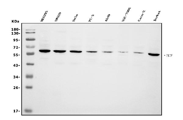
PLTP, Polyclonal Antibody (Cat# AAA19255)
Full Name
Anti-PLTP Antibody
Gene Names
PLTP; BPIFE; HDLCQ9
Reactivity
Human
Applications
WB, IHC-P, ICC, IF, FC/FACS/FCM, EIA
Purity
Immunogen affinity purified.


 FCM (Flow Cytometry)
(Figure 7. Flow Cytometry analysis of U87 cells using anti-PDE6 beta/PDE6B antibody (AAA19261).Overlay histogram showing U87 cells stained with AAA19261 (Blue line). The cells were blocked with 10% normal goat serum. And then incubated with rabbit anti-PDE6 beta/PDE6B Antibody (AAA19261,1μg/1x106 cells) for 30 min at 20 degree C. DyLight®488 conjugated goat anti-rabbit IgG (5-10μg/1x106 cells) was used as secondary antibody for 30 minutes at 20 degree C. Isotype control antibody (Green line) was rabbit IgG (1μg/1x106) used under the same conditions. Unlabelled sample (Red line) was also used as a control.)
FCM (Flow Cytometry)
(Figure 7. Flow Cytometry analysis of U87 cells using anti-PDE6 beta/PDE6B antibody (AAA19261).Overlay histogram showing U87 cells stained with AAA19261 (Blue line). The cells were blocked with 10% normal goat serum. And then incubated with rabbit anti-PDE6 beta/PDE6B Antibody (AAA19261,1μg/1x106 cells) for 30 min at 20 degree C. DyLight®488 conjugated goat anti-rabbit IgG (5-10μg/1x106 cells) was used as secondary antibody for 30 minutes at 20 degree C. Isotype control antibody (Green line) was rabbit IgG (1μg/1x106) used under the same conditions. Unlabelled sample (Red line) was also used as a control.)
PDE6 beta/PDE6B, Polyclonal Antibody (Cat# AAA19261)
Full Name
Anti-PDE6 beta/PDE6B Antibody
Reactivity
Human, Mouse, Rat
Applications
WB, IHC-P, ICC, IF, FC/FACS/FCM, EIA
Purity
Immunogen affinity purified.


 FCM (Flow Cytometry)
(Figure 8. Flow Cytometry analysis of C6 cells using anti-non-muscle Myosin IIB/MYH10 antibody (AAA19262).Overlay histogram showing C6 cells stained with AAA19262 (Blue line). The cells were blocked with 10% normal goat serum. And then incubated with rabbit anti-non-muscle Myosin IIB/MYH10 Antibody (AAA19262,1μg/1x106 cells) for 30 min at 20 degree C. DyLight®488 conjugated goat anti-rabbit IgG (5-10μg/1x106 cells) was used as secondary antibody for 30 minutes at 20 degree C. Isotype control antibody (Green line) was rabbit IgG (1μg/1x106) used under the same conditions. Unlabelled sample (Red line) was also used as a control.)
FCM (Flow Cytometry)
(Figure 8. Flow Cytometry analysis of C6 cells using anti-non-muscle Myosin IIB/MYH10 antibody (AAA19262).Overlay histogram showing C6 cells stained with AAA19262 (Blue line). The cells were blocked with 10% normal goat serum. And then incubated with rabbit anti-non-muscle Myosin IIB/MYH10 Antibody (AAA19262,1μg/1x106 cells) for 30 min at 20 degree C. DyLight®488 conjugated goat anti-rabbit IgG (5-10μg/1x106 cells) was used as secondary antibody for 30 minutes at 20 degree C. Isotype control antibody (Green line) was rabbit IgG (1μg/1x106) used under the same conditions. Unlabelled sample (Red line) was also used as a control.)
non-muscle Myosin IIB/MYH10, Polyclonal Antibody (Cat# AAA19262)
Full Name
Anti-non-muscle Myosin IIB/MYH10 Antibody
Gene Names
MYH10; NMMHCB; NMMHC-IIB
Reactivity
Human, Mouse, Rat
Applications
WB, IHC-P, ICC, IF, FC/FACS/FCM
Purity
Immunogen affinity purified.


 FCM (Flow Cytometry)
(Figure 7. Flow Cytometry analysis of SiHa cells using anti-Aconitase 1/ACO1 antibody (AAA19267).Overlay histogram showing SiHa cells stained with AAA19267 (Blue line). The cells were blocked with 10% normal goat serum. And then incubated with rabbit anti-Aconitase 1/ACO1 Antibody (AAA19267,1μg/1x106 cells) for 30 min at 20 degree C. DyLight®488 conjugated goat anti-rabbit IgG (5-10μg/1x106 cells) was used as secondary antibody for 30 minutes at 20 degree C. Isotype control antibody (Green line) was rabbit IgG (1μg/1x106) used under the same conditions. Unlabelled sample (Red line) was also used as a control.)
FCM (Flow Cytometry)
(Figure 7. Flow Cytometry analysis of SiHa cells using anti-Aconitase 1/ACO1 antibody (AAA19267).Overlay histogram showing SiHa cells stained with AAA19267 (Blue line). The cells were blocked with 10% normal goat serum. And then incubated with rabbit anti-Aconitase 1/ACO1 Antibody (AAA19267,1μg/1x106 cells) for 30 min at 20 degree C. DyLight®488 conjugated goat anti-rabbit IgG (5-10μg/1x106 cells) was used as secondary antibody for 30 minutes at 20 degree C. Isotype control antibody (Green line) was rabbit IgG (1μg/1x106) used under the same conditions. Unlabelled sample (Red line) was also used as a control.)
Aconitase 1/ACO1, Polyclonal Antibody (Cat# AAA19267)
Full Name
Anti-Aconitase 1/ACO1 Antibody
Gene Names
ACO1; IRP1; ACONS; IREB1; IREBP; IREBP1
Reactivity
Human, Mouse, Monkey, Rat
Applications
WB, IHC-P, ICC, IF, FC/FACS/FCM, EIA
Purity
Immunogen affinity purified.


 FCM (Flow Cytometry)
(Figure 7. Flow Cytometry analysis of THP-1 cells using anti-PNPT1 antibody (AAA19306).Overlay histogram showing THP-1 cells stained with AAA19306 (Blue line). The cells were blocked with 10% normal goat serum. And then incubated with rabbit anti-PNPT1 Antibody (AAA19306, 1μg/1x106 cells) for 30 min at 20 degree C. DyLight®488 conjugated goat anti-rabbit IgG (5-10μg/1x106 cells) was used as secondary antibody for 30 minutes at 20 degree C. Isotype control antibody (Green line) was rabbit IgG (1μg/1x106) used under the same conditions. Unlabelled sample (Red line) was also used as a control.)
FCM (Flow Cytometry)
(Figure 7. Flow Cytometry analysis of THP-1 cells using anti-PNPT1 antibody (AAA19306).Overlay histogram showing THP-1 cells stained with AAA19306 (Blue line). The cells were blocked with 10% normal goat serum. And then incubated with rabbit anti-PNPT1 Antibody (AAA19306, 1μg/1x106 cells) for 30 min at 20 degree C. DyLight®488 conjugated goat anti-rabbit IgG (5-10μg/1x106 cells) was used as secondary antibody for 30 minutes at 20 degree C. Isotype control antibody (Green line) was rabbit IgG (1μg/1x106) used under the same conditions. Unlabelled sample (Red line) was also used as a control.)
PNPT1, Polyclonal Antibody (Cat# AAA19306)
Full Name
Anti-PNPT1 Antibody
Gene Names
PNPT1; OLD35; DFNB70; PNPASE; old-35; COXPD13
Reactivity
Human, Mouse, Rat
Applications
WB, IHC-P, ICC, IF, FC/FACS/FCM, EIA
Purity
Immunogen affinity purified.


 WB (Western Blot)
(Figure 1. Western blot analysis of CISD2 using anti-CISD2 antibody (AAA19311).Electrophoresis was performed on a 5-20% SDS-PAGE gel at 70V (Stacking gel) / 90V (Resolving gel) for 2-3 hours. The sample well of each lane was loaded with 50ug of sample under reducing conditions.Lane 1: human HEK293 whole cell lysatesLane 2: human HELA whole cell lysatesLane 3: human MCF-7 whole cell lysatesLane 4: monkey kidney tissue lysatesLane 5: human SW620 whole cell lysatesLane 6: human Raji whole cell lysatesLane 7: rat kidney tissue lysatesLane 8: mouse kidney tissue lysates.After Electrophoresis, proteins were transferred to a Nitrocellulose membrane at 150mA for 50-90 minutes. Blocked the membrane with 5% Non-fat Milk/ TBS for 1. 5 hour at RT. The membrane was incubated with rabbit anti-CISD2 antigen affinity purified polyclonal antibody (Catalog # AAA19311) at 0. 5 μg/mL overnight at 4 degree C, then washed with TBS-0. 1%Tween 3 times with 5 minutes each and probed with a goat anti-rabbit IgG-HRP secondary antibody at a dilution of 1:10000 for 1. 5 hour at RT. The signal is developed using an Enhanced Chemiluminescent detection (ECL) kit (Catalog # with Tanon 5200 system. A specific band was detected for CISD2 at approximately 15KD. The expected band size for CISD2 is at 15KD.)
WB (Western Blot)
(Figure 1. Western blot analysis of CISD2 using anti-CISD2 antibody (AAA19311).Electrophoresis was performed on a 5-20% SDS-PAGE gel at 70V (Stacking gel) / 90V (Resolving gel) for 2-3 hours. The sample well of each lane was loaded with 50ug of sample under reducing conditions.Lane 1: human HEK293 whole cell lysatesLane 2: human HELA whole cell lysatesLane 3: human MCF-7 whole cell lysatesLane 4: monkey kidney tissue lysatesLane 5: human SW620 whole cell lysatesLane 6: human Raji whole cell lysatesLane 7: rat kidney tissue lysatesLane 8: mouse kidney tissue lysates.After Electrophoresis, proteins were transferred to a Nitrocellulose membrane at 150mA for 50-90 minutes. Blocked the membrane with 5% Non-fat Milk/ TBS for 1. 5 hour at RT. The membrane was incubated with rabbit anti-CISD2 antigen affinity purified polyclonal antibody (Catalog # AAA19311) at 0. 5 μg/mL overnight at 4 degree C, then washed with TBS-0. 1%Tween 3 times with 5 minutes each and probed with a goat anti-rabbit IgG-HRP secondary antibody at a dilution of 1:10000 for 1. 5 hour at RT. The signal is developed using an Enhanced Chemiluminescent detection (ECL) kit (Catalog # with Tanon 5200 system. A specific band was detected for CISD2 at approximately 15KD. The expected band size for CISD2 is at 15KD.)
CISD2, Polyclonal Antibody (Cat# AAA19311)
Full Name
Anti-CISD2 Antibody
Gene Names
CISD2; ERIS; WFS2; ZCD2; NAF-1; Miner1
Reactivity
Human, Mouse, Rat, Monkey
Applications
WB, IHC-P, ICC, IF, FC/FACS/FCM, EIA
Purity
Immunogen affinity purified.


 FCM (Flow Cytometry)
(Figure 8. Flow Cytometry analysis of SiHa cells using anti-Astrin/Deepest/SPAG5 antibody (AAA19313).Overlay histogram showing SiHa cells stained with AAA19313 (Blue line). The cells were blocked with 10% normal goat serum. And then incubated with rabbit anti-Astrin/Deepest/SPAG5 Antibody (AAA19313, 1μg/1x106 cells) for 30 min at 20 degree C. DyLight®488 conjugated goat anti-rabbit IgG (5-10μg/1x106 cells) was used as secondary antibody for 30 minutes at 20 degree C. Isotype control antibody (Green line) was rabbit IgG (1μg/1x106) used under the same conditions. Unlabelled sample (Red line) was also used as a control.)
FCM (Flow Cytometry)
(Figure 8. Flow Cytometry analysis of SiHa cells using anti-Astrin/Deepest/SPAG5 antibody (AAA19313).Overlay histogram showing SiHa cells stained with AAA19313 (Blue line). The cells were blocked with 10% normal goat serum. And then incubated with rabbit anti-Astrin/Deepest/SPAG5 Antibody (AAA19313, 1μg/1x106 cells) for 30 min at 20 degree C. DyLight®488 conjugated goat anti-rabbit IgG (5-10μg/1x106 cells) was used as secondary antibody for 30 minutes at 20 degree C. Isotype control antibody (Green line) was rabbit IgG (1μg/1x106) used under the same conditions. Unlabelled sample (Red line) was also used as a control.)
Astrin/Deepest/SPAG5, Polyclonal Antibody (Cat# AAA19313)
Full Name
Anti-Astrin/Deepest/SPAG5 Antibody
Gene Names
SPAG5; MAP126; DEEPEST; hMAP126
Reactivity
Human
Applications
WB, IHC-P, ICC, IF, FC/FACS/FCM, EIA
Purity
Immunogen affinity purified.


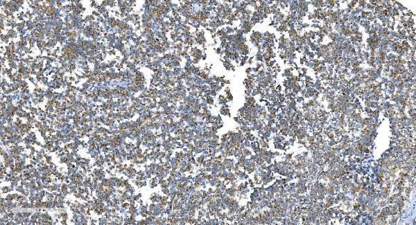
 IHC (Immunohistochemistry)
(Figure 7. IHC analysis of GCHFR using anti-GCHFR antibody (AAA19322).GCHFR was detected in paraffin-embedded section of mouse liver tissue. Heat mediated antigen retrieval was performed in EDTA buffer (pH8. 0, epitope retrieval solution). The tissue section was blocked with 10% goat serum. The tissue section was then incubated with 2μg/ml rabbit anti-GCHFR Antibody (AAA19322) overnight at 4 degree C. Biotinylated goat anti-rabbit IgG was used as secondary antibody and incubated for 30 minutes at 37 degree C. The tissue section was developed using Strepavidin-Biotin-Complex (SABC) (Catalog # with DAB as the chromogen.)
IHC (Immunohistochemistry)
(Figure 7. IHC analysis of GCHFR using anti-GCHFR antibody (AAA19322).GCHFR was detected in paraffin-embedded section of mouse liver tissue. Heat mediated antigen retrieval was performed in EDTA buffer (pH8. 0, epitope retrieval solution). The tissue section was blocked with 10% goat serum. The tissue section was then incubated with 2μg/ml rabbit anti-GCHFR Antibody (AAA19322) overnight at 4 degree C. Biotinylated goat anti-rabbit IgG was used as secondary antibody and incubated for 30 minutes at 37 degree C. The tissue section was developed using Strepavidin-Biotin-Complex (SABC) (Catalog # with DAB as the chromogen.)
GCHFR, Polyclonal Antibody (Cat# AAA19322)
Full Name
Anti-GCHFR Antibody
Gene Names
GCHFR; P35; GFRP; HsT16933
Reactivity
Human, Mouse, Rat
Applications
WB, IHC-P, ICC, IF, FC/FACS/FCM, EIA
Purity
Immunogen affinity purified.


 FCM (Flow Cytometry)
(Figure 6. Flow Cytometry analysis of U937 cells using anti-BDH1 antibody (AAA19328).Overlay histogram showing U937 cells stained with AAA19328 (Blue line). The cells were blocked with 10% normal goat serum. And then incubated with rabbit anti-BDH1 Antibody (AAA19328,1μg/1x106 cells) for 30 min at 20 degree C. DyLight®488 conjugated goat anti-rabbit IgG (5-10μg/1x106 cells) was used as secondary antibody for 30 minutes at 20 degree C. Isotype control antibody (Green line) was rabbit IgG (1μg/1x106) used under the same conditions. Unlabelled sample (Red line) was also used as a control.)
FCM (Flow Cytometry)
(Figure 6. Flow Cytometry analysis of U937 cells using anti-BDH1 antibody (AAA19328).Overlay histogram showing U937 cells stained with AAA19328 (Blue line). The cells were blocked with 10% normal goat serum. And then incubated with rabbit anti-BDH1 Antibody (AAA19328,1μg/1x106 cells) for 30 min at 20 degree C. DyLight®488 conjugated goat anti-rabbit IgG (5-10μg/1x106 cells) was used as secondary antibody for 30 minutes at 20 degree C. Isotype control antibody (Green line) was rabbit IgG (1μg/1x106) used under the same conditions. Unlabelled sample (Red line) was also used as a control.)
BDH1, Polyclonal Antibody (Cat# AAA19328)
Full Name
Anti-BDH1 Antibody
Gene Names
BDH1; BDH; SDR9C1
Reactivity
Human, Rat
Applications
WB, IHC-P, ICC, IF, FC/FACS/FCM, EIA
Purity
Immunogen affinity purified.


 FCM (Flow Cytometry)
(Figure 6. Flow Cytometry analysis of A431 cells using anti-NDUFB5 antibody (AAA19333).Overlay histogram showing A431 cells stained with AAA19333 (Blue line). The cells were blocked with 10% normal goat serum. And then incubated with rabbit anti-NDUFB5 Antibody (AAA19333, 1μg/1x106 cells) for 30 min at 20 degree C. DyLight®488 conjugated goat anti-rabbit IgG (5-10μg/1x106 cells) was used as secondary antibody for 30 minutes at 20 degree C. Isotype control antibody (Green line) was rabbit IgG (1μg/1x106) used under the same conditions. Unlabelled sample (Red line) was also used as a control.)
FCM (Flow Cytometry)
(Figure 6. Flow Cytometry analysis of A431 cells using anti-NDUFB5 antibody (AAA19333).Overlay histogram showing A431 cells stained with AAA19333 (Blue line). The cells were blocked with 10% normal goat serum. And then incubated with rabbit anti-NDUFB5 Antibody (AAA19333, 1μg/1x106 cells) for 30 min at 20 degree C. DyLight®488 conjugated goat anti-rabbit IgG (5-10μg/1x106 cells) was used as secondary antibody for 30 minutes at 20 degree C. Isotype control antibody (Green line) was rabbit IgG (1μg/1x106) used under the same conditions. Unlabelled sample (Red line) was also used as a control.)
NDUFB5, Polyclonal Antibody (Cat# AAA19333)
Full Name
Anti-NDUFB5 Antibody
Gene Names
NDUFB5; SGDH; CISGDH
Reactivity
Human, Mouse, Rat
Applications
WB, IHC-P, ICC, IF, FC/FACS/FCM, EIA
Purity
Immunogen affinity purified.


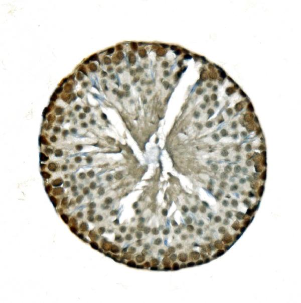
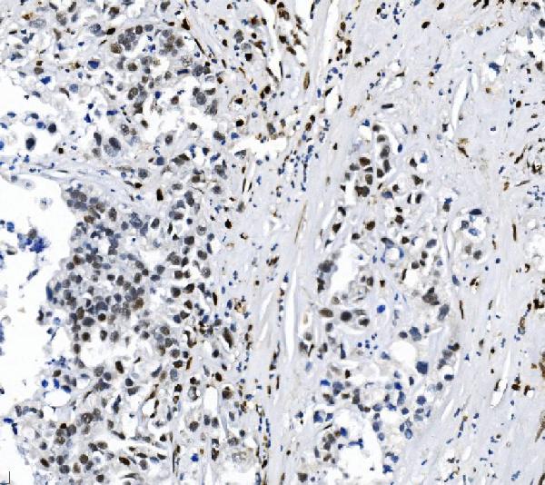
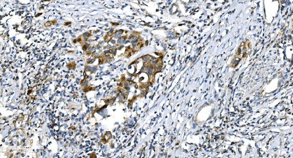
 IHC (Immunohistochemistry)
(Figure 11. IHC analysis of U2AF65/U2AF2 using anti U2AF65/U2AF2 antibody (AAA19376).U2AF65/U2AF2 was detected in paraffin-embedded section of human gallbladder adenocarcinoma tissue. Heat mediated antigen retrieval was performed in EDTA buffer (pH8. 0, epitope retrieval solution). The tissue section was blocked with 10% goat serum. The tissue section was then incubated with 2μg/ml mouse anti-U2AF65/U2AF2 Antibody (AAA19376) overnight at 4 degree C. Biotinylated goat anti-mouse IgG was used as secondary antibody and incubated for 30 minutes at 37 degree C. The tissue section was developed using Strepavidin-Biotin-Complex (SABC) (Catalog # with DAB as the chromogen.)
IHC (Immunohistochemistry)
(Figure 11. IHC analysis of U2AF65/U2AF2 using anti U2AF65/U2AF2 antibody (AAA19376).U2AF65/U2AF2 was detected in paraffin-embedded section of human gallbladder adenocarcinoma tissue. Heat mediated antigen retrieval was performed in EDTA buffer (pH8. 0, epitope retrieval solution). The tissue section was blocked with 10% goat serum. The tissue section was then incubated with 2μg/ml mouse anti-U2AF65/U2AF2 Antibody (AAA19376) overnight at 4 degree C. Biotinylated goat anti-mouse IgG was used as secondary antibody and incubated for 30 minutes at 37 degree C. The tissue section was developed using Strepavidin-Biotin-Complex (SABC) (Catalog # with DAB as the chromogen.)
U2AF65/U2AF2, Monoclonal Antibody (Cat# AAA19376)
Full Name
Anti-U2AF65/U2AF2 Antibody (monoclonal, 3G9)
Gene Names
U2AF2; U2AF65
Reactivity
Human, Mouse, Rat
Applications
WB, IHC-P, ICC, IF, FC/FACS/FCM
Purity
Immunogen affinity purified.


 FCM (Flow Cytometry)
(Figure 7. Flow Cytometry analysis of RH35 cells using anti- EIF4A1 antibody (AAA19380).Overlay histogram showing RH35 cells stained with AAA19380 (Blue line). The cells were blocked with 10% normal goat serum. And then incubated with mouse anti-EIF4A1 Antibody (AAA19380, 1μg/1x106 cells) for 30 min at 20 degree C. DyLight®488 conjugated goat anti-mouse IgG (BA1126, 5-10μg/1x106 cells) was used as secondary antibody for 30 minutes at 20 degree C. Isotype control antibody (Green line) was mouse IgG (1μg/1x106) used under the same conditions. Unlabelled sample (Red line) was also used as a control.)
FCM (Flow Cytometry)
(Figure 7. Flow Cytometry analysis of RH35 cells using anti- EIF4A1 antibody (AAA19380).Overlay histogram showing RH35 cells stained with AAA19380 (Blue line). The cells were blocked with 10% normal goat serum. And then incubated with mouse anti-EIF4A1 Antibody (AAA19380, 1μg/1x106 cells) for 30 min at 20 degree C. DyLight®488 conjugated goat anti-mouse IgG (BA1126, 5-10μg/1x106 cells) was used as secondary antibody for 30 minutes at 20 degree C. Isotype control antibody (Green line) was mouse IgG (1μg/1x106) used under the same conditions. Unlabelled sample (Red line) was also used as a control.)
EIF4A1, Monoclonal Antibody (Cat# AAA19380)
Full Name
Anti-EIF4A1 Antibody (monoclonal, 11B8)
Gene Names
EIF4A1; DDX2A; EIF4A; EIF-4A; eIF4A-I; eIF-4A-I
Reactivity
Human, Mouse, Rat
Applications
WB, IHC-P, ICC, IF, FC/FACS/FCM
Purity
Immunogen affinity purified.


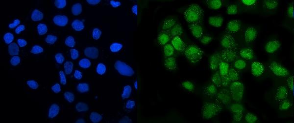
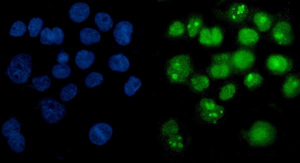
 IF (Immunofluorescence)
(Figure 11. IF analysis of Tubulin alpha using anti- Tubulin alpha antibody (AAA19381).Tubulin alpha was detected in immunocytochemical section of CACO-2 cells. Enzyme antigen retrieval was performed using IHC enzyme antigen retrieval reagent for 15 mins. The cells were blocked with 10% goat serum. And then incubated with 5μg/mL mouse anti- Tubulin alpha Antibody (AAA19381) overnight at 4 degree C. DyLight®594 Conjugated Goat Anti-Mouse IgG (BA1141) was used as secondary antibody at 1:100 dilution and incubated for 30 minutes at 37 degree C. The section was counterstained with DAPI. Visualize using a fluorescence microscope and filter sets appropriate for the label used.)
IF (Immunofluorescence)
(Figure 11. IF analysis of Tubulin alpha using anti- Tubulin alpha antibody (AAA19381).Tubulin alpha was detected in immunocytochemical section of CACO-2 cells. Enzyme antigen retrieval was performed using IHC enzyme antigen retrieval reagent for 15 mins. The cells were blocked with 10% goat serum. And then incubated with 5μg/mL mouse anti- Tubulin alpha Antibody (AAA19381) overnight at 4 degree C. DyLight®594 Conjugated Goat Anti-Mouse IgG (BA1141) was used as secondary antibody at 1:100 dilution and incubated for 30 minutes at 37 degree C. The section was counterstained with DAPI. Visualize using a fluorescence microscope and filter sets appropriate for the label used.)
Tubulin alpha, Monoclonal Antibody (Cat# AAA19381)
Full Name
Anti-Tubulin alpha Antibody (monoclonal, 7B12)
Gene Names
TUBA1A; LIS3; TUBA3; B-ALPHA-1
Reactivity
Human, Mouse, Rat
Applications
WB, IHC-P, ICC, IF, FC/FACS/FCM
Purity
Immunogen affinity purified.


 FCM (Flow Cytometry)
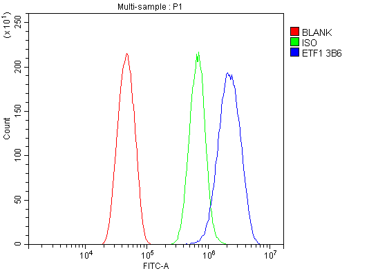
(Figure 11. Flow Cytometry analysis of RH35 cells using anti- eRF1/ETF1 antibody (AAA19382).Overlay histogram showing RH35 cells stained with AAA19382 (Blue line). The cells were blocked with 10% normal goat serum. And then incubated with mouse anti-eRF1/ETF1 Antibody (AAA19382, 1μg/1x106 cells) for 30 min at 20 degree C. DyLight®488 conjugated goat anti-mouse IgG (BA1126, 5-10μg/1x106 cells) was used as secondary antibody for 30 minutes at 20 degree C. Isotype control antibody (Green line) was mouse IgG (1μg/1x106) used under the same conditions. Unlabelled sample (Red line) was also used as a control.)
FCM (Flow Cytometry)
(Figure 11. Flow Cytometry analysis of RH35 cells using anti- eRF1/ETF1 antibody (AAA19382).Overlay histogram showing RH35 cells stained with AAA19382 (Blue line). The cells were blocked with 10% normal goat serum. And then incubated with mouse anti-eRF1/ETF1 Antibody (AAA19382, 1μg/1x106 cells) for 30 min at 20 degree C. DyLight®488 conjugated goat anti-mouse IgG (BA1126, 5-10μg/1x106 cells) was used as secondary antibody for 30 minutes at 20 degree C. Isotype control antibody (Green line) was mouse IgG (1μg/1x106) used under the same conditions. Unlabelled sample (Red line) was also used as a control.)
eRF1/ETF1, Monoclonal Antibody (Cat# AAA19382)
Full Name
Anti-eRF1/ETF1 Antibody (monoclonal, 3B6)
Gene Names
ETF1; ERF; RF1; ERF1; TB3-1; D5S1995; SUP45L1
Reactivity
Human, Mouse, Rat
Applications
WB, IHC-P, ICC, IF, FC/FACS/FCM
Purity
Immunogen affinity purified.


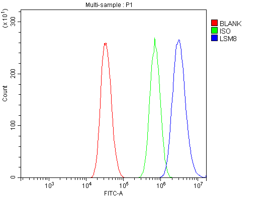 FCM (Flow Cytometry)
(Figure 13. Flow Cytometry analysis of A431 cells using anti-LSM8 antibody (AAA19337).Overlay histogram showing A431 cells stained with AAA19337 (Blue line). The cells were blocked with 10% normal goat serum. And then incubated with rabbit anti-LSM8 Antibody (AAA19337, 1μg/1x106 cells) for 30 min at 20 degree C. DyLight®488 conjugated goat anti-rabbit IgG (5-10μg/1x106 cells) was used as secondary antibody for 30 minutes at 20 degree C. Isotype control antibody (Green line) was rabbit IgG (1μg/1x106) used under the same conditions. Unlabelled sample (Red line) was also used as a control.)
FCM (Flow Cytometry)
(Figure 13. Flow Cytometry analysis of A431 cells using anti-LSM8 antibody (AAA19337).Overlay histogram showing A431 cells stained with AAA19337 (Blue line). The cells were blocked with 10% normal goat serum. And then incubated with rabbit anti-LSM8 Antibody (AAA19337, 1μg/1x106 cells) for 30 min at 20 degree C. DyLight®488 conjugated goat anti-rabbit IgG (5-10μg/1x106 cells) was used as secondary antibody for 30 minutes at 20 degree C. Isotype control antibody (Green line) was rabbit IgG (1μg/1x106) used under the same conditions. Unlabelled sample (Red line) was also used as a control.)
LSM8, Polyclonal Antibody (Cat# AAA19337)
Full Name
Anti-LSM8 Antibody
Reactivity
Human, Mouse, Rat
Applications
WB, IHC-P, ICC, IF, FC/FACS/FCM, EIA
Purity
Immunogen affinity purified.


 FCM (Flow Cytometry)
(Figure 9. Flow Cytometry analysis of HL-60 cells using anti-NDUFB10 antibody (AAA19339).Overlay histogram showing HL-60 cells stained with AAA19339 (Blue line). The cells were blocked with 10% normal goat serum. And then incubated with rabbit anti-NDUFB10 Antibody (AAA19339, 1μg/1x106 cells) for 30 min at 20 degree C. DyLight®488 conjugated goat anti-rabbit IgG (5-10μg/1x106 cells) was used as secondary antibody for 30 minutes at 20 degree C. Isotype control antibody (Green line) was rabbit IgG (1μg/1x106) used under the same conditions. Unlabelled sample (Red line) was also used as a control.)
FCM (Flow Cytometry)
(Figure 9. Flow Cytometry analysis of HL-60 cells using anti-NDUFB10 antibody (AAA19339).Overlay histogram showing HL-60 cells stained with AAA19339 (Blue line). The cells were blocked with 10% normal goat serum. And then incubated with rabbit anti-NDUFB10 Antibody (AAA19339, 1μg/1x106 cells) for 30 min at 20 degree C. DyLight®488 conjugated goat anti-rabbit IgG (5-10μg/1x106 cells) was used as secondary antibody for 30 minutes at 20 degree C. Isotype control antibody (Green line) was rabbit IgG (1μg/1x106) used under the same conditions. Unlabelled sample (Red line) was also used as a control.)
NDUFB10, Polyclonal Antibody (Cat# AAA19339)
Full Name
Anti-NDUFB10 Antibody
Gene Names
NDUFB10; PDSW
Reactivity
Human, Mouse, Rat
Applications
WB, IHC-P, ICC, IF, FC/FACS/FCM, EIA
Purity
Immunogen affinity purified.


 FCM (Flow Cytometry)
(Figure 7. Flow Cytometry analysis of HL-60 cells using anti-PARP antibody (AAA19346).Overlay histogram showing HL-60 cells stained with AAA19346 (Blue line). The cells were blocked with 10% normal goat serum. And then incubated with mouse anti- PARP Antibody (AAA19346, 1μg/1x106 cells) for 30 min at 20 degree C. DyLight®488 conjugated goat anti-mouse IgG (BA1126, 5-10μg/1x106 cells) was used as secondary antibody for 30 minutes at 20 degree C. Isotype control antibody (Green line) was mouse IgG (1μg/1x106) used under the same conditions. Unlabelled sample (Red line) was also used as a control.)
FCM (Flow Cytometry)
(Figure 7. Flow Cytometry analysis of HL-60 cells using anti-PARP antibody (AAA19346).Overlay histogram showing HL-60 cells stained with AAA19346 (Blue line). The cells were blocked with 10% normal goat serum. And then incubated with mouse anti- PARP Antibody (AAA19346, 1μg/1x106 cells) for 30 min at 20 degree C. DyLight®488 conjugated goat anti-mouse IgG (BA1126, 5-10μg/1x106 cells) was used as secondary antibody for 30 minutes at 20 degree C. Isotype control antibody (Green line) was mouse IgG (1μg/1x106) used under the same conditions. Unlabelled sample (Red line) was also used as a control.)
PARP, Monoclonal Antibody (Cat# AAA19346)
Full Name
Anti-PARP Antibody (monoclonal, 10G9)
Gene Names
PARP1; PARP; PPOL; ADPRT; ARTD1; ADPRT1; PARP-1; ADPRT 1; pADPRT-1
Reactivity
Human, Mouse, Rat
Applications
WB, IHC-P, ICC, IF, FC/FACS/FCM
Purity
Immunogen affinity purified.


 FCM (Flow Cytometry)
(Figure 11. Flow Cytometry analysis of A431 cells using anti-SAMHD1 antibody (AAA19355).Overlay histogram showing A431 cells stained with AAA19355 (Blue line). The cells were blocked with 10% normal goat serum. And then incubated with mouse anti- SAMHD1 Antibody (AAA19355, 1μg/1x106 cells) for 30 min at 20 degree C. DyLight®488 conjugated goat anti-mouse IgG (BA1126, 5-10μg/1x106 cells) was used as secondary antibody for 30 minutes at 20 degree C. Isotype control antibody (Green line) was mouse IgG (1μg/1x106) used under the same conditions. Unlabelled sample (Red line) was also used as a control.)
FCM (Flow Cytometry)
(Figure 11. Flow Cytometry analysis of A431 cells using anti-SAMHD1 antibody (AAA19355).Overlay histogram showing A431 cells stained with AAA19355 (Blue line). The cells were blocked with 10% normal goat serum. And then incubated with mouse anti- SAMHD1 Antibody (AAA19355, 1μg/1x106 cells) for 30 min at 20 degree C. DyLight®488 conjugated goat anti-mouse IgG (BA1126, 5-10μg/1x106 cells) was used as secondary antibody for 30 minutes at 20 degree C. Isotype control antibody (Green line) was mouse IgG (1μg/1x106) used under the same conditions. Unlabelled sample (Red line) was also used as a control.)
SAMHD1, Monoclonal Antibody (Cat# AAA19355)
Full Name
Anti-SAMHD1 Antibody (monoclonal, 3B9)
Gene Names
SAMHD1; DCIP; CHBL2; HDDC1; MOP-5; SBBI88
Reactivity
Human
Applications
WB, IHC-P, ICC, IF, FC/FACS/FCM
Purity
Immunogen affinity purified.


 FCM (Flow Cytometry)
(Figure 9. Flow Cytometry analysis of A549 cells using anti-JAB1 antibody (AAA19365).Overlay histogram showing A549 cells stained with AAA19365 (Blue line). The cells were blocked with 10% normal goat serum. And then incubated with mouse anti-JAB1 Antibody (AAA19365, 1μg/1x106 cells) for 30 min at 20 degree C. DyLight®488 conjugated goat anti-mouse IgG (BA1126, 5-10μg/1x106 cells) was used as secondary antibody for 30 minutes at 20 degree C. Isotype control antibody (Green line) was mouse IgG (1μg/1x106) used under the same conditions. Unlabelled sample (Red line) was also used as a control.)
FCM (Flow Cytometry)
(Figure 9. Flow Cytometry analysis of A549 cells using anti-JAB1 antibody (AAA19365).Overlay histogram showing A549 cells stained with AAA19365 (Blue line). The cells were blocked with 10% normal goat serum. And then incubated with mouse anti-JAB1 Antibody (AAA19365, 1μg/1x106 cells) for 30 min at 20 degree C. DyLight®488 conjugated goat anti-mouse IgG (BA1126, 5-10μg/1x106 cells) was used as secondary antibody for 30 minutes at 20 degree C. Isotype control antibody (Green line) was mouse IgG (1μg/1x106) used under the same conditions. Unlabelled sample (Red line) was also used as a control.)
JAB1, Monoclonal Antibody (Cat# AAA19365)
Full Name
Anti-JAB1 Antibody (monoclonal, 4G9)
Gene Names
COPS5; CSN5; JAB1; SGN5; MOV-34
Reactivity
Human, Mouse, Rat
Applications
WB, IHC-P, ICC, IF, FC/FACS/FCM
Purity
Immunogen affinity purified.


 FCM (Flow Cytometry)
(Figure 12. Flow Cytometry analysis of U251 cells using anti-HP1 alpha/CBX5 antibody (AAA19373).Overlay histogram showing U251 cells stained with AAA19373 (Blue line). The cells were blocked with 10% normal goat serum. And then incubated with mouse anti- HP1 alpha/CBX5 Antibody (AAA19373, 1μg/1x106 cells) for 30 min at 20 degree C. DyLight®488 conjugated goat anti-mouse IgG (BA1126, 5-10μg/1x106 cells) was used as secondary antibody for 30 minutes at 20 degree C. Isotype control antibody (Green line) was mouse IgG (1μg/1x106) used under the same conditions. Unlabelled sample (Red line) was also used as a control.)
FCM (Flow Cytometry)
(Figure 12. Flow Cytometry analysis of U251 cells using anti-HP1 alpha/CBX5 antibody (AAA19373).Overlay histogram showing U251 cells stained with AAA19373 (Blue line). The cells were blocked with 10% normal goat serum. And then incubated with mouse anti- HP1 alpha/CBX5 Antibody (AAA19373, 1μg/1x106 cells) for 30 min at 20 degree C. DyLight®488 conjugated goat anti-mouse IgG (BA1126, 5-10μg/1x106 cells) was used as secondary antibody for 30 minutes at 20 degree C. Isotype control antibody (Green line) was mouse IgG (1μg/1x106) used under the same conditions. Unlabelled sample (Red line) was also used as a control.)
HP1 alpha/CBX5, Monoclonal Antibody (Cat# AAA19373)
Full Name
Anti-HP1 alpha/CBX5 Antibody (monoclonal, 8G6)
Gene Names
CBX5; HP1; HP1A
Reactivity
Human, Mouse, Rat
Applications
WB, IHC-P, ICC, IF, FC/FACS/FCM
Purity
Immunogen affinity purified.


 FCM (Flow Cytometry)
(Figure 8. Flow Cytometry analysis of THP-1 cells using anti-NOX2/gp91phox/CYBB antibody (AAA19218).Overlay histogram showing THP-1 cells stained with AAA19218 (Blue line). The cells were blocked with 10% normal goat serum. And then incubated with rabbit anti-NOX2/gp91phox/CYBB Antibody (AAA19218, 1μg/1x106 cells) for 30 min at 20 degree C. DyLight®488 conjugated goat anti-rabbit IgG (5-10μg/1x106 cells) was used as secondary antibody for 30 minutes at 20 degree C. Isotype control antibody (Green line) was rabbit IgG (1μg/1x106) used under the same conditions. Unlabelled sample (Red line) was also used as a control.)
FCM (Flow Cytometry)
(Figure 8. Flow Cytometry analysis of THP-1 cells using anti-NOX2/gp91phox/CYBB antibody (AAA19218).Overlay histogram showing THP-1 cells stained with AAA19218 (Blue line). The cells were blocked with 10% normal goat serum. And then incubated with rabbit anti-NOX2/gp91phox/CYBB Antibody (AAA19218, 1μg/1x106 cells) for 30 min at 20 degree C. DyLight®488 conjugated goat anti-rabbit IgG (5-10μg/1x106 cells) was used as secondary antibody for 30 minutes at 20 degree C. Isotype control antibody (Green line) was rabbit IgG (1μg/1x106) used under the same conditions. Unlabelled sample (Red line) was also used as a control.)
NOX2/gp91phox/CYBB, Polyclonal Antibody (Cat# AAA19218)
Full Name
Anti-NOX2/gp91phox/CYBB Antibody
Gene Names
CYBB; CGD; NOX2; AMCBX2; GP91-1; GP91PHOX; p91-PHOX; GP91-PHOX
Reactivity
Human
Applications
WB, IHC-P, ICC, IF, FC/FACS/FCM, EIA
Purity
Immunogen affinity purified.


 FCM (Flow Cytometry)
(Figure 9. Flow Cytometry analysis of THP-1 cells using anti-Ku70 antibody (AAA19362).Overlay histogram showing THP-1 cells stained with AAA19362 (Blue line). The cells were blocked with 10% normal goat serum. And then incubated with mouse anti- Ku70 Antibody (AAA19362, 1μg/1x106 cells) for 30 min at 20 degree C. DyLight®488 conjugated goat anti-mouse IgG (BA1126, 5-10μg/1x106 cells) was used as secondary antibody for 30 minutes at 20 degree C. Isotype control antibody (Green line) was mouse IgG (1μg/1x106) used under the same conditions. Unlabelled sample (Red line) was also used as a control.)
FCM (Flow Cytometry)
(Figure 9. Flow Cytometry analysis of THP-1 cells using anti-Ku70 antibody (AAA19362).Overlay histogram showing THP-1 cells stained with AAA19362 (Blue line). The cells were blocked with 10% normal goat serum. And then incubated with mouse anti- Ku70 Antibody (AAA19362, 1μg/1x106 cells) for 30 min at 20 degree C. DyLight®488 conjugated goat anti-mouse IgG (BA1126, 5-10μg/1x106 cells) was used as secondary antibody for 30 minutes at 20 degree C. Isotype control antibody (Green line) was mouse IgG (1μg/1x106) used under the same conditions. Unlabelled sample (Red line) was also used as a control.)
Ku70, Monoclonal Antibody (Cat# AAA19362)
Full Name
Anti-Ku70 Antibody (monoclonal, 3D7)
Gene Names
XRCC6; ML8; KU70; TLAA; CTC75; CTCBF; G22P1
Reactivity
Human
Applications
WB, IHC-P, ICC, IF, FC/FACS/FCM
Purity
Immunogen affinity purified.


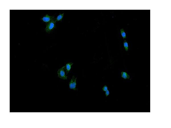
 IF (Immunofluorescence)
(Figure 9. IF analysis of P4HB using anti- P4HB antibody (AAA19258).P4HB was detected in immunocytochemical section of T-47D cells. Enzyme antigen retrieval was performed using IHC enzyme antigen retrieval reagent for 15 mins. The cells were blocked with 10% goat serum. And then incubated with 5μg/mL rabbit anti- P4HB Antibody (AAA19258) overnight at 4 degree C. DyLight®488 Conjugated Goat Anti-Rabbit IgG was used as secondary antibody at 1:100 dilution and incubated for 30 minutes at 37 degree C. The section was counterstained with DAPI. Visualize using a fluorescence microscope and filter sets appropriate for the label used.)
IF (Immunofluorescence)
(Figure 9. IF analysis of P4HB using anti- P4HB antibody (AAA19258).P4HB was detected in immunocytochemical section of T-47D cells. Enzyme antigen retrieval was performed using IHC enzyme antigen retrieval reagent for 15 mins. The cells were blocked with 10% goat serum. And then incubated with 5μg/mL rabbit anti- P4HB Antibody (AAA19258) overnight at 4 degree C. DyLight®488 Conjugated Goat Anti-Rabbit IgG was used as secondary antibody at 1:100 dilution and incubated for 30 minutes at 37 degree C. The section was counterstained with DAPI. Visualize using a fluorescence microscope and filter sets appropriate for the label used.)
P4HB, Polyclonal Antibody (Cat# AAA19258)
Full Name
Anti-P4HB Antibody
Gene Names
P4HB; DSI; GIT; PDI; PHDB; PDIA1; PO4DB; PO4HB; PROHB; ERBA2L; P4Hbeta
Reactivity
Human, Monkey, Mouse, Rat
Applications
WB, IHC-P, ICC, IF, FC/FACS/FCM, EIA
Purity
Immunogen affinity purified.


 FCM (Flow Cytometry)
(Figure 13. Flow Cytometry analysis of SiHa cells using anti-DRP1/DNM1L antibody (AAA19220).Overlay histogram showing SiHa cells stained with AAA19220 (Blue line). The cells were blocked with 10% normal goat serum. And then incubated with rabbit anti-DRP1/DNM1L Antibody (AAA19220, 1μg/1x106 cells) for 30 min at 20 degree C. DyLight®488 conjugated goat anti-rabbit IgG (5-10μg/1x106 cells) was used as secondary antibody for 30 minutes at 20 degree C. Isotype control antibody (Green line) was rabbit IgG (1μg/1x106) used under the same conditions. Unlabelled sample (Red line) was also used as a control.)
FCM (Flow Cytometry)
(Figure 13. Flow Cytometry analysis of SiHa cells using anti-DRP1/DNM1L antibody (AAA19220).Overlay histogram showing SiHa cells stained with AAA19220 (Blue line). The cells were blocked with 10% normal goat serum. And then incubated with rabbit anti-DRP1/DNM1L Antibody (AAA19220, 1μg/1x106 cells) for 30 min at 20 degree C. DyLight®488 conjugated goat anti-rabbit IgG (5-10μg/1x106 cells) was used as secondary antibody for 30 minutes at 20 degree C. Isotype control antibody (Green line) was rabbit IgG (1μg/1x106) used under the same conditions. Unlabelled sample (Red line) was also used as a control.)
DRP1/DNM1L, Polyclonal Antibody (Cat# AAA19220)
Full Name
Anti-DRP1/DNM1L Antibody
Gene Names
DNM1L; DLP1; DRP1; DVLP; EMPF; DYMPLE; HDYNIV
Reactivity
Human, Mouse, Rat
Applications
WB, IHC-P, ICC, IF, FC/FACS/FCM, EIA
Purity
Immunogen affinity purified.


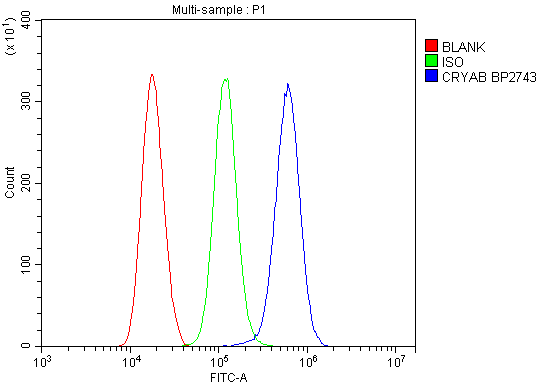 FCM (Flow Cytometry)
(Figure 13. Flow Cytometry analysis of THP-1 cells using anti-Alpha B Crystallin/CRYAB antibody (AAA19277).Overlay histogram showing THP-1 cells stained with AAA19277 (Blue line). The cells were blocked with 10% normal goat serum. And then incubated with rabbit anti-Alpha B Crystallin/CRYAB Antibody (AAA19277, 1μg/1x106 cells) for 30 min at 20 degree C. DyLight®488 conjugated goat anti-rabbit IgG (5-10μg/1x106 cells) was used as secondary antibody for 30 minutes at 20 degree C. Isotype control antibody (Green line) was rabbit IgG (1μg/1x106) used under the same conditions. Unlabelled sample (Red line) was also used as a control.)
FCM (Flow Cytometry)
(Figure 13. Flow Cytometry analysis of THP-1 cells using anti-Alpha B Crystallin/CRYAB antibody (AAA19277).Overlay histogram showing THP-1 cells stained with AAA19277 (Blue line). The cells were blocked with 10% normal goat serum. And then incubated with rabbit anti-Alpha B Crystallin/CRYAB Antibody (AAA19277, 1μg/1x106 cells) for 30 min at 20 degree C. DyLight®488 conjugated goat anti-rabbit IgG (5-10μg/1x106 cells) was used as secondary antibody for 30 minutes at 20 degree C. Isotype control antibody (Green line) was rabbit IgG (1μg/1x106) used under the same conditions. Unlabelled sample (Red line) was also used as a control.)
Alpha B Crystallin/CRYAB, Polyclonal Antibody (Cat# AAA19277)
Full Name
Anti-Alpha B Crystallin/CRYAB Antibody
Gene Names
CRYAB; CRYA2; CTPP2; HSPB5; CMD1II
Reactivity
Human, Mouse, Rat, Monkey
Applications
WB, IHC-P, ICC, IF, FC/FACS/FCM, EIA
Purity
Immunogen affinity purified.


 IF (Immunofluorescence)
(Figure 15. IF analysis of LSM2 using anti- LSM2 antibody (AAA19316).LSM2 was detected in immunocytochemical section of MCF-7 cells. Enzyme antigen retrieval was performed using IHC enzyme antigen retrieval reagent for 15 mins. The cells were blocked with 10% goat serum. And then incubated with 5μg/mL rabbit anti- LSM2 Antibody (AAA19316) overnight at 4 degree C. DyLight®488 Conjugated Goat Anti-Rabbit IgG was used as secondary antibody at 1:100 dilution and incubated for 30 minutes at 37 degree C. The section was counterstained with DAPI. Visualize using a fluorescence microscope and filter sets appropriate for the label used.)
IF (Immunofluorescence)
(Figure 15. IF analysis of LSM2 using anti- LSM2 antibody (AAA19316).LSM2 was detected in immunocytochemical section of MCF-7 cells. Enzyme antigen retrieval was performed using IHC enzyme antigen retrieval reagent for 15 mins. The cells were blocked with 10% goat serum. And then incubated with 5μg/mL rabbit anti- LSM2 Antibody (AAA19316) overnight at 4 degree C. DyLight®488 Conjugated Goat Anti-Rabbit IgG was used as secondary antibody at 1:100 dilution and incubated for 30 minutes at 37 degree C. The section was counterstained with DAPI. Visualize using a fluorescence microscope and filter sets appropriate for the label used.)
LSM2, Polyclonal Antibody (Cat# AAA19316)
Full Name
Anti-LSM2 Antibody
Gene Names
LSM2; G7B; snRNP; C6orf28; YBL026W
Reactivity
Human, Mouse, Rat, Monkey
Applications
WB, IHC-P, ICC, IF, FC/FACS/FCM
Purity
Immunogen affinity purified.


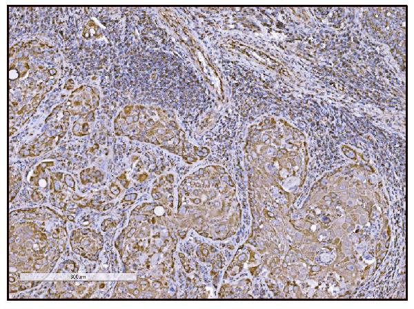


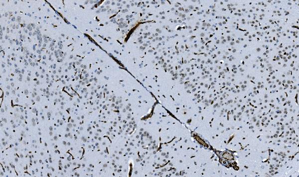
 IHC (Immunohistochemistry)
(Figure 14. IHC analysis of FABP-1/GOT2 using anti-FABP-1/GOT2 antibody (AAA19309).FABP-1/GOT2 was detected in paraffin-embedded section of rat lymph node tissue. Heat mediated antigen retrieval was performed in EDTA buffer (pH8. 0, epitope retrieval solution). The tissue section was blocked with 10% goat serum. The tissue section was then incubated with 2μg/ml rabbit anti-FABP-1/GOT2 Antibody (AAA19309) overnight at 4 degree C. Biotinylated goat anti-rabbit IgG was used as secondary antibody and incubated for 30 minutes at 37 degree C. The tissue section was developed using Strepavidin-Biotin-Complex (SABC) (Catalog # with DAB as the chromogen.)
IHC (Immunohistochemistry)
(Figure 14. IHC analysis of FABP-1/GOT2 using anti-FABP-1/GOT2 antibody (AAA19309).FABP-1/GOT2 was detected in paraffin-embedded section of rat lymph node tissue. Heat mediated antigen retrieval was performed in EDTA buffer (pH8. 0, epitope retrieval solution). The tissue section was blocked with 10% goat serum. The tissue section was then incubated with 2μg/ml rabbit anti-FABP-1/GOT2 Antibody (AAA19309) overnight at 4 degree C. Biotinylated goat anti-rabbit IgG was used as secondary antibody and incubated for 30 minutes at 37 degree C. The tissue section was developed using Strepavidin-Biotin-Complex (SABC) (Catalog # with DAB as the chromogen.)
FABP-1/GOT2, Polyclonal Antibody (Cat# AAA19309)
Full Name
Anti-FABP-1/GOT2 Antibody
Gene Names
GOT2; KAT4; KATIV; mitAAT
Reactivity
Human, Mouse, Rat
Applications
WB, IHC-P, ICC, IF, FC/FACS/FCM, EIA
Purity
Immunogen affinity purified.


 FCM (Flow Cytometry)
(Figure 7. Flow Cytometry analysis of THP-1 cells using anti-Ku70 antibody (AAA19361).Overlay histogram showing THP-1 cells stained with AAA19361 (Blue line). The cells were blocked with 10% normal goat serum. And then incubated with mouse anti- Ku70 Antibody (AAA19361, 1μg/1x106 cells) for 30 min at 20 degree C. DyLight®488 conjugated goat anti-mouse IgG (BA1126, 5-10μg/1x106 cells) was used as secondary antibody for 30 minutes at 20 degree C. Isotype control antibody (Green line) was mouse IgG (1μg/1x106) used under the same conditions. Unlabelled sample (Red line) was also used as a control.)
FCM (Flow Cytometry)
(Figure 7. Flow Cytometry analysis of THP-1 cells using anti-Ku70 antibody (AAA19361).Overlay histogram showing THP-1 cells stained with AAA19361 (Blue line). The cells were blocked with 10% normal goat serum. And then incubated with mouse anti- Ku70 Antibody (AAA19361, 1μg/1x106 cells) for 30 min at 20 degree C. DyLight®488 conjugated goat anti-mouse IgG (BA1126, 5-10μg/1x106 cells) was used as secondary antibody for 30 minutes at 20 degree C. Isotype control antibody (Green line) was mouse IgG (1μg/1x106) used under the same conditions. Unlabelled sample (Red line) was also used as a control.)
Ku70, Monoclonal Antibody (Cat# AAA19361)
Full Name
Anti-Ku70 Antibody (monoclonal, 9B6)
Gene Names
XRCC6; ML8; KU70; TLAA; CTC75; CTCBF; G22P1
Reactivity
Human
Applications
WB, IHC-P, ICC, IF, FC/FACS/FCM
Purity
Immunogen affinity purified.


 FCM (Flow Cytometry)
(Figure 11. Flow Cytometry analysis of THP-1 cells using anti-EPRS1/PARS antibody (AAA19268).Overlay histogram showing THP-1 cells stained with AAA19268 (Blue line). The cells were blocked with 10% normal goat serum. And then incubated with rabbit anti-EPRS1/PARS Antibody (AAA19268, 1μg/1x106 cells) for 30 min at 20 degree C. DyLight®488 conjugated goat anti-rabbit IgG (5-10μg/1x106 cells) was used as secondary antibody for 30 minutes at 20 degree C. Isotype control antibody (Green line) was rabbit IgG (1μg/1x106) used under the same conditions. Unlabelled sample (Red line) was also used as a control.)
FCM (Flow Cytometry)
(Figure 11. Flow Cytometry analysis of THP-1 cells using anti-EPRS1/PARS antibody (AAA19268).Overlay histogram showing THP-1 cells stained with AAA19268 (Blue line). The cells were blocked with 10% normal goat serum. And then incubated with rabbit anti-EPRS1/PARS Antibody (AAA19268, 1μg/1x106 cells) for 30 min at 20 degree C. DyLight®488 conjugated goat anti-rabbit IgG (5-10μg/1x106 cells) was used as secondary antibody for 30 minutes at 20 degree C. Isotype control antibody (Green line) was rabbit IgG (1μg/1x106) used under the same conditions. Unlabelled sample (Red line) was also used as a control.)
EPRS1/PARS, Polyclonal Antibody (Cat# AAA19268)
Full Name
Anti-EPRS1/PARS Antibody
Gene Names
EPRS; EARS; PARS; QARS; QPRS; PIG32; GLUPRORS
Reactivity
Human, Mouse, Rat
Applications
WB, IHC-P, ICC, IF, FC/FACS/FCM, EIA
Purity
Immunogen affinity purified.