Highly validated and characterized monoclonal/polyclonal
antibodies and recombinant
proteins
The majority of AAA Biotech’s antibodies are highly validated and can be use in multiple
applications such as ELISA, FC,
ICC, IF, IHC, IP, WB, etc. We have antibodies available for rare species, in multiple conjugated
forms or recombinant
antibodies.
As for our high quality proteins, the majority have 90% purity, detected by SDS-PAGE while some are
available in
different tags such as Flag, GST, His, MBP, etc. We also carry high quality native and biologically
active proteins.
AAA Biotech is constantly working to expand our capacity to provide recombinant proteins and
antibodies to most
target proteins.
SELECT `options_values_price` as `price`, `products_options_values_name` as `package`
FROM `products_attributes`
JOIN `products_options_values` ON `products_options_values`.`products_options_values_id` = `products_attributes`.`options_values_id`
WHERE `products_attributes`.`products_id` = '23923'
Query
Database
1.53 ms
SELECT `options_values_price` as `price`, `products_options_values_name` as `package`
FROM `products_attributes`
JOIN `products_options_values` ON `products_options_values`.`products_options_values_id` = `products_attributes`.`options_values_id`
WHERE `products_attributes`.`products_id` = '19143'
Query
Database
1.51 ms
SELECT `options_values_price` as `price`, `products_options_values_name` as `package`
FROM `products_attributes`
JOIN `products_options_values` ON `products_options_values`.`products_options_values_id` = `products_attributes`.`options_values_id`
WHERE `products_attributes`.`products_id` = '19154'
Query
Database
3.62 ms
SELECT `options_values_price` as `price`, `products_options_values_name` as `package`
FROM `products_attributes`
JOIN `products_options_values` ON `products_options_values`.`products_options_values_id` = `products_attributes`.`options_values_id`
WHERE `products_attributes`.`products_id` = '19131'
Query
Database
1.52 ms
SELECT `options_values_price` as `price`, `products_options_values_name` as `package`
FROM `products_attributes`
JOIN `products_options_values` ON `products_options_values`.`products_options_values_id` = `products_attributes`.`options_values_id`
WHERE `products_attributes`.`products_id` = '28639'
View: search.php
Views
102.73 ms
View: template.php
Views
102.09 ms
After Filters
Timer
0.14 ms
Database (5 total Queries, 5 of them unique across 2 Connections)
Time
Query String
2.61 ms
SELECT `options_values_price` as `price`, `products_options_values_name` as `package`
FROM `products_attributes`
JOIN `products_options_values` ON `products_options_values`.`products_options_values_id` = `products_attributes`.`options_values_id`
WHERE `products_attributes`.`products_id` = '23923'
SELECT `options_values_price` as `price`, `products_options_values_name` as `package`
FROM `products_attributes`
JOIN `products_options_values` ON `products_options_values`.`products_options_values_id` = `products_attributes`.`options_values_id`
WHERE `products_attributes`.`products_id` = '19143'
SELECT `options_values_price` as `price`, `products_options_values_name` as `package`
FROM `products_attributes`
JOIN `products_options_values` ON `products_options_values`.`products_options_values_id` = `products_attributes`.`options_values_id`
WHERE `products_attributes`.`products_id` = '19154'
SELECT `options_values_price` as `price`, `products_options_values_name` as `package`
FROM `products_attributes`
JOIN `products_options_values` ON `products_options_values`.`products_options_values_id` = `products_attributes`.`options_values_id`
WHERE `products_attributes`.`products_id` = '19131'
SELECT `options_values_price` as `price`, `products_options_values_name` as `package`
FROM `products_attributes`
JOIN `products_options_values` ON `products_options_values`.`products_options_values_id` = `products_attributes`.`options_values_id`
WHERE `products_attributes`.`products_id` = '28639'
Keratin 15 is a type I keratin which is expressed only in basal keratinocytes in stratified epithelia and does not appear to have a natural type II expression partner. Keratin 15 is down regulated in activated keratinocytes. Cytokeratin 15 is a specific marker of stem cells of the hair-follicle bulge and may be a useful marker for between basal cell carcinoma (BCC) and trichoepithelioma. Trichoblastoma are benign neoplasms of follicular differentiation frequently found in nevus sebaceous. Many morphologic features are shared with nodular basal cell carcinoma, sometimes rendering a difficult. Trichoblastoma and BCC show variable expression of Cytokeratin 15 and Cytokeratin 19, and absence of hair keratins.
⇄⧉purity => string (91) "Purified Ab with BSA and Azide at 200ug/ml OR Purified Ab WITHOUT BSA and Az...
$value['hits']['hits'][0]['_source']['purity']
Purified Ab with BSA and Azide at 200ug/ml OR Purified Ab WITHOUT BSA and Azide at 1.0mg/ml
⇄⧉form => string (166) "200ug/ml of Ab Purified from Bioreactor Concentrate by Protein A/G. Prepared...
$value['hits']['hits'][0]['_source']['form']
200ug/ml of Ab Purified from Bioreactor Concentrate by Protein A/G. Prepared in 10mM PBS with 0.05% BSA & 0.05% azide. Also available WITHOUT BSA & azide at 1.0mg/ml.
Antibody with azide: store at 2 to 8 degree C.<br>Antibody without azide: store at -20 to -80 degree C.<br>Antibody is stable for 24 months. Non-hazardous. No MSDS required.
⇄⧉app_tested => string (105) "Flow Cytometry (FC/FACS), Immunofluorescence (IF), Western Blot (WB), Immuno...
FC/FACS: 1-2ug/million cells<br>IF: 1-2ug/ml<br>WB: 1-2ug/ml<br>Immunohistochemistry (Formalin-fixed) (1-2ug/ml for 30 min at RT) (Staining of formalin-fixed tissues requires boiling tissue sections in 10mM Citrate Buffer, pH 6.0, for 10-20 min followed by cooling at RT for 20 minutes)
⇄⧉testing_protocols => string (2490) "Application Data||Analysis of Protein Array containing more than 19,000 full...
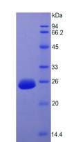
Application Data||Analysis of Protein Array containing more than 19,000 full-length human proteins using Cytokeratin 15 Mouse Monoclonal Antibody (KRT15/2957). Z- and S- Score: The Z-score represents the strength of a signal that a monoclonal antibody (MAb) (in combination with a fluorescently-tagged anti-IgG secondary antibody) produces when binding to a particular protein on the HuProtTM array. Z-scores are described in units of standard deviations (SD's) above the mean value of all signals generated on that array. If targets on HuProtTM are arranged in descending order of the Z-score, the S-score is the difference (also in units of SD's) between the Z-score. S-score therefore represents the relative target specificity of a MAb to its intended target. A MAb is considered to specific to its intended target, if the MAb has an S-score of at least 2.5. For example, if a MAb binds to protein X with a Z-score of 43 and to protein Y with a Z-score of 14, then the S-score for the binding of that MAb to protein X is equal to 29.||AAA23923_APP9.jpg!!WB (Western Blot)||Western Blot Analysis of HCT116 cell lysate using Cytokeratin 15 Mouse Monoclonal Antibody (KRT15/2957)||AAA23923_WB8.jpg!!FCM (Flow Cytometry)||Flow Cytometric Analysis of PFA-fixed HeLa cells using Cytokeratin 15 Mouse MAb (KRT15/2957) followed by Goat anti-Mouse IgG-CF488 (Blue); Isotype Control (Red).||AAA23923_FCM7.jpg!!SDS-PAGE||SDS-PAGE Analysis Purified Cytokeratin 15 Mouse Monoclonal Antibody (KRT15/2957). Confirmation of Purity and Integrity of Antibody||AAA23923_SDS_PAGE6.jpg!!WB (Western Blot)||Western Blot Analysis of Human Thymus tissue lysate using Cytokeratin 15 Mouse Monoclonal Antibody (KRT15/2957)||AAA23923_WB5.jpg!!IF (Immunofluorescence)||Immunofluorescence Analysis of methanol-fixed human MCF-7 cells. Cytokeratin 15 Mouse Monoclonal Antibody (KRT15/2957) followed by Goat anti-Mouse IgG-CF488 (Green). The nuclear counterstain is Reddot (Red)||AAA23923_IF4.jpg!!IHC (Immunohistochemistry)||Formalin-fixed, paraffin-embedded human Colon Carcinoma stained with Cytokeratin 15 Mouse Monoclonal Antibody (KRT15/2957)||AAA23923_IHC3.jpg!!IHC (Immunohistochemistry)||Formalin-fixed, paraffin-embedded human Basal Cell Carcinoma stained with Cytokeratin 15 Mouse Monoclonal Antibody (KRT15/2957).||AAA23923_IHC2.jpg!!IHC (Immunohistochemistry)||Formalin-fixed, paraffin-embedded human Prostate Carcinoma stained with Cytokeratin 15 Mouse Monoclonal Antibody (KRT15/2957)||AAA23923_IHC.jpg
Yu-Hong Shen, Cui-Ping Xu, Zhi-Meng Shi, Yan-Jiao Zhang, Ya-Guang Qiao, He-Ping Zhao. Cytokeratin 15 is an Effective Indicator for Progression and Malignancy of Esophageal Squamous Cell Carcinomas. Asian Pac J Cancer Prev, 17 (9), 4217-4222.
aaa23923 mouse human monoclonal igg2c kappa krt15 2957 purified ab with bsa and azide at 200ug ml or without 1.0mg of from bioreactor concentrate by protein a g prepared in 10mm pbs 0.05 also available keratin 15 is type i which expressed only basal keratinocytes stratified epithelia does not appear to have natural ii expression partner down regulated activated cytokeratin specific marker stem cells the hair follicle bulge may be useful for between cell carcinoma bcc trichoepithelioma trichoblastoma are benign neoplasms follicular differentiation frequently found nevus sebaceous many morphologic features shared nodular sometimes rendering difficult show variable 19 absence keratins flow cytometry fc facs immunofluorescence if western blot wb immunohistochemistry ihc paraffin 1 2ug million formalin fixed 30 min rt staining tissues requires boiling tissue sections citrate buffer ph 6.0 10 20 followed cooling minutes embedded prostate stained antibody aaa23923_ihc aaa23923_ihc2 colon aaa23923_ihc3 analysis methanol mcf 7 goat anti igg cf488 green nuclear counterstain reddot red aaa23923_if4 thymus lysate using aaa23923_wb5 sds page confirmation purity integrity aaa23923_sds6 cytometric pfa hela mab blue isotype control aaa23923_fc7 hct116 aaa23923_wb8 testing data array containing more than 19,000 full length proteins z s score represents strength signal that combination fluorescently tagged secondary produces when binding particular on huprottm scores described units standard deviations sd's above mean value all signals generated targets arranged descending order difference therefore relative target specificity its intended considered has an least 2.5 example binds x 43 y 14 then equal 29 aaa23923_td9 esophageal squamous ck15 k1co ka15 basic beta complex acidic gene cytoskeletal krtb krtl15 k15 52kda ck k1c15_human 24430190 np_002266.2 p19012 148030 positive a431 skin immunogen recombinant cellular localization cytoplasmic hu chromosome location 17q21.2 keratin15 variable19 paraffin1 fixed30 ph6.0 mcf7 least2.5 x43 y14 equal29
Store at -20 degree C for one year. After reconstitution, at 4 degree C for one month. It can also be aliquotted and stored frozen at -20 degree C for a longer time. Avoid repeated freezing and thawing.
FCM (Flow Cytometry)||Figure 6. Flow Cytometry analysis of Hela cells using anti-PDE4D antibody (AAA19143).<br>Overlay histogram showing Hela cells stained with AAA19143 (Blue line).The cells were blocked with 10% normal goat serum. And then incubated with rabbit anti-PDE4D Antibody (AAA19143,1ug/1x10^6 cells) for 30 min at 20 degree C. DyLight®488 conjugated goat anti-rabbit IgG (5-10ug/1x10^6 cells) was used as secondary antibody for 30 minutes at 20 degree C. Isotype control antibody (Green line) was rabbit IgG (1ug/1x106) used under the same conditions. Unlabelled sample (Red line) was also used as a control.||AAA19143_FCM6.jpg!!FCM (Flow Cytometry)||Figure 5. Flow Cytometry analysis of A549 cells using anti-PDE4D antibody (AAA19143).<br>Overlay histogram showing A549 cells stained with AAA19143 (Blue line).The cells were blocked with 10% normal goat serum. And then incubated with rabbit anti-PDE4D Antibody (AAA19143,1ug/1x10^6 cells) for 30 min at 20 degree C. DyLight®488 conjugated goat anti-rabbit IgG (5-10ug/1x10^6 cells) was used as secondary antibody for 30 minutes at 20 degree C. Isotype control antibody (Green line) was rabbit IgG (1ug/1x106) used under the same conditions. Unlabelled sample (Red line) was also used as a control.||AAA19143_FCM5.jpg!!IHC (Immunohistochemistry)||Figure 4. IHC analysis of PDE4D using anti-PDE4D antibody (AAA19143).<br>PDE4D was detected in paraffin-embedded section of rat lung tissue. Heat mediated antigen retrieval was performed in citrate buffer (pH6, epitope retrieval solution) for 20 mins. The tissue section was blocked with 10% goat serum. The tissue section was then incubated with 1ug/ml rabbit anti-PDE4D Antibody (AAA19143) overnight at 4 degree C. Biotinylated goat anti-rabbit IgG was used as secondary antibody and incubated for 30 minutes at 37 degree C. The tissue section was developed using Strepavidin-Biotin-Complex (SABC) with DAB as the chromogen. <br>||AAA19143_IHC4.jpg!!IHC (Immunohistochemistry)||Figure 3. IHC analysis of PDE4D using anti-PDE4D antibody (AAA19143).<br>PDE4D was detected in paraffin-embedded section of human placenta tissue. Heat mediated antigen retrieval was performed in citrate buffer (pH6, epitope retrieval solution) for 20 mins. The tissue section was blocked with 10% goat serum. The tissue section was then incubated with 1ug/ml rabbit anti-PDE4D Antibody (AAA19143) overnight at 4 degree C. Biotinylated goat anti-rabbit IgG was used as secondary antibody and incubated for 30 minutes at 37 degree C. The tissue section was developed using Strepavidin-Biotin-Complex (SABC) with DAB as the chromogen.<br>||AAA19143_IHC3.jpg!!IHC (Immunohistochemistry)||Figure 2. IHC analysis of PDE4D using anti-PDE4D antibody (AAA19143).<br>PDE4D was detected in paraffin-embedded section of mouse lung tissue. Heat mediated antigen retrieval was performed in citrate buffer (pH6, epitope retrieval solution) for 20 mins. The tissue section was blocked with 10% goat serum. The tissue section was then incubated with 1ug/ml rabbit anti-PDE4D Antibody (AAA19143) overnight at 4 degree C. Biotinylated goat anti-rabbit IgG was used as secondary antibody and incubated for 30 minutes at 37 degree C. The tissue section was developed using Strepavidin-Biotin-Complex (SABC) with DAB as the chromogen.<br>||AAA19143_IHC2.jpg!!WB (Western Blot)||Figure 1. Western blot analysis of PDE4D using anti-PDE4D antibody (AAA19143).<br>Electrophoresis was performed on a 5-20% SDS-PAGE gel at 70V (Stacking gel) / 90V (Resolving gel) for 2-3 hours. The sample well of each lane was loaded with 50ug of sample under reducing conditions.<br>Lane 1: human Hela whole cell lysates.<br>After Electrophoresis, proteins were transferred to a Nitrocellulose membrane at 150mA for 50-90 minutes. Blocked the membrane with 5% Non-fat Milk/ TBS for 1.5 hour at RT. The membrane was incubated with rabbit anti-PDE4D antigen affinity purified polyclonal antibody at 0.5ug/mL overnight at 4 degree C, then washed with TBS-0.1%Tween 3 times with 5 minutes each and probed with a goat anti-rabbit IgG-HRP secondary antibody at a dilution of 1:10000 for 1.5 hour at RT. The signal is developed using an Enhanced Chemiluminescent detection (ECL) kit with Tanon 5200 system. A specific band was detected for PDE4D at approximately 91KD. The expected band size for PDE4D is at 91KD.||AAA19143_WB.jpg
⇄⧉etc_term1 => string (397) "Immunogen||E Coli-derived human PDE4D recombinant protein (Position: H466-Q7...
$value['hits']['hits'][1]['_source']['etc_term1']
Immunogen||E Coli-derived human PDE4D recombinant protein (Position: H466-Q709).!!Subcellular Localization||Apical cell membrane.!!Tissue Specificity||Expressed in colonic epithelial cells (at protein level). Widespread; most abundant in skeletal muscle. Isoform 6 is detected in brain. Isoform 8 is detected in brain, placenta, lung and kidney. Isoform 7 is detected in heart and skeletal muscle.
⇄⧉etc_term2 => string (169) "Reconstitution||Add 0.2ml of distilled water will yield a concentration of 5...
$value['hits']['hits'][1]['_source']['etc_term2']
Reconstitution||Add 0.2ml of distilled water will yield a concentration of 500ug/ml.!!Contents||Each vial contains 4mg Trehalose, 0.9mg NaCl, 0.2mg Na2HPO4, 0.05mg NaN3.
Description: cAMP-specific 3',5'-cyclic phosphodiesterase 4D is an enzyme that in humans is encoded by the PDE4D gene. This gene encodes one of four mammalian counterparts to the fruit fly 'dunce' gene. The encoded protein has 3',5'-cyclic-AMP phosphodiesterase activity and degrades cAMP, which acts as a signal transduction molecule in multiple cell types. This gene uses different promoters to generate multiple alternatively spliced transcript variants that encode functional proteins.<br>Protein Function: Hydrolyzes the second messenger cAMP, which is a key regulator of many important physiological processes.
aaa19143 rabbit human mouse rat no cross reactivity with other proteins polyclonal lyophilized elisa eia flow cytometry fc facs immunohistochemistry ihc immunocytochemistry icc western blot wb 0.1 0.5mug ml paraffin embedded section 0.5 1mug frozen 1 3g 1x10 6 cells direct figure analysis of pde4d using anti antibody electrophoresis was performed on a 5 20 sds page gel at 70v stacking 90v resolving for 2 3 hours the sample well each lane loaded 50ug under reducing conditions hela whole cell lysates after were transferred to nitrocellulose membrane 150ma 50 90 minutes blocked non fat milk tbs 1.5 hour rt incubated antigen affinity purified 0.5ug overnight 4 degree c then washed tween times and probed goat igg hrp secondary dilution 1:10000 signal is developed an enhanced chemiluminescent detection ecl kit tanon 5200 system specific band detected approximately 91kd expected size aaa19143_wb in lung tissue heat mediated retrieval citrate buffer ph6 epitope solution mins 10 serum 1ug biotinylated used as 30 37 strepavidin biotin complex sabc dab chromogen aaa19143_ihc2 placenta aaa19143_ihc3
aaa19143_ihc4 a549 overlay histogram showing stained blue line normal aaa19143,1ug 1x10^6 min dylight® 488 conjugated 10ug isotype control green 1x10⁶ same unlabelled red also aaa19143_fc5 aaa19143_fc6 picoband camp 3',5' cyclic phosphodiesterase 4d dpde3 pde43 isoform pde4d4 strk1 acrdys2 hspde4d pde4dn2 24,429 da 157277988 np_001098101.1 q08499 nm_001104631.1 o43433 q13549 q13550 q13551 q7z2l8 q8iv84 q8iva9 q8ivd2 q8ivd3 q96hl4 q9hcx7 l20970 mrna immunogen e coli derived recombinant protein position h466 q709 subcellular localization apical specificity expressed colonic epithelial level widespread most abundant skeletal muscle brain 8 kidney 7 heart reconstitution add 0.2ml distilled water will yield concentration 500ug contents vial contains 4mg trehalose 0.9mg nacl 0.2mg na2hpo4 0.05mg nan3 wb0.1 section0.5 frozen1 a5 for2 150ma50 tbs1.5 overnight4 mins10 as30 dylight®488 brain8 kidney7
Store at -20 degree C for one year. After reconstitution, at 4 degree C for one month. It can also be aliquotted and stored frozen at -20 degree C for a longer time. Avoid repeated freezing and thawing.
⇄app_tested => string (58) "ELISA (EIA), Immunohistochemistry (IHC), Western Blot (WB)"
IHC (Immunohistochemistry)||Figure 7. IHC analysis of MYLK using anti-MYLK antibody (AAA19154).<br>MYLK was detected in paraffin-embedded section of rat lung tissue. Heat mediated antigen retrieval was performed in citrate buffer (pH6, epitope retrieval solution) for 20 mins. The tissue section was blocked with 10% goat serum. The tissue section was then incubated with 1ug/ml rabbit anti-MYLK Antibody (AAA19154) overnight at 4 degree C. Biotinylated goat anti-rabbit IgG was used as secondary antibody and incubated for 30 minutes at 37 degree C. The tissue section was developed using Strepavidin-Biotin-Complex (SABC) with DAB as the chromogen. <br>||AAA19154_IHC7.jpg!!IHC (Immunohistchemistry)||Figure 6. IHC analysis of MYLK using anti-MYLK antibody (AAA19154).<br>MYLK was detected in paraffin-embedded section of human rectal cancer tissue. Heat mediated antigen retrieval was performed in citrate buffer (pH6, epitope retrieval solution) for 20 mins. The tissue section was blocked with 10% goat serum. The tissue section was then incubated with 1ug/ml rabbit anti-MYLK Antibody (AAA19154) overnight at 4 degree C. Biotinylated goat anti-rabbit IgG was used as secondary antibody and incubated for 30 minutes at 37 degree C. The tissue section was developed using Strepavidin-Biotin-Complex (SABC) with DAB as the chromogen.<br>||AAA19154_IHC6.jpg!!IHC (Immunohistochemistry)||Figure 5. IHC analysis of MYLK using anti-MYLK antibody (AAA19154).<br>MYLK was detected in paraffin-embedded section of human placenta tissue. Heat mediated antigen retrieval was performed in citrate buffer (pH6, epitope retrieval solution) for 20 mins. The tissue section was blocked with 10% goat serum. The tissue section was then incubated with 1ug/ml rabbit anti-MYLK Antibody (AAA19154) overnight at 4 degree C. Biotinylated goat anti-rabbit IgG was used as secondary antibody and incubated for 30 minutes at 37 degree C. The tissue section was developed using Strepavidin-Biotin-Complex (SABC) with DAB as the chromogen.<br>||AAA19154_IHC5.jpg!!IHC (Immunohistochemistry)||Figure 4. IHC analysis of MYLK using anti-MYLK antibody (AAA19154).<br>MYLK was detected in paraffin-embedded section of rat small intestine tissue. Heat mediated antigen retrieval was performed in citrate buffer (pH6, epitope retrieval solution) for 20 mins. The tissue section was blocked with 10% goat serum. The tissue section was then incubated with 1ug/ml rabbit anti-MYLK Antibody (AAA19154) overnight at 4 degree C. Biotinylated goat anti-rabbit IgG was used as secondary antibody and incubated for 30 minutes at 37 degree C. The tissue section was developed using Strepavidin-Biotin-Complex (SABC) with DAB as the chromogen.<br>||AAA19154_IHC4.jpg!!IHC (Immunohistochemistry)||Figure 3. IHC analysis of MYLK using anti-MYLK antibody (AAA19154).<br>MYLK was detected in paraffin-embedded section of mouse small intestine tissue. Heat mediated antigen retrieval was performed in citrate buffer (pH6, epitope retrieval solution) for 20 mins. The tissue section was blocked with 10% goat serum. The tissue section was then incubated with 1ug/ml rabbit anti-MYLK Antibody (AAA19154) overnight at 4 degree C. Biotinylated goat anti-rabbit IgG was used as secondary antibody and incubated for 30 minutes at 37 degree C. The tissue section was developed using Strepavidin-Biotin-Complex (SABC) with DAB as the chromogen.<br>||AAA19154_IHC3.jpg!!IHC (Immunohistochemistry)||Figure 2. IHC analysis of MYLK using anti-MYLK antibody (AAA19154).<br>MYLK was detected in paraffin-embedded section of mouse lung tissue. Heat mediated antigen retrieval was performed in citrate buffer (pH6, epitope retrieval solution) for 20 mins. The tissue section was blocked with 10% goat serum. The tissue section was then incubated with 1ug/ml rabbit anti-MYLK Antibody (AAA19154) overnight at 4 degree C. Biotinylated goat anti-rabbit IgG was used as secondary antibody and incubated for 30 minutes at 37 degree C. The tissue section was developed using Strepavidin-Biotin-Complex (SABC) with DAB as the chromogen.<br>||AAA19154_IHC2.jpg!!WB (Western Blot)||Figure 1. Western blot analysis of MYLK using anti-MYLK antibody (AAA19154).<br>Electrophoresis was performed on a 5-20% SDS-PAGE gel at 70V (Stacking gel) / 90V (Resolving gel) for 2-3 hours. The sample well of each lane was loaded with 50ug of sample under reducing conditions.<br>Lane 1: human SGC-7901 whole cell lysates,<br>Lane 2: rat spleen tissue lysates,<br>Lane 3: mouse spleen tissue lysates.<br>After Electrophoresis, proteins were transferred to a Nitrocellulose membrane at 150mA for 50-90 minutes. Blocked the membrane with 5% Non-fat Milk/ TBS for 1.5 hour at RT. The membrane was incubated with rabbit anti-MYLK antigen affinity purified polyclonal antibody at 0.5ug/mL overnight at 4 degree C, then washed with TBS-0.1%Tween 3 times with 5 minutes each and probed with a goat anti-rabbit IgG-HRP secondary antibody at a dilution of 1:10000 for 1.5 hour at RT. The signal is developed using an Enhanced Chemiluminescent detection (ECL) kit with Tanon 5200 system. A specific band was detected for MYLK at approximately 250KD. The expected band size for MYLK is at 211KD.||AAA19154_WB.jpg
⇄⧉etc_term1 => string (784) "Immunogen||E Coli-derived human MYLK recombinant protein (Position: D1441-D1...
$value['hits']['hits'][2]['_source']['etc_term1']
Immunogen||E Coli-derived human MYLK recombinant protein (Position: D1441-D1709).!!Subcellular Localization||Cytoplasm. Cell projection, lamellipodium. Cleavage furrow. Cytoplasm, cytoskeleton. Localized to stress fibers during interphase and to the cleavage furrow during mitosis.!!Tissue Specificity||Smooth muscle and non-muscle isozymes are expressed in a wide variety of adult and fetal tissues and in cultured endothelium with qualitative expression appearing to be neither tissue- nor development-specific. Non-muscle isoform 2 is the dominant splice variant expressed in various tissues. Telokin has been found in a wide variety of adult and fetal tissues. Accumulates in well differentiated enterocytes of the intestinal epithelium in response to tumor necrosis factor (TNF).
⇄⧉etc_term2 => string (169) "Reconstitution||Add 0.2ml of distilled water will yield a concentration of 5...
$value['hits']['hits'][2]['_source']['etc_term2']
Reconstitution||Add 0.2ml of distilled water will yield a concentration of 500ug/ml.!!Contents||Each vial contains 4mg Trehalose, 0.9mg NaCl, 0.2mg Na2HPO4, 0.05mg NaN3.
Description: Myosin light chain kinase, smooth muscle also known as kinase-related protein (KRP) or telokin is an enzyme that in humans is encoded by the MYLK gene. This gene, a muscle member of the immunoglobulin gene superfamily, encodes myosin light chain kinase which is a calcium/calmodulin dependent enzyme. This kinase phosphorylates myosin regulatory light chains to facilitate myosin interaction with actin filaments to produce contractile activity. This gene encodes both smooth muscle and nonmuscle isoforms. In addition, using a separate promoter in an intron in the 3' region, it encodes telokin, a small protein identical in sequence to the C-terminus of myosin light chain kinase, that is independently expressed in smooth muscle and functions to stabilize unphosphorylated myosin filaments. A pseudogene is located on the p arm of chromosome 3. Four transcript variants that produce four isoforms of the calcium/calmodulin dependent enzyme have been identified as well as two transcripts that produce two isoforms of telokin. <br>Protein Function: Calcium/calmodulin-dependent myosin light chain kinase implicated in smooth muscle contraction via phosphorylation of myosin light chains (MLC). Also regulates actin-myosin interaction through a non-kinase activity. Phosphorylates PTK2B/PYK2 and myosin light-chains. Involved in the inflammatory response (e.g. apoptosis, vascular permeability, leukocyte diapedesis), cell motility and morphology, airway hyperreactivity and other activities relevant to asthma. Required for tonic airway smooth muscle contraction that is necessary for physiological and asthmatic airway resistance. Necessary for gastrointestinal motility. Implicated in the regulation of endothelial as well as vascular permeability, probably via the regulation of cytoskeletal rearrangements. In the nervous system it has been shown to control the growth initiation of astrocytic processes in culture and to participate in transmitter release at synapses formed between cultured sympathetic ganglion cells. Critical participant in signaling sequences that result in fibroblast apoptosis. Plays a role in the regulation of epithelial cell survival. Required for epithelial wound healing, especially during actomyosin ring contraction during purse-string wound closure. Mediates RhoA- dependent membrane blebbing. Triggers TRPC5 channel activity in a calcium-dependent signaling, by inducing its subcellular localization at the plasma membrane. Promotes cell migration (including tumor cells) and tumor metastasis. PTK2B/PYK2 activation by phosphorylation mediates ITGB2 activation and is thus essential to trigger neutrophil transmigration during acute lung injury (ALI). May regulate optic nerve head astrocyte migration. Probably involved in mitotic cytoskeletal regulation. Regulates tight junction probably by modulating ZO-1 exchange in the perijunctional actomyosin ring. Mediates burn-induced microvascular barrier injury; triggers endothelial contraction in the development of microvascular hyperpermeability by phosphorylating MLC. Essential for intestinal barrier dysfunction. Mediates Giardia spp.-mediated reduced epithelial barrier function during giardiasis intestinal infection via reorganization of cytoskeletal F-actin and tight junctional ZO-1. Necessary for hypotonicity-induced Ca(2+) entry and subsequent activation of volume-sensitive organic osmolyte/anion channels (VSOAC) in cervical cancer cells. Responsible for high proliferative ability of breast cancer cells through anti-apoptosis.
aaa19154 rabbit human mouse rat no cross reactivity with other proteins polyclonal lyophilized elisa eia immunohistochemistry ihc western blot wb 0.1 0.5mug ml paraffin embedded section 0.5 1mug direct figure 1 analysis of mylk using anti antibody electrophoresis was performed on a 5 20 sds page gel at 70v stacking 90v resolving for 2 3 hours the sample well each lane loaded 50ug under reducing conditions sgc 7901 whole cell lysates spleen tissue after were transferred to nitrocellulose membrane 150ma 50 90 minutes blocked non fat milk tbs 1.5 hour rt incubated antigen affinity purified 0.5ug overnight 4 degree c then washed tween times and probed goat igg hrp secondary dilution 1:10000 signal is developed an enhanced chemiluminescent detection ecl kit tanon 5200 system specific band detected approximately 250kd expected size 211kd aaa19154_wb in lung heat mediated retrieval citrate buffer ph6 epitope solution mins 10 serum 1ug biotinylated used as 30 37 strepavidin biotin complex sabc dab chromogen aaa19154_ihc2 small intestine aaa19154_ihc3 aaa19154_ihc4 placenta aaa19154_ihc5 6 rectal cancer aaa19154_ihc6 7
 picoband myosin light chain kinase smooth muscle mlck smmlck related protein krp telokin deglutamylated form mlck1 mylk1 isoform 9 aat7 mlck108 mlck210 mstp083 110,076 da 1008806537 np_001308238.1 q15746 nm_001321309.1 o95796 o95797 o95798 o95799 q14844 q16794 q17s15 q3zcp9 q5my99 b4due3 d3dn97 x85337 mrna immunogen e coli derived recombinant position d1441 d1709 subcellular localization cytoplasm projection lamellipodium cleavage furrow cytoskeleton localized stress fibers during interphase mitosis specificity isozymes are expressed wide variety adult fetal tissues cultured endothelium qualitative expression appearing be neither nor development dominant splice variant various has been found accumulates differentiated enterocytes intestinal epithelium response tumor necrosis factor tnf reconstitution add 0.2ml distilled water will yield concentration 500ug contents vial contains 4mg trehalose 0.9mg nacl 0.2mg na2hpo4 0.05mg nan3 wb0.1 section0.5 figure1 a5 for2 150ma50 tbs1.5 overnight4 mins10 as30 aaa19154_ihc56 aaa19154_ihc67 isoform9
Store at -20 degree C for one year. After reconstitution, at 4 degree C for one month. It can also be aliquotted and stored frozen at -20 degree C for a longer time. Avoid repeated freezing and thawing.
⇄app_tested => string (45) "Immunohistochemistry (IHC), Western Blot (WB)"
WB (Western Blot)||Figure 1. Western blot analysis of Ran using anti-Ran antibody (AAA19131). <br>Electrophoresis was performed on a 5-20% SDS-PAGE gel at 70V (Stacking gel) / 90V (Resolving gel) for 2-3 hours. The sample well of each lane was loaded with 50ug of sample under reducing conditions. <br>Lane 1: rat brain tissue lysate,<br>Lane 2: rat testis tissue lysate,<br>Lane 3: rat thymus tissue lysate,<br>Lane 4: mouse brain tissue lysate,<br>Lane 5: mouse testis tissue lysate,<br>Lane 6: mouse thymus tissue lysate,<br>Lane 7: human A549 whole cell lysate,<br>Lane 8: human 22RV1 whole cell lysate,<br>Lane 9: human Hela whole cell lysate. <br>After Electrophoresis, proteins were transferred to a Nitrocellulose membrane at 150mA for 50-90 minutes. Blocked the membrane with 5% Non-fat Milk/ TBS for 1.5 hour at RT. The membrane was incubated with rabbit anti-Ran antigen affinity purified polyclonal antibody at 0.5ug/mL overnight at 4 degree C, then washed with TBS-0.1%Tween 3 times with 5 minutes each and probed with a goat anti-rabbit IgG-HRP secondary antibody at a dilution of 1:10000 for 1.5 hour at RT. The signal is developed using an Enhanced Chemiluminescent detection (ECL) kit with Tanon 5200 system. A specific band was detected for Ran at approximately 24KD. The expected band size for Ran is at 24KD.||AAA19131_WB6.jpg!!IHC (Immunohistochemistry)||Figure 3. IHC analysis of Ran using anti-Ran antibody (AAA19131).<br>Ran was detected in paraffin-embedded section of human lung cancer tissue. Heat mediated antigen retrieval was performed in citrate buffer (pH6, epitope retrieval solution) for 20 mins. The tissue section was blocked with 10% goat serum. The tissue section was then incubated with 1ug/ml rabbit anti-Ran Antibody (AAA19131) overnight at 4 degree C. Biotinylated goat anti-rabbit IgG was used as secondary antibody and incubated for 30 minutes at 37 degree C. The tissue section was developed using Strepavidin-Biotin-Complex (SABC) with DAB as the chromogen. <br>||AAA19131_IHC5.jpg!!IHC (Immunohistochemistry)||Figure 2. IHC analysis of Ran using anti-Ran antibody (AAA19131).<br>Ran was detected in paraffin-embedded section of human intestinal cancer tissue. Heat mediated antigen retrieval was performed in citrate buffer (pH6, epitope retrieval solution) for 20 mins. The tissue section was blocked with 10% goat serum. The tissue section was then incubated with 1ug/ml rabbit anti-Ran Antibody (AAA19131) overnight at 4 degree C. Biotinylated goat anti-rabbit IgG was used as secondary antibody and incubated for 30 minutes at 37 degree C. The tissue section was developed using Strepavidin-Biotin-Complex (SABC) with DAB as the chromogen.||AAA19131_IHC4.jpg!!IHC (Immunohistochemistry)||Figure 4. IHC analysis of Ran using anti-Ran antibody (AAA19131).<br>Ran was detected in paraffin-embedded section of human mammary cancer tissue. Heat mediated antigen retrieval was performed in citrate buffer (pH6, epitope retrieval solution) for 20 mins. The tissue section was blocked with 10% goat serum. The tissue section was then incubated with 1ug/ml rabbit anti-Ran Antibody (AAA19131) overnight at 4 degree C. Biotinylated goat anti-rabbit IgG was used as secondary antibody and incubated for 30 minutes at 37 degree C. The tissue section was developed using Strepavidin-Biotin-Complex (SABC) with DAB as the chromogen. <br>||AAA19131_IHC3.jpg!!IHC (Immunohistochemistry)||Figure 6. IHC analysis of Ran using anti-Ran antibody (AAA19131).<br>Ran was detected in paraffin-embedded section of rat small intestine tissue. Heat mediated antigen retrieval was performed in citrate buffer (pH6, epitope retrieval solution) for 20 mins. The tissue section was blocked with 10% goat serum. The tissue section was then incubated with 1ug/ml rabbit anti-Ran Antibody (AAA19131) overnight at 4 degree C. Biotinylated goat anti-rabbit IgG was used as secondary antibody and incubated for 30 minutes at 37 degree C. The tissue section was developed using Strepavidin-Biotin-Complex (SABC) with DAB as the chromogen. <br>||AAA19131_IHC2.jpg!!IHC (Immunohistochemistry)||Figure 5. IHC analysis of Ran using anti-Ran antibody (AAA19131).<br>Ran was detected in paraffin-embedded section of mouse small intestine tissue. Heat mediated antigen retrieval was performed in citrate buffer (pH6, epitope retrieval solution) for 20 mins. The tissue section was blocked with 10% goat serum. The tissue section was then incubated with 1ug/ml rabbit anti-Ran Antibody (AAA19131) overnight at 4 degree C. Biotinylated goat anti-rabbit IgG was used as secondary antibody and incubated for 30 minutes at 37 degree C. The tissue section was developed using Strepavidin-Biotin-Complex (SABC) with DAB as the chromogen. <br>||AAA19131_IHC.jpg
⇄⧉etc_term1 => string (540) "Immunogen||E Coli-derived human Ran recombinant protein (Position: A2-L216)....
$value['hits']['hits'][3]['_source']['etc_term1']
Immunogen||E Coli-derived human Ran recombinant protein (Position: A2-L216). Human Ran shares 100% amino acid (aa) sequence identity with both mouse and rat Ran.!!Subcellular Localization||Nucleus. Cytoplasm. Melanosome. Nucleus envelope. Predominantly nuclear during interphase. Becomes dispersed throughout the cytoplasm during mitosis. Colocalizes with NEMP1 at the nuclear envelope (By similarity). Identified by mass spectrometry in melanosome fractions from stage I to stage IV.!!Tissue Specificity||Expressed in a variety of tissues.
⇄⧉etc_term2 => string (163) "Reconstitution||Add 0.2ml of distilled water will yield a concentration of 5...
$value['hits']['hits'][3]['_source']['etc_term2']
Reconstitution||Add 0.2ml of distilled water will yield a concentration of 500ug/ml.!!Contents||Each vial contains 5mg BSA, 0.9mg NaCl, 0.2mg Na2HPO4, 0.05mg NaN3.
Description: RAN (ras-related nuclear protein) is a small GTP binding protein belonging to the RAS superfamily that is essential for the translocation of RNA and proteins through the nuclear pore complex. The RAN protein is also involved in control of DNA synthesis and cell cycle progression. Nuclear localization of RAN requires the presence of regulator of chromosome condensation 1 (RCC1). Mutations in RAN disrupt DNA synthesis. Because of its many functions, it is likely that RAN interacts with several other proteins. RAN regulates formation and organization of the microtubule network independently of its role in the nucleus-cytosol exchange of macromolecules. RAN could be a key signaling molecule regulating microtubule polymerization during mitosis. RCC1 generates a high local concentration of RAN-GTP around chromatin which, in turn, induces the local nucleation of microtubules. RAN is an androgen receptor (AR) coactivator that binds differentially with different lengths of polyglutamine within the androgen receptor. Polyglutamine repeat expansion in the AR is linked to Kennedy's disease (X-linked spinal and bulbar muscular atrophy). RAN coactivation of the AR diminishes with polyglutamine expansion within the AR, and this weak coactivation may lead to partial androgen insensitivity during the development of Kennedy's disease.<br>Protein Function: GTP-binding protein involved in nucleocytoplasmic transport. Required for the import of protein into the nucleus and also for RNA export. Involved in chromatin condensation and control of cell cycle (By similarity). The complex with BIRC5/ survivin plays a role in mitotic spindle formation by serving as a physical scaffold to help deliver the RAN effector molecule TPX2 to microtubules. Acts as a negative regulator of the kinase activity of VRK1 and VRK2.
Cell Biology; Cell Cycle; Cell Division; spindle; Signal Transduction; Signaling Pathway; G Protein Signaling; Small G Proteins; RAS Family; Protein Trafficking; nuclear import/export; Epigenetics and Nuclear Signaling; Nuclear Signaling Pathways; Nuclear
⇄ncbi_full_name => string (41) "GTP-binding nuclear protein Ran isoform 2"
aaa19131 rabbit human mouse rat no cross reactivity with other proteins polyclonal immunogen affinity purified lyophilized immunohistochemistry ihc western blot wb paraffin embedded section 0.5 1mug ml by heat 0.1 0.5mug figure 5 analysis of ran using anti antibody
 was detected in small intestine tissue mediated antigen retrieval performed citrate buffer ph6 epitope solution for 20 mins the blocked 10 goat serum then incubated 1ug overnight at 4 degree c biotinylated igg used as secondary and 30 minutes 37 developed strepavidin biotin complex sabc dab chromogen aaa19131_ihc 6 aaa19131_ihc2 mammary cancer aaa19131_ihc3 2 intestinal aaa19131_ihc4 3 lung aaa19131_ihc5 1 electrophoresis on a sds page gel 70v stacking 90v resolving hours sample well each lane loaded 50ug under reducing conditions brain lysate testis thymus 7 a549 whole cell 8 22rv1 9 hela after were transferred to nitrocellulose membrane 150ma 50 90 non fat milk tbs 1.5 hour rt 0.5ug washed tween times probed hrp dilution 1:10000 signal is an enhanced chemiluminescent detection ecl kit tanon 5200 system specific band approximately 24kd expected size aaa19131_wb6 picoband gtp binding nuclear protein androgen receptor associated 24 gtpase ras like tc4 related ara24 ok sw cl.81 isoform member oncogene family gsp1 24423 mw 663855578 np_001287725.1 p62826 nm_001300796.1 p17080 p28746 p28747 q6ipb2 q86v08 q8ni90 q9csp3 q9cwi7 q9cza2 q9udj5 a8k3z8 m31469 mrna biology cycle division spindle transduction signaling pathway g trafficking import export epigenetics pathways e coli derived recombinant position a2 l216 shares 100 amino acid aa sequence identity both subcellular localization nucleus cytoplasm melanosome envelope predominantly during interphase becomes dispersed throughout mitosis colocalizes nemp1 similarity identified mass spectrometry fractions from stage i iv specificity expressed variety tissues reconstitution add 0.2ml distilled water will yield concentration 500ug contents vial contains 5mg bsa 0.9mg nacl 0.2mg na2hpo4 0.05mg nan3 section0.5 heat0.1 figure5 for20 blocked10 at4 and30 minutes37 aaa19131_ihc6 aaa19131_ihc32 aaa19131_ihc43 aaa19131_ihc51 thymus7 cell8 22rv19 150ma50 tbs1.5 associated24 shares100
WB (Western Blot)||FG Pancreatic Carcinoma Cell Lines stably expressing vector along (FG-V) the b3 integrin subunit (FG-b3) or a b3 truncation mutant (FG-759x). Src Mab (AAA28639) was diluted 1:500 in 1% BSA/TBST and incubated Overnight at 4 degree C. After washing 3x 5 min. with TBST the blots were incubated with 1:5000 Goat anti-mouse or Goat anti-rabbit secondary antibody for 1 hr at Room temperature. The blots were again washed 3x 5 min. with TBST and developed using ECL reagent.Data and protocol kindly provided by Dr. Weis of Cheresh Lab, UCSD.||AAA28639_WB6.jpg!!WB (Western Blot)||The anti-Src Mab is used in Western blot to detect Src in Jurkat cell lysate.||AAA28639_WB5.jpg!!IF (Immunofluorescence)||Fluorescent image of A549 cell stained with SRC Antibody. A549 cells were fixed with 4% PFA (20 min), permeabilized with Triton X-100 (0.1%, 10 min), then incubated with SRC primary antibody (1:25, 1 h at 37 degree). For secondary antibody, Alexa Fluor 488 conjugated donkey anti-mouse antibody (green) was used (1:400, 50 min at 37 degree).Cytoplasmic actin was counterstained with Alexa Fluor 555 (red) conjugated Phalloidin (7units/ml, 1 h at 37 degree).SRC immunoreactivity is localized to Cytoplasm significantly.||AAA28639_IF4.jpg!!WB (Western Blot)||Western blot analysis of lysates from HT29, Jurkat cell line (from left to right), using SRC Antibody. AAA28639 was diluted at 1:1000 at each lane. A goat anti-mouse IgG H&L(HRP) at 1:3000 dilution was used as the secondary antibody. Lysates at 35ug per lane.||AAA28639_WB3.jpg!!WB (Western Blot)||Anti-SRC Antibody at 1:500 dilution + HT-29 whole cell lysate Lysates/proteins at 20 ug per lane. <br>Secondary Goat Anti-Rabbit IgG, (H+L), Peroxidase conjugated at 1/10000 dilution.<br> Predicted band size :60 kDa Blocking/Dilution buffer:5% NFDM/TBST.||AAA28639_WB2.jpg!!WB (Western Blot)||Anti-SRC Antibody at 1:2000 dilution + HT-29 whole cell lysate Lysates/proteins at 20 ug per lane. <br>Secondary Goat Anti-Rabbit IgG, (H+L), Peroxidase conjugated at 1/10000 dilution.<br> Predicted band size :60 kDa Blocking/Dilution buffer:5% NFDM/TBST.||AAA28639_WB.jpg
Cellular Location||Cell membrane; Lipid-anchor. Mitochondrion inner membrane. Nucleus. Cytoplasm, cytoskeleton. Cytoplasm, perinuclear region. Cell junction, focal adhesion. Note=Localizes to focal adhesion sites following integrin engagement (PubMed:22801373). Localization to focal adhesion sites requires myristoylation and the SH3 domain (PubMed:7525268). Colocalizes with PDLIM4 at the perinuclear region, but not at focal adhesions (PubMed:19307596)!!Tissue Location||Expressed ubiquitously. Platelets, neurons and osteoclasts express 5-fold to 200-fold higher levels than most other tissues [Isoform 2]: Expressed in brain.
Background: This gene is highly similar to the v-src gene of Rous sarcoma virus. This proto-oncogene may play a role in the regulation of embryonic development and cell growth. The protein encoded by this gene is a tyrosine-protein kinase whose activity can be inhibited by phosphorylation by c-SRC kinase. Mutations in this gene could be involved in the malignant progression of colon cancer. Two transcript variants encoding the same protein have been found for this gene.<br><br><br>Function: Non-receptor protein tyrosine kinase which is activated following engagement of many different classes of cellular receptors including immune response receptors, integrins and other adhesion receptors, receptor protein tyrosine kinases, G protein-coupled receptors as well as cytokine receptors. Participates in signaling pathways that control a diverse spectrum of biological activities including gene transcription, immune response, cell adhesion, cell cycle progression, apoptosis, migration, and transformation. Due to functional redundancy between members of the SRC kinase family, identification of the specific role of each SRC kinase is very difficult. SRC appears to be one of the primary kinases activated following engagement of receptors and plays a role in the activation of other protein tyrosine kinase (PTK) families. Receptor clustering or dimerization leads to recruitment of SRC to the receptor complexes where it phosphorylates the tyrosine residues within the receptor cytoplasmic domains. Plays an important role in the regulation of cytoskeletal organization through phosphorylation of specific substrates such as AFAP1. Phosphorylation of AFAP1 allows the SRC SH2 domain to bind AFAP1 and to localize to actin filaments. Cytoskeletal reorganization is also controlled through the phosphorylation of cortactin (CTTN) (Probable). When cells adhere via focal adhesions to the extracellular matrix, signals are transmitted by integrins into the cell resulting in tyrosine phosphorylation of a number of focal adhesion proteins, including PTK2/FAK1 and paxillin (PXN) (PubMed:21411625). In addition to phosphorylating focal adhesion proteins, SRC is also active at the sites of cell-cell contact adherens junctions and phosphorylates substrates such as beta-catenin (CTNNB1), delta-catenin (CTNND1), and plakoglobin (JUP). Another type of cell- cell junction, the gap junction, is also a target for SRC, which phosphorylates connexin-43 (GJA1). SRC is implicated in regulation of pre-mRNA-processing and phosphorylates RNA-binding proteins such as KHDRBS1 (Probable). Also plays a role in PDGF-mediated tyrosine phosphorylation of both STAT1 and STAT3, leading to increased DNA binding activity of these transcription factors (By similarity). Involved in the RAS pathway through phosphorylation of RASA1 and RASGRF1 (PubMed:11389730). Plays a role in EGF-mediated calcium- activated chloride channel activation (PubMed:18586953). Required for epidermal growth factor receptor (EGFR) internalization through phosphorylation of clathrin heavy chain (CLTC and CLTCL1) at 'Tyr- 1477'. Involved in beta-arrestin (ARRB1 and ARRB2) desensitization through phosphorylation and activation of GRK2, leading to beta- arrestin phosphorylation and internalization. Has a critical role in the stimulation of the CDK20/MAPK3 mitogen-activated protein kinase cascade by epidermal growth factor (Probable). Might be involved not only in mediating the transduction of mitogenic signals at the level of the plasma membrane but also in controlling progression through the cell cycle via interaction with regulatory proteins in the nucleus (PubMed:7853507). Plays an important role in osteoclastic bone resorption in conjunction with PTK2B/PYK2. Both the formation of a SRC- PTK2B/PYK2 complex and SRC kinase activity are necessary for this function. Recruited to activated integrins by PTK2B/PYK2, thereby phosphorylating CBL, which in turn induces the activation and recruitment of phosphatidylinositol 3-kinase to the cell membrane in a signaling pathway that is critical for osteoclast function (PubMed:8755529, PubMed:14585963). Promotes energy production in osteoclasts by activating mitochondrial cytochrome C oxidase (PubMed:12615910). Phosphorylates DDR2 on tyrosine residues, thereby promoting its subsequent autophosphorylation (PubMed:16186108). Phosphorylates RUNX3 and COX2 on tyrosine residues, TNK2 on 'Tyr-284' and CBL on 'Tyr-731' (PubMed:20100835, PubMed:21309750). Enhances DDX58/RIG-I-elicited antiviral signaling (PubMed:19419966). Phosphorylates PDPK1 at 'Tyr-9', 'Tyr-373' and 'Tyr-376' (PubMed:14585963). Phosphorylates BCAR1 at 'Tyr-128' (PubMed:22710723). Phosphorylates CBLC at multiple tyrosine residues, phosphorylation at 'Tyr-341' activates CBLC E3 activity (PubMed:20525694). Phosphorylates synaptic vesicle protein synaptophysin (SYP) (By similarity). Involved in anchorage-independent cell growth (PubMed:19307596). Required for podosome formation (By similarity). Mediates IL6 signaling by activating YAP1-NOTCH pathway to induce inflammation-induced epithelial regeneration (PubMed:25731159)
⇄⧉products_references => string (1157) "Microexon-based regulation of ITSN1 and Src SH3 domains specificity relies o...
Microexon-based regulation of ITSN1 and Src SH3 domains specificity relies on introduction of charged amino acids into the interaction interface. Dergai M, et al. Biochem Biophys Res Commun, 2010 Aug 20. PMID 20659428. Human phosphatidylethanolamine-binding protein 4 promotes transactivation of estrogen receptor alpha (ERalpha) in human cancer cells by inhibiting proteasome-dependent ERalpha degradation via association with Src. Liu H, et al. J Biol Chem, 2010 Jul 16. PMID 20460377. Expression of a Src family kinase in chronic myelogenous leukemia cells induces resistance to imatinib in a kinase-dependent manner. Pene-Dumitrescu T, et al. J Biol Chem, 2010 Jul 9. PMID 20452982. Hydrogen peroxide activates focal adhesion kinase and c-Src by a phosphatidylinositol 3 kinase-dependent mechanism and promotes cell migration in Caco-2 cell monolayers. Basuroy S, et al. Am J Physiol Gastrointest Liver Physiol, 2010 Jul. PMID 20378826. Protein kinase G type Ialpha activity in human ovarian cancer cells significantly contributes to enhanced Src activation and DNA synthesis/cell proliferation. Leung EL, et al. Mol Cancer Res, 2010 Apr. PMID 20371672.