Filters
Clonality
Type
Reactivity
Gene Name
Isotype
Host
Application
Clone
39 results for "Primary Antibodies Rabbit Polyclonal Antibodies" - showing 1-39

 WB (Western Blot)
(Figure 1, Wstern blot: primary antibody, anti-S1(MERS-CoV) rabbit polyclonal antibody (1:2000 dilution); secondary antibody, HRPconjugated goat anti-rabbit IgGA, full length S1 protein of MERS-CoVB, receptor binding domain of S1 (MERS-CoV) protein)
WB (Western Blot)
(Figure 1, Wstern blot: primary antibody, anti-S1(MERS-CoV) rabbit polyclonal antibody (1:2000 dilution); secondary antibody, HRPconjugated goat anti-rabbit IgGA, full length S1 protein of MERS-CoVB, receptor binding domain of S1 (MERS-CoV) protein)
S1 (MERS-CoV), Antibody (Cat# AAA13694)
Full Name
Anti-S1 (MERS-CoV) Rabbit Polyclonal Antibody
Applications
WB, EIA
Purity
Protein A sepharose purified IgG

 WB (Western Blot)
(WB of AAA24032 with 1:500 antibody dilution in DiluObuffer . Apparent MW is 78kDa.)
WB (Western Blot)
(WB of AAA24032 with 1:500 antibody dilution in DiluObuffer . Apparent MW is 78kDa.)
SPNS2, Polyclonal Antibody (Cat# AAA24032)
Full Name
SPNS2 Antibody
Reactivity
D. melanogaster, Monkey, Mouse, Rat
Applications
EIA, IHC, IP, WB


 WB (Western Blot)
(WB Suggested Anti-THOC1 Antibody Titration: 0.2-1 ug/mlELISA Titer: 1:312500Positive Control: HepG2 cell lysate)
WB (Western Blot)
(WB Suggested Anti-THOC1 Antibody Titration: 0.2-1 ug/mlELISA Titer: 1:312500Positive Control: HepG2 cell lysate)
THOC1, Polyclonal Antibody (Cat# AAA23479)
Full Name
THOC1 Antibody - C-terminal region
Gene Names
THOC1; P84; HPR1; P84N5
Reactivity
Cow, Dog, Guinea Pig, Horse, Human, Mouse, Rabbit, Rat, Yeast, Zebrafish
Applications
IHC, WB
Purity
Affinity Purified
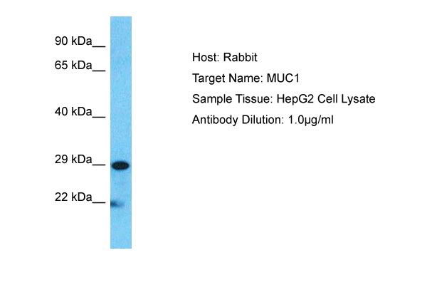


 WB (Western Blot)
(Host: RabbitTarget Name: MUC1Sample Type: HepG2Lane A: Primary AntibodyLane B: Primary Antibody + Blocking PeptidePrimary Antibody Concentration: 1ug/mlPeptide Concentration: 5ug/mlLysate Quantity: 25ug/lane/laneGel Concentration: 0.12)
WB (Western Blot)
(Host: RabbitTarget Name: MUC1Sample Type: HepG2Lane A: Primary AntibodyLane B: Primary Antibody + Blocking PeptidePrimary Antibody Concentration: 1ug/mlPeptide Concentration: 5ug/mlLysate Quantity: 25ug/lane/laneGel Concentration: 0.12)
MUC1, Polyclonal Antibody (Cat# AAA23492)
Full Name
MUC1 antibody - C-terminal region
Gene Names
MUC1; EMA; MCD; PEM; PUM; KL-6; MAM6; MCKD; PEMT; CD227; H23AG; MCKD1; MUC-1; ADMCKD; ADMCKD1; CA 15-3; MUC-1/X; MUC1/ZD; MUC-1/SEC
Reactivity
Cow, Dog, Guinea Pig, Horse, Human, Mouse, Rabbit, Rat, Pig
Applications
IHC, WB
Purity
Affinity Purified


 WB (Western Blot)
(Western blot analysis of Bid (arrow) using rabbit polyclonal Bid Antibody (BH3). 293 cell lysates (2 ug/lane) either nontransfected (Lane 1) or transiently transfected (Lane 2) with the Bid gene.)
WB (Western Blot)
(Western blot analysis of Bid (arrow) using rabbit polyclonal Bid Antibody (BH3). 293 cell lysates (2 ug/lane) either nontransfected (Lane 1) or transiently transfected (Lane 2) with the Bid gene.)
Bid, Polyclonal Antibody (Cat# AAA28769)
Full Name
Bid Antibody (BH3 Domain Specific)
Gene Names
BID; FP497
Reactivity
Human, mouse
Applications
WB, EIA, IHC
Purity
Purified Rabbit Polyclonal Antibody (Pab)
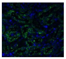


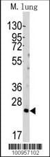
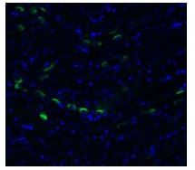
 IF (Immunofluorescence)
(Figure 7 Immunofluorescence Validation of TMEM41B in Rat LungImmunofluorescent analysis of 4% paraformaldehyde-fixed rat lung labeling TMEM41B at 20ug/mL, followed by goat anti-rabbit IgG secondary antibody at 1/500 dilution (green) and DAPI staining (blue).)
IF (Immunofluorescence)
(Figure 7 Immunofluorescence Validation of TMEM41B in Rat LungImmunofluorescent analysis of 4% paraformaldehyde-fixed rat lung labeling TMEM41B at 20ug/mL, followed by goat anti-rabbit IgG secondary antibody at 1/500 dilution (green) and DAPI staining (blue).)
TMEM41B, Polyclonal Antibody (Cat# AAA11036)
Full Name
TMEM41B (NT) Antibody
Reactivity
Human, Mouse, Rat
Predicted species reactivity based on immunogen sequence: Horse (100%); Chimpanzee (100%).
Predicted species reactivity based on immunogen sequence: Horse (100%); Chimpanzee (100%).
Applications
EIA, IF, WB
Purity
TMEM41B Antibody is affinity chromatography purified via peptide column.


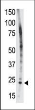
 IF (Immunofluorescence)
(Figure 6 Immunofluorescence Validation of TMEM41B in Rat LungImmunofluorescent analysis of 4% paraformaldehyde-fixed rat lung labeling TMEM41B at 20ug/mL, followed by goat anti-rabbit IgG secondary antibody at 1/500 dilution (green) and DAPI staining (blue).)
IF (Immunofluorescence)
(Figure 6 Immunofluorescence Validation of TMEM41B in Rat LungImmunofluorescent analysis of 4% paraformaldehyde-fixed rat lung labeling TMEM41B at 20ug/mL, followed by goat anti-rabbit IgG secondary antibody at 1/500 dilution (green) and DAPI staining (blue).)
TMEM41B, Polyclonal Antibody (Cat# AAA11035)
Full Name
TMEM41B (CT) Antibody
Reactivity
Human, Mouse, Rat
Predicted species reactivity based on immunogen sequence: Bovine (100%); Chicken (100%); Chimpanzee (100%).
Predicted species reactivity based on immunogen sequence: Bovine (100%); Chicken (100%); Chimpanzee (100%).
Applications
EIA, IF, WB
Purity
TMEM41B Antibody is affinity chromatography purified via peptide column.


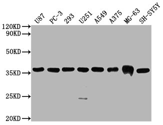
 WB (Western Blot)
(Western blot of Latexin (arrow) in 293 cell lysates (2 ug/lane) either nontransfected (Lane 1) or transiently transfected with the LXN gene (Lane 2))
WB (Western Blot)
(Western blot of Latexin (arrow) in 293 cell lysates (2 ug/lane) either nontransfected (Lane 1) or transiently transfected with the LXN gene (Lane 2))
Latexin / MUM, Polyclonal Antibody (Cat# AAA12323)
Full Name
Rabbit Polyclonal to Human Latexin / MUM
Gene Names
LXN; ECI; TCI
Reactivity
Human
Applications
IHC - Paraffin, WB, FC/FACS
Purity
Immunoaffinity Purified


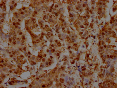
 WB (Western Blot)
(Formalin-fixed and paraffin-embedded human cancer tissue reacted with the primary antibody, which was peroxidase-conjugated to the secondary antibody, followed by AEC staining. This data demonstrates the use of this antibody for immunohistochemistry; clinical relevance has not been evaluated. BC = breast carcinoma; HC = hepatocarcinoma.)
WB (Western Blot)
(Formalin-fixed and paraffin-embedded human cancer tissue reacted with the primary antibody, which was peroxidase-conjugated to the secondary antibody, followed by AEC staining. This data demonstrates the use of this antibody for immunohistochemistry; clinical relevance has not been evaluated. BC = breast carcinoma; HC = hepatocarcinoma.)
SIRT3, Polyclonal Antibody (Cat# AAA28669)
Full Name
SIRT3 Antibody (C-term)
Gene Names
SIRT3; SIR2L3
Reactivity
Human, mouse
Applications
WB, EIA, IHC
Purity
Purified Rabbit Polyclonal Antibody (Pab)


 IHC (Immunohistchemistry)
(Formalin-fixed and paraffin-embedded human cancer tissue reacted with the primary antibody, which was peroxidase-conjugated to the secondary antibody, followed by DAB staining. This data demonstrates the use of this antibody for immunohistochemistry; clinical relevance has not been evaluated. BC = breast carcinoma; HC = hepatocarcinoma.)
IHC (Immunohistchemistry)
(Formalin-fixed and paraffin-embedded human cancer tissue reacted with the primary antibody, which was peroxidase-conjugated to the secondary antibody, followed by DAB staining. This data demonstrates the use of this antibody for immunohistochemistry; clinical relevance has not been evaluated. BC = breast carcinoma; HC = hepatocarcinoma.)
PACSIN2, Polyclonal Antibody (Cat# AAA28758)
Full Name
PACSIN2 Antibody (C-term)
Gene Names
PACSIN2; SDPII
Reactivity
Human, mouse
Applications
EIA, IHC, WB, IF
Purity
Purified Rabbit Polyclonal Antibody (Pab)


 WB (Western Blot)
(Formalin-fixed and paraffin-embedded human cancer tissue reacted with the primary antibody, which was peroxidase-conjugated to the secondary antibody, followed by DAB staining. This data demonstrates the use of this antibody for immunohistochemistry; clinical relevance has not been evaluated. BC = breast carcinoma; HC = hepatocarcinoma.)
WB (Western Blot)
(Formalin-fixed and paraffin-embedded human cancer tissue reacted with the primary antibody, which was peroxidase-conjugated to the secondary antibody, followed by DAB staining. This data demonstrates the use of this antibody for immunohistochemistry; clinical relevance has not been evaluated. BC = breast carcinoma; HC = hepatocarcinoma.)
SPAK, Polyclonal Antibody (Cat# AAA28754)
Full Name
SPAK Antibody (Center)
Gene Names
STK39; DCHT; PASK; SPAK
Reactivity
Human, mouse (Predicted Reactivity: Rat)
Applications
WB, EIA, IHC
Purity
Purified Rabbit Polyclonal Antibody (Pab)


 FCM (Flow Cytometry)
(SEPT9 Antibody (A555) flow cytometric analysis of HepG2 cells (right histogram) compared to a negative control cell (left histogram).FITC-conjugated goat-anti-rabbit secondary antibodies were used for the analysis.)
FCM (Flow Cytometry)
(SEPT9 Antibody (A555) flow cytometric analysis of HepG2 cells (right histogram) compared to a negative control cell (left histogram).FITC-conjugated goat-anti-rabbit secondary antibodies were used for the analysis.)
SEPT9, Polyclonal Antibody (Cat# AAA28793)
Full Name
SEPT9 Antibody (C-term)
Gene Names
SEPT9; MSF; MSF1; NAPB; SINT1; PNUTL4; SeptD1; AF17q25
Reactivity
Human
Applications
WB, EIA, IHC, FC/FACS
Purity
Purified Rabbit Polyclonal Antibody (Pab)

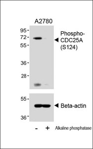


 IHC (Immunohistchemistry)
(Immunohistochemical analysis of (AAA28653) on paraffin-embedded Human breastcarcinoma tissue. Tissue was fixed withformaldehyde at room temperature. Heatinduced epitope retrieval was performed byEDTA buffer (pH9. 0). Samples wereincubated with primary antibody(1:100) for 1hour at room temperature. Undiluted CRFAnti-Polyvalent HRP Polymer antibody wasused as the secondary antibody.)
IHC (Immunohistchemistry)
(Immunohistochemical analysis of (AAA28653) on paraffin-embedded Human breastcarcinoma tissue. Tissue was fixed withformaldehyde at room temperature. Heatinduced epitope retrieval was performed byEDTA buffer (pH9. 0). Samples wereincubated with primary antibody(1:100) for 1hour at room temperature. Undiluted CRFAnti-Polyvalent HRP Polymer antibody wasused as the secondary antibody.)
Phospho-CDC25A (S124), Polyclonal Antibody (Cat# AAA28653)
Full Name
Phospho-CDC25A (S124) Antibody
Gene Names
CDC25A; CDC25A2
Reactivity
Human (Predicted Reactivity: Rat)
Applications
WB, EIA, IHC
Purity
Peptide Affinity Purified Rabbit Polyclonal Antibody (Pab)


 IF (Immunofluorescence)
(Figure 7 Immunofluorescence Validation of TMPRSS2 in Rat TestisImmunofluorescent analysis of 4% paraformaldehyde-fixed rat Testis labeling TMPRSS2 with 9569 at 20ug/mL, followed by goat anti-rabbit IgG secondary antibody at 1/500 dilution (green) and DAPI staining (blue).)
IF (Immunofluorescence)
(Figure 7 Immunofluorescence Validation of TMPRSS2 in Rat TestisImmunofluorescent analysis of 4% paraformaldehyde-fixed rat Testis labeling TMPRSS2 with 9569 at 20ug/mL, followed by goat anti-rabbit IgG secondary antibody at 1/500 dilution (green) and DAPI staining (blue).)
TMPRSS2, Polyclonal Antibody (Cat# AAA11037)
Full Name
TMPRSS2 (CT) Antibody
Gene Names
TMPRSS2; PP9284; PRSS10
Reactivity
Human, Mouse, Rat
Predicted species reactivity based on immunogen sequence: Monkey (100%); Gorilla (100%); Cat (92%).
Predicted species reactivity based on immunogen sequence: Monkey (100%); Gorilla (100%); Cat (92%).
Applications
EIA, IF, WB
Purity
TMPRSS2 Antibody is affinity chromatography purified via peptide column.


 IHC (Immunohistchemistry)
(Formalin-fixed and paraffin-embedded human testis tissue reacted with PARK2 (Parkin) antibody (C-term) , which was peroxidase-conjugated to the secondary antibody, followed by DAB staining. This data demonstrates the use of this antibody for immunohistochemistry; clinical relevance has not been evaluated.)
IHC (Immunohistchemistry)
(Formalin-fixed and paraffin-embedded human testis tissue reacted with PARK2 (Parkin) antibody (C-term) , which was peroxidase-conjugated to the secondary antibody, followed by DAB staining. This data demonstrates the use of this antibody for immunohistochemistry; clinical relevance has not been evaluated.)
Parkin, Polyclonal Antibody (Cat# AAA28668)
Full Name
Parkin Antibody (C-term)
Gene Names
PARK2; PDJ; PRKN; AR-JP; LPRS2
Reactivity
Human, mouse
Applications
WB, EIA, IF, FC/FACS, IHC
Purity
Purified Rabbit Polyclonal Antibody (Pab)


 IF (Immunofluorescence)
(Figure 7 Immunofluorescence Validation of TMPRSS2 in Rat BrainImmunofluorescent analysis of 4% paraformaldehyde-fixed rat brain labeling TMPRSS2 at 20ug/mL, followed by goat anti-rabbit IgG secondary antibody at 1/500 dilution (green) and DAPI staining (blue).)
IF (Immunofluorescence)
(Figure 7 Immunofluorescence Validation of TMPRSS2 in Rat BrainImmunofluorescent analysis of 4% paraformaldehyde-fixed rat brain labeling TMPRSS2 at 20ug/mL, followed by goat anti-rabbit IgG secondary antibody at 1/500 dilution (green) and DAPI staining (blue).)
TMPRSS2, Polyclonal Antibody (Cat# AAA11038)
Full Name
TMPRSS2 (IN) Antibody
Gene Names
TMPRSS2; PP9284; PRSS10
Reactivity
Human, Mouse, Rat
Predicted species reactivity based on immunogen sequence: Horse (100%); Rabbit (100%); Monkey (100%); Sheep (100%); Gorilla (100%); Cat (100%).
Predicted species reactivity based on immunogen sequence: Horse (100%); Rabbit (100%); Monkey (100%); Sheep (100%); Gorilla (100%); Cat (100%).
Applications
EIA, IF, WB
Purity
TMPRSS2 Antibody is affinity chromatography purified via peptide column.


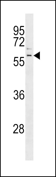
 WB (Western Blot)
(Western blot analysis of LIN28B Antibody (N-term) in HL60 cell line lysates (35 ug/lane). LIN28B (arrow) was detected using the purified Pab.)
WB (Western Blot)
(Western blot analysis of LIN28B Antibody (N-term) in HL60 cell line lysates (35 ug/lane). LIN28B (arrow) was detected using the purified Pab.)
LIN28B, Polyclonal Antibody (Cat# AAA12320)
Full Name
Rabbit Polyclonal to Human LIN28B
Gene Names
LIN28B; CSDD2
Reactivity
Human
Applications
IHC - Paraffin, IF, WB, FC/FACS
Purity
Ammonium sulfate precipitation


 DB (Dot Blot)
(Formalin-fixed and paraffin-embedded human cancer tissue reacted with the primary antibody, which was peroxidase-conjugated to the secondary antibody, followed by AEC staining. This data demonstrates the use of this antibody for immunohistochemistry; clinical relevance has not been evaluated. BC = breast carcinoma; HC = hepatocarcinoma.)
DB (Dot Blot)
(Formalin-fixed and paraffin-embedded human cancer tissue reacted with the primary antibody, which was peroxidase-conjugated to the secondary antibody, followed by AEC staining. This data demonstrates the use of this antibody for immunohistochemistry; clinical relevance has not been evaluated. BC = breast carcinoma; HC = hepatocarcinoma.)
Phospho-HIST1H3B3 (S10), Polyclonal Antibody (Cat# AAA28670)
Full Name
Phospho-HIST1H3B3 (S10) Antibody
Gene Names
HIST1H3A; H3/A; H3FA
Reactivity
Human
Applications
DB, EIA, IHC, WB
Purity
Peptide Affinity Purified Rabbit Polyclonal Antibody (Pab)


 IF (Immunofluorescence)
(Western blot analysis of lysate from RPMI 8226 cell line, using CD38 Antibody (C-term). AAA28702 was diluted at 1:1000. A goat anti-rabbit IgG H&L(HRP) at 1:5000 dilution was used as the secondary antibody. Lysate at 35ug.)
IF (Immunofluorescence)
(Western blot analysis of lysate from RPMI 8226 cell line, using CD38 Antibody (C-term). AAA28702 was diluted at 1:1000. A goat anti-rabbit IgG H&L(HRP) at 1:5000 dilution was used as the secondary antibody. Lysate at 35ug.)
CD38, Polyclonal Antibody (Cat# AAA28702)
Full Name
CD38 Antibody (C-term)
Gene Names
CD38; T10; ADPRC 1
Reactivity
Human (Predicted Reactivity: Monkey)
Applications
WB, EIA, IHC, IF, FC/FACS
Purity
Purified Rabbit Polyclonal Antibody (Pab)

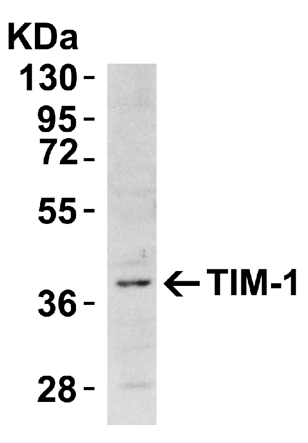
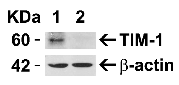


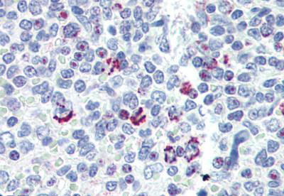
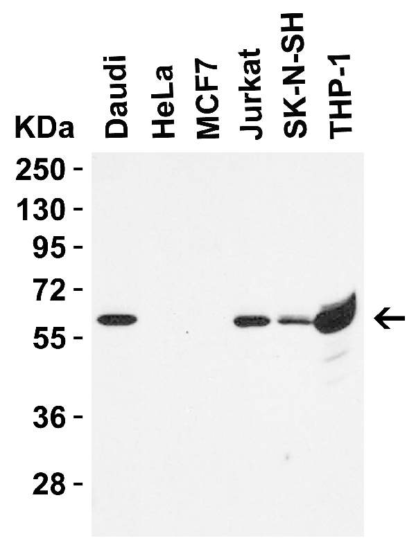
 IHC (Immunohistchemistry)
(Figure 5 Immunohistochemistry Validation of TIM-1Immunohistochemical analysis of paraffin-embedded human uterus tissue using anti-TIM-1 antibody (3809) at 10 μg/ml. Tissue was fixed with formaldehyde and blocked with 10% serum for 1 h at RT; antigen retrieval was by heat mediation with a citrate buffer (pH6). Samples were incubated with primary antibody overnight at 4˚ C. A goat anti-rabbit IgG H&L (HRP) at 1/250 was used as secondary. Counter stained with Hematoxylin.)
IHC (Immunohistchemistry)
(Figure 5 Immunohistochemistry Validation of TIM-1Immunohistochemical analysis of paraffin-embedded human uterus tissue using anti-TIM-1 antibody (3809) at 10 μg/ml. Tissue was fixed with formaldehyde and blocked with 10% serum for 1 h at RT; antigen retrieval was by heat mediation with a citrate buffer (pH6). Samples were incubated with primary antibody overnight at 4˚ C. A goat anti-rabbit IgG H&L (HRP) at 1/250 was used as secondary. Counter stained with Hematoxylin.)
TIM-1, Polyclonal Antibody (Cat# AAA10935)
Full Name
TIM-1 Antibody
Gene Names
HAVCR1; TIM; KIM1; TIM1; HAVCR; KIM-1; TIM-1; TIMD1; TIMD-1; HAVCR-1
Reactivity
Human, Mouse
Applications
EIA, WB, IHC, IF
Purity
TIM-1 Antibody is affinity chromatography purified via peptide column.


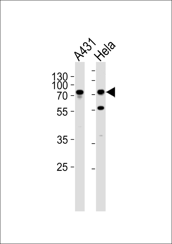
 IF (Immunofluorescence)
(Confocal immunofluorescent analysis of GAPDH Antibody (N-term) with Hela cell followed by Alexa Fluor 488-conjugated goat anti-rabbit lgG (green). Actin filaments have been labeled with Alexa Fluor 555 phalloidin (red).DAPI was used to stain the cell nuclear (blue).)
IF (Immunofluorescence)
(Confocal immunofluorescent analysis of GAPDH Antibody (N-term) with Hela cell followed by Alexa Fluor 488-conjugated goat anti-rabbit lgG (green). Actin filaments have been labeled with Alexa Fluor 555 phalloidin (red).DAPI was used to stain the cell nuclear (blue).)
GAPDH, Polyclonal Antibody (Cat# AAA28776)
Full Name
GAPDH Antibody (N-term)
Gene Names
GAPDH; G3PD; GAPD; HEL-S-162eP
Reactivity
Human
Applications
IF, EIA, WB, IHC
Purity
Purified Rabbit Polyclonal Antibody (Pab)


 FCM (Flow Cytometry)
(CDK4 Antibody (C-term) flow cytometric analysis of HL60 cells (bottom histogram) compared to a negative control cell (top histogram).FITC-conjugated goat-anti-rabbit secondary antibodies were used for the analysis.)
FCM (Flow Cytometry)
(CDK4 Antibody (C-term) flow cytometric analysis of HL60 cells (bottom histogram) compared to a negative control cell (top histogram).FITC-conjugated goat-anti-rabbit secondary antibodies were used for the analysis.)
CDK4, Polyclonal Antibody (Cat# AAA28747)
Full Name
CDK4 Antibody (C-term)
Gene Names
CDK4; CMM3; PSK-J3
Reactivity
Human
Applications
FC/FACS, EIA, IHC, WB, IF
Purity
Purified Rabbit Polyclonal Antibody (Pab)


 IF (Immunofluorescence)
(Fluorescent confocal image of Hela cell stained with HDAC2 Antibody (C-term) .Hela cells were fixed with 4% PFA (20 min), permeabilized with Triton X-100 (0.1%, 10 min), then incubated with HDAC2 primary antibody (1:25, 1 h at 37 degree C). For secondary antibody, Alexa Fluor 488 conjugated donkey anti-rabbit antibody (green) was used (1:400, 50 min at 37 degree C).Cytoplasmic actin was counterstained with Alexa Fluor 555 (red) conjugated Phalloidin (7units/ml, 1 h at 37 degree C). Nuclei were counterstained with DAPI (blue) (10 ug/ml, 10 min). hHDAC2 immunoreactivity is localized to Nucleus significantly.)
IF (Immunofluorescence)
(Fluorescent confocal image of Hela cell stained with HDAC2 Antibody (C-term) .Hela cells were fixed with 4% PFA (20 min), permeabilized with Triton X-100 (0.1%, 10 min), then incubated with HDAC2 primary antibody (1:25, 1 h at 37 degree C). For secondary antibody, Alexa Fluor 488 conjugated donkey anti-rabbit antibody (green) was used (1:400, 50 min at 37 degree C).Cytoplasmic actin was counterstained with Alexa Fluor 555 (red) conjugated Phalloidin (7units/ml, 1 h at 37 degree C). Nuclei were counterstained with DAPI (blue) (10 ug/ml, 10 min). hHDAC2 immunoreactivity is localized to Nucleus significantly.)
HDAC2, Polyclonal Antibody (Cat# AAA28801)
Full Name
HDAC2 Antibody (C-term)
Gene Names
HDAC2; HD2; RPD3; YAF1
Reactivity
Human
Applications
WB, IHC, IF, EIA


 IHC (Immunohistchemistry)
(Figure 9 Immunohistochemistry Validation of hRIP3 in Human Colon Tissue Immunohistochemical analysis of paraffin-embedded human colon tissue using anti-hRIP3 antibody (8963) at 2 μg/ml. Tissue was fixed with formaldehyde and blocked with 10% serum for 1 h at RT; antigen retrieval was by heat mediation with a citrate buffer (pH6). Samples were incubated with primary antibody overnight at 4˚C. A goat anti-rabbit IgG H&L (HRP) at 1/250 was used as secondary. Counter stained with Hematoxylin.)
IHC (Immunohistchemistry)
(Figure 9 Immunohistochemistry Validation of hRIP3 in Human Colon Tissue Immunohistochemical analysis of paraffin-embedded human colon tissue using anti-hRIP3 antibody (8963) at 2 μg/ml. Tissue was fixed with formaldehyde and blocked with 10% serum for 1 h at RT; antigen retrieval was by heat mediation with a citrate buffer (pH6). Samples were incubated with primary antibody overnight at 4˚C. A goat anti-rabbit IgG H&L (HRP) at 1/250 was used as secondary. Counter stained with Hematoxylin.)
hRIP3, Polyclonal Antibody (Cat# AAA11031)
Full Name
hRIP3 Antibody
Gene Names
RIPK3; RIP3
Reactivity
Human
Applications
EIA, IF, IHC, WB
Purity
hRIP3 Antibody is Protein A purified.


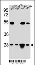 WB (Western Blot)
(TFAM Antibody (C-term) western blot analysis in Hela,Jurkat,K562,MCF-7 cell line lysates (35ug/lane).This demonstrates the TFAM antibody detected the TFAM protein (arrow).)
WB (Western Blot)
(TFAM Antibody (C-term) western blot analysis in Hela,Jurkat,K562,MCF-7 cell line lysates (35ug/lane).This demonstrates the TFAM antibody detected the TFAM protein (arrow).)
TFAM, Polyclonal Antibody (Cat# AAA28696)
Full Name
TFAM Antibody (C-term)
Gene Names
TFAM; TCF6; MTTF1; MTTFA; TCF6L1; TCF6L2; TCF6L3
Reactivity
Human
Applications
EIA, IHC, FC/FACS, IF, WB
Purity
Peptide Affinity Purified Rabbit Polyclonal Antibody (Pab)


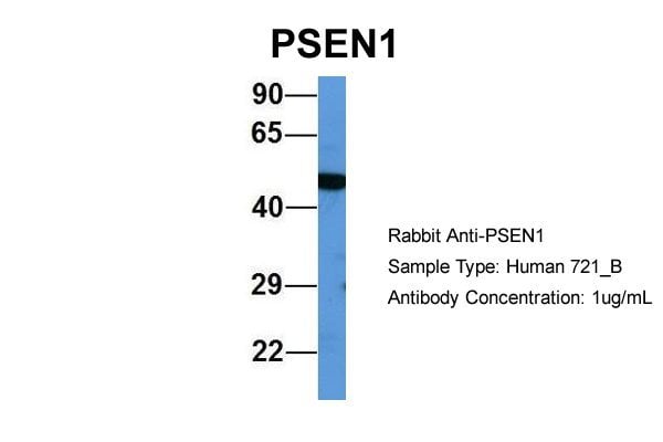


 WB (Western Blot)
(WB Suggested Anti-PSEN1 Antibody Titration: 0.2-1 ug/mlELISA Titer: 1:312500Positive Control: Human brain)
WB (Western Blot)
(WB Suggested Anti-PSEN1 Antibody Titration: 0.2-1 ug/mlELISA Titer: 1:312500Positive Control: Human brain)
PSEN1, Polyclonal Antibody (Cat# AAA23585)
Full Name
PSEN1 antibody - middle region
Gene Names
PSEN1; AD3; FAD; PS1; PS-1; S182; ACNINV3
Reactivity
Cow, Dog, Guinea Pig, Horse, Human, Mouse, Rabbit, Rat
Applications
IHC, IP, WB
Purity
Affinity Purified


 WB (Western Blot)
(Figure 9 KD Validation of PUMA (Han et al., 2010)Immunoblot analyses of Tet-induced p53 cells treated with NOXA, Puma, Bim or non-targeting siRNAs that were utilized in this experiment. PUMA protein levels were markedly reduced in PUMA KD cells detected by anti-PUMA antibodies (3041).)
WB (Western Blot)
(Figure 9 KD Validation of PUMA (Han et al., 2010)Immunoblot analyses of Tet-induced p53 cells treated with NOXA, Puma, Bim or non-targeting siRNAs that were utilized in this experiment. PUMA protein levels were markedly reduced in PUMA KD cells detected by anti-PUMA antibodies (3041).)
PUMA, Polyclonal Antibody (Cat# AAA10943)
Full Name
PUMA Antibody
Gene Names
BBC3; JFY1; PUMA; JFY-1
Reactivity
Human, Mouse
Applications
EIA, WB, ICC, IF
Purity
PUMA Antibody is affinity chromatography purified via peptide column.


 FCM (Flow Cytometry)
(GTF2I Antibody (C-term) flow cytometric analysis of k562 cells (bottom histogram) compared to a negative control cell (top histogram).FITC-conjugated goat-anti-rabbit secondary antibodies were used for the analysis.)
FCM (Flow Cytometry)
(GTF2I Antibody (C-term) flow cytometric analysis of k562 cells (bottom histogram) compared to a negative control cell (top histogram).FITC-conjugated goat-anti-rabbit secondary antibodies were used for the analysis.)
GTF2I, Polyclonal Antibody (Cat# AAA28707)
Full Name
GTF2I Antibody (C-term)
Gene Names
GTF2I; WBS; DIWS; SPIN; IB291; BAP135; BTKAP1; TFII-I; WBSCR6; GTFII-I
Reactivity
Human (Predicted Reactivity: Rat)
Applications
IF, EIA, IHC, FC/FACS, WB
Purity
Peptide Affinity Purified Rabbit Polyclonal Antibody (Pab)


 IP (Immunoprecipitation)
(Immunoprecipitating GAPDH in Hela whole cell lysateLane 1: Mouse control IgG instead of in Hela whole cell lysate. Lane 2: (5ul) + Hela whole cell lysate (500ug)Lane 3: Hela whole cell lysate (10ug)For western blotting, the blot was detected at 1:5000, and a HRP-conjugated Protein G antibody was used as the secondary antibody at 1:2000)
IP (Immunoprecipitation)
(Immunoprecipitating GAPDH in Hela whole cell lysateLane 1: Mouse control IgG instead of in Hela whole cell lysate. Lane 2: (5ul) + Hela whole cell lysate (500ug)Lane 3: Hela whole cell lysate (10ug)For western blotting, the blot was detected at 1:5000, and a HRP-conjugated Protein G antibody was used as the secondary antibody at 1:2000)
GAPDH, Monoclonal Antibody (Cat# AAA27042)
Full Name
GAPDH Monoclonal Antibody
Reactivity
Human, Mouse, Rabbit
Applications
EIA, WB, IHC, IP, IF
Purity
>95%, Protein G purified


 IP (Immunoprecipitation)
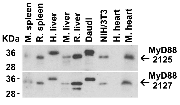
(Figure 11 Immunoprecipitation Validation in HEK293 cells (Kawai et al., 2004)HEK293 cells were transiently transfected with DYKDDDDK-IRF7. Ccell lysates were immunoprecipitated with control rabbit anti-mouse immunoglobulin serum (IgG) or anti-MyD88 (Ab1 and Ab2), followed by immunoblotting with anti-DYKDDDDK.)
IP (Immunoprecipitation)
(Figure 11 Immunoprecipitation Validation in HEK293 cells (Kawai et al., 2004)HEK293 cells were transiently transfected with DYKDDDDK-IRF7. Ccell lysates were immunoprecipitated with control rabbit anti-mouse immunoglobulin serum (IgG) or anti-MyD88 (Ab1 and Ab2), followed by immunoblotting with anti-DYKDDDDK.)
MYD88, Polyclonal Antibody (Cat# AAA10944)
Full Name
MYD88 Antibody
Gene Names
MYD88; MYD88D
Reactivity
Human, Mouse, Rat
Applications
EIA, WB
Purity
MYD88 Antibody is affinity chromatography purified via peptide column.


 IF (Immunofluorescence)
(Fluorescent confocal image of Hela cell stained with XRCC6 Antibody (C-term). Hela cells were fixed with 4% PFA (20 min), permeabilized with Triton X-100 (0.1%, 10 min), then incubated with XRCC6 primary antibody (1:25, 1 h at 37 degree). For secondary antibody, Alexa Fluor 488 conjugated donkey anti-rabbit antibody (green) was used (1:400, 50 min at 37 degree).Cytoplasmic actin was counterstained with Alexa Fluor 555 (red) conjugated Phalloidin (7units/ml, 1 h at 37 degree). Nuclei were counterstained with DAPI (blue) (10 ug/ml, 10 min). XRCC6 immunoreactivity is localized to nucleus significantly and Cytoplasm weakly.)
IF (Immunofluorescence)
(Fluorescent confocal image of Hela cell stained with XRCC6 Antibody (C-term). Hela cells were fixed with 4% PFA (20 min), permeabilized with Triton X-100 (0.1%, 10 min), then incubated with XRCC6 primary antibody (1:25, 1 h at 37 degree). For secondary antibody, Alexa Fluor 488 conjugated donkey anti-rabbit antibody (green) was used (1:400, 50 min at 37 degree).Cytoplasmic actin was counterstained with Alexa Fluor 555 (red) conjugated Phalloidin (7units/ml, 1 h at 37 degree). Nuclei were counterstained with DAPI (blue) (10 ug/ml, 10 min). XRCC6 immunoreactivity is localized to nucleus significantly and Cytoplasm weakly.)
XRCC6, Polyclonal Antibody (Cat# AAA28768)
Full Name
XRCC6 Antibody (C-term)
Gene Names
XRCC6; ML8; KU70; TLAA; CTC75; CTCBF; G22P1
Reactivity
Human
Applications
WB, EIA, IHC, FC/FACS, IF
Purity
Peptide Affinity Purified Rabbit Polyclonal Antibody (Pab)


 FCM (Flow Cytometry)
(Overlay Peak curve showing Hela cells stained with AAA28066 (red line) at 1:200. The cells were fixed in 4% formaldehyde and permeated by 0.2% TritonX-100. Then 10% normal goat serum was Incubated to block non-specific protein-protein interactions followed by the antibody (1?g/1*106cells) for 1 h at 4 degree C. The secondary antibody used was FITC-conjugated Goat Anti-Mouse IgG(H+L) at 1/100 dilution for 30min at 4 degree C. Isotype control antibody (green line) was mouse IgG2b (1?g/1*106cells) used under the same conditions. Acquisition of >10,000 events was performed.)
FCM (Flow Cytometry)
(Overlay Peak curve showing Hela cells stained with AAA28066 (red line) at 1:200. The cells were fixed in 4% formaldehyde and permeated by 0.2% TritonX-100. Then 10% normal goat serum was Incubated to block non-specific protein-protein interactions followed by the antibody (1?g/1*106cells) for 1 h at 4 degree C. The secondary antibody used was FITC-conjugated Goat Anti-Mouse IgG(H+L) at 1/100 dilution for 30min at 4 degree C. Isotype control antibody (green line) was mouse IgG2b (1?g/1*106cells) used under the same conditions. Acquisition of >10,000 events was performed.)
ACTB, Monoclonal Antibody (Cat# AAA28066)
Full Name
ACTB Monoclonal Antibody
Reactivity
Human, Mouse, Rat, Rabbit
Applications
EIA, WB, IHC, IF, FC/FACS, IP
Purity
>95%, Protein A purified


 IP (Immunoprecipitation)
(Immunoprecipitating GAPDH in Hela whole cell lysate Lane 1: Mouse control IgG instead of AAA27043 in Hela whole cell lysate. Lane 2: AAA27043 (5ul) + Hela whole cell lysate (500ug) Lane 3: Hela whole cell lysate (10ug) For western blotting, the blot was detected with AAA27043 at 1:5000, and a HRPconjugated Protein G antibody was used as the secondary antibody at 1:2000)
IP (Immunoprecipitation)
(Immunoprecipitating GAPDH in Hela whole cell lysate Lane 1: Mouse control IgG instead of AAA27043 in Hela whole cell lysate. Lane 2: AAA27043 (5ul) + Hela whole cell lysate (500ug) Lane 3: Hela whole cell lysate (10ug) For western blotting, the blot was detected with AAA27043 at 1:5000, and a HRPconjugated Protein G antibody was used as the secondary antibody at 1:2000)
GAPDH, Monoclonal Antibody (Cat# AAA27043)
Full Name
GAPDH Monoclonal Antibody
Reactivity
Human, Rat, Rabbit, Mouse
Applications
EIA, WB, IHC, IP, IF
Purity
>95%, Protein G purified


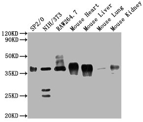
 WB (Western Blot)
(PLD6 Antibody (Center) (Cat# AAA28684) western blot analysis in mouse testis tissue lysates (35ug/lane).This demonstrates the PLD6 antibody detected the PLD6 protein (arrow).)
WB (Western Blot)
(PLD6 Antibody (Center) (Cat# AAA28684) western blot analysis in mouse testis tissue lysates (35ug/lane).This demonstrates the PLD6 antibody detected the PLD6 protein (arrow).)
PLD6, Polyclonal Antibody (Cat# AAA28684)
Full Name
PLD6 Antibody (Center)
Gene Names
PLD6; ZUC
Reactivity
Human, Mouse
Applications
WB, EIA
Purity
This antibody is purified through a protein A column, followed by peptide affinity purification.


 IHC (Immunohistchemistry)
(Validation of PKR in Rat Lung Immunohistochemical analysis of paraffin-embedded rat lung tissue using anti-PKR antibody (AAA10917) at 2.5 ug/ml. Tissue was fixed with formaldehyde and blocked with 10% serum for 1h at RT; antigen retrieval was by heat mediation with a citrate buffer (pH6). Samples were incubated with primary antibody overnight at 4 degree C. A goat anti-rabbit IgG H&L (HRP) at 1/250 was used as secondary. Counter stained with Hematoxylin.)
IHC (Immunohistchemistry)
(Validation of PKR in Rat Lung Immunohistochemical analysis of paraffin-embedded rat lung tissue using anti-PKR antibody (AAA10917) at 2.5 ug/ml. Tissue was fixed with formaldehyde and blocked with 10% serum for 1h at RT; antigen retrieval was by heat mediation with a citrate buffer (pH6). Samples were incubated with primary antibody overnight at 4 degree C. A goat anti-rabbit IgG H&L (HRP) at 1/250 was used as secondary. Counter stained with Hematoxylin.)
PKR, Polyclonal Antibody (Cat# AAA10917)
Full Name
PKR Antibody
Gene Names
EIF2AK2; PKR; PRKR; EIF2AK1; PPP1R83
Reactivity
Human, Mouse, Rat
Applications
EIA, WB, IHC, IF
Purity
PKR Antibody is affinity chromatography purified via peptide column.


 IP (Immunoprecipitation)
(Figure 13 Immunoprecipitation Validation in HEK293 cells (Kawai et al., 2004)HEK293 cells were transiently transfected with DYKDDDDK-IRF7. Ccell lysates were immunoprecipitated with control rabbit anti-mouse immunoglobulin serum (IgG) or anti-MyD88 (Ab1 and Ab2), followed by immunoblotting with anti-DYKDDDDK.)
IP (Immunoprecipitation)
(Figure 13 Immunoprecipitation Validation in HEK293 cells (Kawai et al., 2004)HEK293 cells were transiently transfected with DYKDDDDK-IRF7. Ccell lysates were immunoprecipitated with control rabbit anti-mouse immunoglobulin serum (IgG) or anti-MyD88 (Ab1 and Ab2), followed by immunoblotting with anti-DYKDDDDK.)
MYD88, Polyclonal Antibody (Cat# AAA10930)
Full Name
MYD88 Antibody
Gene Names
MYD88; MYD88D
Reactivity
Human, Mouse
Applications
EIA, WB, IHC, IF
Purity
MYD88 Antibody is affinity chromatography purified via peptide column.


 IHC (Immunohistochemistry)
(Immunohistochemical analysis of paraffin-embedded Human brain section using Pink 1 (AAA28759). AAA28759 was diluted at 1:1000 dilution. A undiluted binotinylated goat polyvalent antibody was used as the secondary, followed by DAB staining)
IHC (Immunohistochemistry)
(Immunohistochemical analysis of paraffin-embedded Human brain section using Pink 1 (AAA28759). AAA28759 was diluted at 1:1000 dilution. A undiluted binotinylated goat polyvalent antibody was used as the secondary, followed by DAB staining)
S100B, Polyclonal Antibody (Cat# AAA28759)
Full Name
S100B Antibody
Gene Names
S100B; NEF; S100; S100-B; S100beta
Reactivity
Human, Mouse, Rat (Predicted: Rabbit)
Applications
WB, IHC-P-Leica, FC/FACS, IHC, EIA
Purity
Peptide Affinity Purified Rabbit Polyclonal Antibody (Pab)


 IF (Immunofluorescence)
(Fluorescent confocal image of MCF-7 cell stained with BHLH3 Antibody (N-term). MCF-7 cells were fixed with 4% PFA (20 min), permeabilized with Triton X-100 (0.1%, 10 min), then incubated with BHLH3 primary antibody (1:25, 1 h at 37 degree). For secondary antibody, Alexa Fluor 488 conjugated donkey anti-rabbit antibody (green) was used (1:400, 50 min at 37 degree).Cytoplasmic actin was counterstained with Alexa Fluor 555 (red) conjugated Phalloidin (7units/ml, 1 h at 37 degree). Nuclei were counterstained with DAPI (blue) (10 ug/ml, 10 min).BHLH3 immunoreactivity is localized to nucleus significantly.)
IF (Immunofluorescence)
(Fluorescent confocal image of MCF-7 cell stained with BHLH3 Antibody (N-term). MCF-7 cells were fixed with 4% PFA (20 min), permeabilized with Triton X-100 (0.1%, 10 min), then incubated with BHLH3 primary antibody (1:25, 1 h at 37 degree). For secondary antibody, Alexa Fluor 488 conjugated donkey anti-rabbit antibody (green) was used (1:400, 50 min at 37 degree).Cytoplasmic actin was counterstained with Alexa Fluor 555 (red) conjugated Phalloidin (7units/ml, 1 h at 37 degree). Nuclei were counterstained with DAPI (blue) (10 ug/ml, 10 min).BHLH3 immunoreactivity is localized to nucleus significantly.)
BHLH3, Polyclonal Antibody (Cat# AAA28734)
Full Name
BHLH3 Antibody (N-term)
Gene Names
BHLHE41; DEC2; hDEC2; BHLHB3; SHARP1
Reactivity
Human, Mouse
Applications
FC, IF, WB, EIA
Purity
This antibody is purified through a protein A column, followed by peptide affinity purification.


 Application Data
(Figure 10 KD Validation in HeLa cells (Horinaka et al., 2005)HeLa cells were transfected with DR4siRNA or LacZ control siRNA. At 24 h after transfection, the cells were treated with or without 20 ?M luteolin for 24 h. Western blot analysis was carried out with anti-DR4 antibodies (1139). DR4 expression was markedly reduced after DR4 knockdown.)
Application Data
(Figure 10 KD Validation in HeLa cells (Horinaka et al., 2005)HeLa cells were transfected with DR4siRNA or LacZ control siRNA. At 24 h after transfection, the cells were treated with or without 20 ?M luteolin for 24 h. Western blot analysis was carried out with anti-DR4 antibodies (1139). DR4 expression was markedly reduced after DR4 knockdown.)
DR4, Polyclonal Antibody (Cat# AAA10952)
Full Name
DR4 Antibody
Gene Names
TNFRSF10A; DR4; APO2; CD261; TRAILR1; TRAILR-1
Reactivity
Human
Applications
EIA, WB, ICC
Purity
DR4 Antibody is Antibody is affinity chromatography purified via peptide column.