Filters
Clonality
Type
Reactivity
Gene Name
Isotype
Host
Application
Clone
44 results for " Media Cell Culture" - showing 1-44
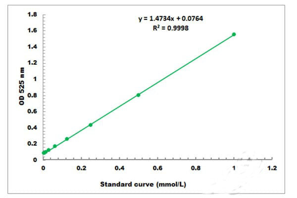
 Standard Curve (Sample)
Alcohol Dehydrogenase, Assay Kit (Cat# AAA27843)
Full Name
Alcohol Dehydrogenase Microplate Assay Kit
Reactivity
General
Applications
Functional Assay
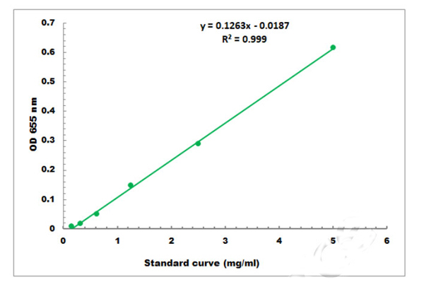
 Standard Curve (Sample)
Polyphenol Oxidase, Assay Kit (Cat# AAA17847)
Full Name
Polyphenol Oxidase Assay Kit
Reactivity
General
Applications
Functional Assay
 Standard Curve (Sample)
Nitric Oxide, Assay Kit (Cat# AAA27833)
Full Name
Nitric Oxide Microplate Assay Kit
Reactivity
General
Applications
Functional Assay
 Standard Curve (Sample)
Acetyl-CoA Carboxylase, Assay Kit (Cat# AAA27886)
Full Name
Acetyl-CoA Carboxylase Microplate Assay Kit
 Standard Curve (Sample)
Total Antioxidant Capacity, Assay Kit (Cat# AAA27830)
Full Name
Total Antioxidant Capacity Microplate Assay Kit
Reactivity
General
Applications
Functional Assay
 Standard Curve (Sample)
NAD/NADH, Assay Kit (Cat# AAA17848)
Full Name
NAD/NADH Assay Kit
Reactivity
General
Applications
Functional Assay
 Standard Curve (Sample)
Selenium, Assay Kit (Cat# AAA27898)
Full Name
Selenium Microplate Assay Kit
Reactivity
General
Applications
FA
 Standard Curve (Sample)
Hydroxyproline, Assay Kit (Cat# AAA27829)
Full Name
Hydroxyproline Microplate Assay Kit
Reactivity
General
Applications
Functional Assay
 Standard Curve (Sample)
Glutathione Reductase, Assay Kit (Cat# AAA27822)
Full Name
Glutathione Reductase Microplate Assay Kit
Gene Names
GR; ATGR2; EMB2360; glutathione reductase; GR
Reactivity
General
Applications
Functional Assay
 Standard Curve (Sample)
Pectinase, Assay Kit (Cat# AAA27819)
Full Name
Pectinase Microplate Assay Kit
Reactivity
General
Applications
Functional Assay
 Standard Curve (Sample)
Hydrogen Peroxide, Assay Kit (Cat# AAA17853)
Full Name
Hydrogen Peroxide Assay Kit
Reactivity
General
Applications
Functional Assay
 Standard Curve (Sample)
Chitinase, Assay Kit (Cat# AAA27826)
Full Name
Chitinase Microplate Assay Kit
Reactivity
General
Applications
Functional Assay
 Standard Curve (Sample)
Lactate Dehydrogenase, Assay Kit (Cat# AAA17850)
Full Name
Lactate Dehydrogenase Assay Kit
Reactivity
General
Applications
Functional Assay
 Standard Curve (Sample)
Trehalose, Assay Kit (Cat# AAA27853)
Full Name
Trehalose Microplate Assay Kit
Reactivity
General
Applications
Functional Assay
 Standard Curve (Sample)
Superoxide Anion, Assay Kit (Cat# AAA27890)
Full Name
Superoxide Anion Microplate Assay Kit
Reactivity
General
Applications
Functional Assay
 Standard Curve (Sample)
Succinate Dehydrogenase, Assay Kit (Cat# AAA27836)
Full Name
Succinate Dehydrogenase Microplate Assay Kit
Reactivity
General
Applications
Functional Assay
 Standard Curve (Sample)
Proline, Assay Kit (Cat# AAA27887)
Full Name
Proline Microplate Assay Kit
Reactivity
General
Applications
Functional Assay
 Standard Curve (Sample)
Acetolactate Synthase, Assay Kit (Cat# AAA27891)
Full Name
Acetolactate Synthase Microplate Assay Kit
Reactivity
General
Applications
Functional Assay
 Standard Curve (Sample)
Lipid Peroxidation (MDA), Assay Kit (Cat# AAA17852)
Full Name
Malondialdehyde Microplate Assay Kit
Reactivity
General
Applications
Functional Assay
 Standard Curve (Sample)
Pepsin, Assay Kit (Cat# AAA27849)
Full Name
Pepsin Microplate Assay Kit
Reactivity
General
Applications
Functional Assay
 Standard Curve (Sample)
ATP Synthase, Assay Kit (Cat# AAA27888)
Full Name
ATP Synthase Microplate Assay Kit
Reactivity
General
Applications
Functional Assay
 Standard Curve (Sample)
Triglyceride, Assay Kit (Cat# AAA27848)
Full Name
Triglyceride Microplate Assay Kit
Reactivity
General
Applications
Functional Assay
 Standard Curve (Sample)
Cysteine, Assay Kit (Cat# AAA27897)
Full Name
Cysteine Microplate Assay Kit
Reactivity
General
Applications
FA
 Standard Curve (Sample)
Diamine Oxidase, Assay Kit (Cat# AAA27821)
Full Name
Diamine Oxidase Microplate Assay Kit
Reactivity
General
Applications
Functional Assay
 Standard Curve (Sample)
Catalase, Assay Kit (Cat# AAA27850)
Full Name
Catalase Microplate Assay Kit
Reactivity
General
Applications
Functional Assay
 Standard Curve (Sample)
Glycogen Synthase, Assay Kit (Cat# AAA27903)
Full Name
Glycogen Synthase Microplate Assay Kit
Reactivity
General
Applications
FA
 Standard Curve (Sample)
Aldehyde Dehydrogenase, Assay Kit (Cat# AAA27847)
Full Name
Aldehyde Dehydrogenase Microplate Assay Kit
Gene Names
Aldh3a2; Aldh4; FALDH
Reactivity
General
Applications
Functional Assay
 Standard Curve (Sample)
Phosphoenolpyruvate Carboxylase, Assay Kit (Cat# AAA27842)
Full Name
Phosphoenolpyruvate Carboxylase Microplate Assay Kit
Reactivity
General
Applications
Functional Assay
 Standard Curve (Sample)
Glycolate Oxidase, Assay Kit (Cat# AAA27835)
Full Name
Glycolate Oxidase Microplate Assay Kit
Gene Names
HAO1; GOX; GOX1; HAOX1
Reactivity
General
Applications
Functional Assay
 Standard Curve (Sample)
Cellulase, Assay Kit (Cat# AAA27825)
Full Name
Cellulase Microplate Assay Kit
Reactivity
General
Applications
Functional Assay
 Standard Curve (Sample)
Sorbitol, Assay Kit (Cat# AAA27827)
Full Name
Sorbitol Microplate Assay Kit
Reactivity
General
Applications
Functional Assay
 Standard Curve (Sample)
Na+/K+ ATPase, Assay Kit (Cat# AAA27840)
Full Name
Na+/K+ ATPase Microplate Assay Kit
Reactivity
General
Applications
Functional Assay
 Standard Curve (Sample)
Isocitrate Lyase, Assay Kit (Cat# AAA27832)
Full Name
Isocitrate Lyase Microplate Assay Kit
Gene Names
ICL; isocitrate lyase
Reactivity
General
Applications
Functional Assay
 Standard Curve (Sample)
Phosphofructokinase, Assay Kit (Cat# AAA27818)
Full Name
Phosphofructokinase Microplate Assay Kit
Gene Names
Pfk; BcDNA.GH12192; BcDNA:GH12192; CG4001; DmelCG4001; l(2)06339; pfk; PFK; PFK1
Reactivity
General
Applications
Functional Assay
 Standard Curve (Sample)
Beta-Galactosidase, Assay Kit (Cat# AAA27852)
Full Name
Beta-Galactosidase Microplate Assay Kit
Gene Names
GLB1; EBP; ELNR1; MPS4B
Reactivity
General
Applications
Functional Assay
 Standard Curve (Sample)
Ca2+/Mg2+ ATPase, Assay Kit (Cat# AAA27824)
Full Name
Ca2+/Mg2+ ATPase Microplate Assay Kit
Gene Names
ATP2B1; PMCA1
Reactivity
General
Applications
Functional Assay
 Standard Curve (Sample)
IFN gamma, ELISA Kit (Cat# AAA13472)
Full Name
Human IFN gamma HS ELISA Kit
Gene Names
IFNG; IFG; IFI
Reactivity
Human. No cross reactivity with other human cytokines.
RPMI 1640 Medium Modified w/o L-Glutamine, w/o Amino acids, Glucose, Culture Media (Cat# AAA14817)
Full Name
RPMI 1640 Medium Modified w/o L-Glutamine, w/o Amino acids, Glucose (Powder)
RPMI 1640 Medium Modified w/L-Glutamine w/o Phenol Red, Biotin, Culture Media (Cat# AAA14832)
Full Name
RPMI 1640 Medium Modified w/L-Glutamine w/o Phenol Red, Biotin (Powder)


 Application Data
(Immunoperoxidase staining of rat lymph node cryosection with Mouse anti Rat CD11 antibody, clone ED8 followed by horseradish peroxidase conjugated Goat anti Mouse IgG1 as a detection reagent. High power)
Application Data
(Immunoperoxidase staining of rat lymph node cryosection with Mouse anti Rat CD11 antibody, clone ED8 followed by horseradish peroxidase conjugated Goat anti Mouse IgG1 as a detection reagent. High power)
CD11b, Monoclonal Antibody (Cat# AAA12238)
Full Name
MOUSE ANTI RAT CD11b
Gene Names
ITGAM; CD11B
Applications
Immunohistology Frozen*


 IF (Immunofluorescence)
(Figure 10 Immunofluorescence images of PUMA in the P14 rat retina (Wakabayashi et al., 2012)PUMA expression in the rat retina detected by anti-PUMA antibodies (3043). The specimens were counterstained with Hoechst 33258 to visualize nuclei (+DNA). GCL, ganglion cell layer; INL, inner nuclear layer; IPL, inner plexiform layer; ONL, outer nuclear layer; OPL, outer plexiform layer; P, postnatal day.)
IF (Immunofluorescence)
(Figure 10 Immunofluorescence images of PUMA in the P14 rat retina (Wakabayashi et al., 2012)PUMA expression in the rat retina detected by anti-PUMA antibodies (3043). The specimens were counterstained with Hoechst 33258 to visualize nuclei (+DNA). GCL, ganglion cell layer; INL, inner nuclear layer; IPL, inner plexiform layer; ONL, outer nuclear layer; OPL, outer plexiform layer; P, postnatal day.)
PUMA, Polyclonal Antibody (Cat# AAA10934)
Full Name
PUMA Antibody
Gene Names
BBC3; JFY1; PUMA; JFY-1
Reactivity
Human
Applications
EIA, WB, IHC, IF
Purity
PUMA Antibody is affinity chromatography purified via peptide column.


 Application Data
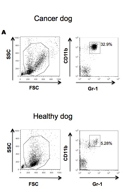
(Published customer image: Effects of EDTA versus media storage of patient blood samples on flow cytometric results. Peripheral blood cells were (a) stained immediate following collection with CD11b and CADO48A or maintained in EDTA collection tubes for (b) 24 or (c) 48 hours or in media for (d) 24 or (e) 48 hours prior to staining with CD11b and CADO48A. Both EDTA and media samples were kept refrigerated. These findings show a decrease over time of population distinction in EDTA with minimal changes when cells are kept in media up to 48 hours. All cells were gated on P1.From: Sherger et al. BMC Veterinary Research 2012 8:209)
Application Data
(Published customer image: Effects of EDTA versus media storage of patient blood samples on flow cytometric results. Peripheral blood cells were (a) stained immediate following collection with CD11b and CADO48A or maintained in EDTA collection tubes for (b) 24 or (c) 48 hours or in media for (d) 24 or (e) 48 hours prior to staining with CD11b and CADO48A. Both EDTA and media samples were kept refrigerated. These findings show a decrease over time of population distinction in EDTA with minimal changes when cells are kept in media up to 48 hours. All cells were gated on P1.From: Sherger et al. BMC Veterinary Research 2012 8:209)
CD11b, Monoclonal Antibody (Cat# AAA12092)
Full Name
MOUSE ANTI DOG CD11b
Gene Names
ITGAM; CD11B
Applications
FC/FACS, IP


 Application Data
(Published customer image Infiltration of GFP+ BM-cells in infarct and peri-infarct regions. (A-B) Dot plots of viable macrophages/granulocytes (CD11b+CD45high, top right quadrants) and microglia (CD11b+CD45dim, bottom right quadrants) in cortex from BM-chimeric unmanipulated mice and mice exposed to pMCAO. (C) Bar graph showing mean numbers of CD11b+CD45dim microglia and CD11b+CD45high macrophages/granulocytes in BM-chimeric mice 24 hours after pMCAO, subdivided based on expression of GFP (n = 5). Approximately 92% of of the CD45high population were GFP+. (D) Estimation and comparison of mean numbers of CD11b+CD45dim microglia in non-chimeric (n = 10) versus BM-chimeric mice (n = 5) 24 hours after of pMCAO shows significantly fewer CD11b+CD45dim microglial cells in irradiated mice. (E) Overview, showing distribution of infiltrating GFP+ BM-derived cells into infarct (IF) and peri-infarct (P-IF) regions 24 hours after pMCAO. (E-G) By 24 hours, GFP+ single cells (F) and vessel-associated aggregates of GFP+ cells (arrows in G) were observed in infarct and peri-infarct regions. Some of the vessel-associated cells were round, leukocyte-like cells (arrows) while others were elongated cells lining the vasculature (arrow heads in G and in insert). (H) Bar graph showing mean numbers of single GFP+ cells and vessel-associated aggregates of GFP+ cells in ipsi- and contralateral cortex 24 hours after surgery (n = 10). (I-P) Immunohistochemical staining of CD45.1 (I, K), CD45.2 (J, L), IgG2a (M, O) and CD45 (N, P) in ischemic tissue in BM-chimeric (I, J, M, N) and non-chimeric mice (K, L, O, P) 24 hours after pMCAO. N.D, none detected. Scale bars: 200 um (A), 10 um (B, C). 50 um (I-P) *P < 0.05, **P < 0.01, and ***P < 0.001.From: Clausen BH, Lambertsen KL, Babcock AA, Holm TH, Dagnaes-Hansen F, Finsen B. Interleukin-1beta and tumor necrosis factor-alpha are expressed by different subsets of microglia and macrophages after ischemic stroke in mice. J Neuroinflammation. 2008 Oct 23;5:46.)
Application Data
(Published customer image Infiltration of GFP+ BM-cells in infarct and peri-infarct regions. (A-B) Dot plots of viable macrophages/granulocytes (CD11b+CD45high, top right quadrants) and microglia (CD11b+CD45dim, bottom right quadrants) in cortex from BM-chimeric unmanipulated mice and mice exposed to pMCAO. (C) Bar graph showing mean numbers of CD11b+CD45dim microglia and CD11b+CD45high macrophages/granulocytes in BM-chimeric mice 24 hours after pMCAO, subdivided based on expression of GFP (n = 5). Approximately 92% of of the CD45high population were GFP+. (D) Estimation and comparison of mean numbers of CD11b+CD45dim microglia in non-chimeric (n = 10) versus BM-chimeric mice (n = 5) 24 hours after of pMCAO shows significantly fewer CD11b+CD45dim microglial cells in irradiated mice. (E) Overview, showing distribution of infiltrating GFP+ BM-derived cells into infarct (IF) and peri-infarct (P-IF) regions 24 hours after pMCAO. (E-G) By 24 hours, GFP+ single cells (F) and vessel-associated aggregates of GFP+ cells (arrows in G) were observed in infarct and peri-infarct regions. Some of the vessel-associated cells were round, leukocyte-like cells (arrows) while others were elongated cells lining the vasculature (arrow heads in G and in insert). (H) Bar graph showing mean numbers of single GFP+ cells and vessel-associated aggregates of GFP+ cells in ipsi- and contralateral cortex 24 hours after surgery (n = 10). (I-P) Immunohistochemical staining of CD45.1 (I, K), CD45.2 (J, L), IgG2a (M, O) and CD45 (N, P) in ischemic tissue in BM-chimeric (I, J, M, N) and non-chimeric mice (K, L, O, P) 24 hours after pMCAO. N.D, none detected. Scale bars: 200 um (A), 10 um (B, C). 50 um (I-P) *P < 0.05, **P < 0.01, and ***P < 0.001.From: Clausen BH, Lambertsen KL, Babcock AA, Holm TH, Dagnaes-Hansen F, Finsen B. Interleukin-1beta and tumor necrosis factor-alpha are expressed by different subsets of microglia and macrophages after ischemic stroke in mice. J Neuroinflammation. 2008 Oct 23;5:46.)
CD11b, Monoclonal Antibody (Cat# AAA12181)
Full Name
RAT ANTI MOUSE CD11b
Gene Names
Itgam; CR3; CR3A; MAC1; Cd11b; Ly-40; Mac-1; Mac-1a; CD11b/CD18; F730045J24Rik
Reactivity
Human
Applications
FC/FACS, IF, IP


 Application Data
(Published customer image Infiltration of GFP+ BM-cells in infarct and peri-infarct regions. (A-B) Dot plots of viable macrophages/granulocytes (CD11b+CD45high, top right quadrants) and microglia (CD11b+CD45dim, bottom right quadrants) in cortex from BM-chimeric unmanipulated mice and mice exposed to pMCAO. (C) Bar graph showing mean numbers of CD11b+CD45dim microglia and CD11b+CD45high macrophages/granulocytes in BM-chimeric mice 24 hours after pMCAO, subdivided based on expression of GFP (n = 5). Approximately 92% of of the CD45high population were GFP+. (D) Estimation and comparison of mean numbers of CD11b+CD45dim microglia in non-chimeric (n = 10) versus BM-chimeric mice (n = 5) 24 hours after of pMCAO shows significantly fewer CD11b+CD45dim microglial cells in irradiated mice. (E) Overview, showing distribution of infiltrating GFP+ BM-derived cells into infarct (IF) and peri-infarct (P-IF) regions 24 hours after pMCAO. (E-G) By 24 hours, GFP+ single cells (F) and vessel-associated aggregates of GFP+ cells (arrows in G) were observed in infarct and peri-infarct regions. Some of the vessel-associated cells were round, leukocyte-like cells (arrows) while others were elongated cells lining the vasculature (arrow heads in G and in insert). (H) Bar graph showing mean numbers of single GFP+ cells and vessel-associated aggregates of GFP+ cells in ipsi- and contralateral cortex 24 hours after surgery (n = 10). (I-P) Immunohistochemical staining of CD45.1 (I, K), CD45.2 (J, L), IgG2a (M, O) and CD45 (N, P) in ischemic tissue in BM-chimeric (I, J, M, N) and non-chimeric mice (K, L, O, P) 24 hours after pMCAO. N.D, none detected. Scale bars: 200 um (A), 10 um (B, C). 50 um (I-P) *P < 0.05, **P < 0.01, and ***P < 0.001.From: Clausen BH, Lambertsen KL, Babcock AA, Holm TH, Dagnaes-Hansen F, Finsen B. Interleukin-1beta and tumor necrosis factor-alpha are expressed by different subsets of microglia and macrophages after ischemic stroke in mice. J Neuroinflammation. 2008 Oct 23;5:46.)
Application Data
(Published customer image Infiltration of GFP+ BM-cells in infarct and peri-infarct regions. (A-B) Dot plots of viable macrophages/granulocytes (CD11b+CD45high, top right quadrants) and microglia (CD11b+CD45dim, bottom right quadrants) in cortex from BM-chimeric unmanipulated mice and mice exposed to pMCAO. (C) Bar graph showing mean numbers of CD11b+CD45dim microglia and CD11b+CD45high macrophages/granulocytes in BM-chimeric mice 24 hours after pMCAO, subdivided based on expression of GFP (n = 5). Approximately 92% of of the CD45high population were GFP+. (D) Estimation and comparison of mean numbers of CD11b+CD45dim microglia in non-chimeric (n = 10) versus BM-chimeric mice (n = 5) 24 hours after of pMCAO shows significantly fewer CD11b+CD45dim microglial cells in irradiated mice. (E) Overview, showing distribution of infiltrating GFP+ BM-derived cells into infarct (IF) and peri-infarct (P-IF) regions 24 hours after pMCAO. (E-G) By 24 hours, GFP+ single cells (F) and vessel-associated aggregates of GFP+ cells (arrows in G) were observed in infarct and peri-infarct regions. Some of the vessel-associated cells were round, leukocyte-like cells (arrows) while others were elongated cells lining the vasculature (arrow heads in G and in insert). (H) Bar graph showing mean numbers of single GFP+ cells and vessel-associated aggregates of GFP+ cells in ipsi- and contralateral cortex 24 hours after surgery (n = 10). (I-P) Immunohistochemical staining of CD45.1 (I, K), CD45.2 (J, L), IgG2a (M, O) and CD45 (N, P) in ischemic tissue in BM-chimeric (I, J, M, N) and non-chimeric mice (K, L, O, P) 24 hours after pMCAO. N.D, none detected. Scale bars: 200 um (A), 10 um (B, C). 50 um (I-P) *P < 0.05, **P < 0.01, and ***P < 0.001.From: Clausen BH, Lambertsen KL, Babcock AA, Holm TH, Dagnaes-Hansen F, Finsen B. Interleukin-1beta and tumor necrosis factor-alpha are expressed by different subsets of microglia and macrophages after ischemic stroke in mice. J Neuroinflammation. 2008 Oct 23;5:46.)
CD11b, Monoclonal Antibody (Cat# AAA12231)
Full Name
RAT ANTI MOUSE CD11b:Low Endotoxin
Gene Names
Itgam; CR3; CR3A; MAC1; Cd11b; Ly-40; Mac-1; Mac-1a; CD11b/CD18; F730045J24Rik
Applications
FC/FACS, FN, IF, IP