Highly validated and characterized monoclonal/polyclonal
antibodies and recombinant
proteins
The majority of AAA Biotech’s antibodies are highly validated and can be use in multiple
applications such as ELISA, FC,
ICC, IF, IHC, IP, WB, etc. We have antibodies available for rare species, in multiple conjugated
forms or recombinant
antibodies.
As for our high quality proteins, the majority have 90% purity, detected by SDS-PAGE while some are
available in
different tags such as Flag, GST, His, MBP, etc. We also carry high quality native and biologically
active proteins.
AAA Biotech is constantly working to expand our capacity to provide recombinant proteins and
antibodies to most
target proteins.
SELECT `p`.*, `pd`.*, IFNULL(pdns.ncbi_summary, "N/A") as ncbi_summary_pdns, IFNULL(pdns.sp_comments, "N/A") as sp_comments_pdns, IFNULL(pdns.ncbi_research_articles, "N/A") as ncbi_research_articles_pdns, IFNULL(pe.products_description_extra, "N/A") as products_description_extra
FROM (`products`, `products` as `p`)
LEFT OUTER JOIN `products_description` as `pd` ON `p`.`products_id` = `pd`.`products_id`
LEFT OUTER JOIN `products_description_ncbi_sp` as `pdns` ON `p`.`products_id` = `pdns`.`products_id`
LEFT OUTER JOIN `products_extra` as `pe` ON `p`.`products_id` = `pe`.`products_id`
WHERE `p`.`products_id` = '19716'
AND `pd`.`language_id` = 1
LIMIT 1
Query
Database
2.00 ms
select p.*, pd.*,
ifnull(pdns.ncbi_summary, 'N/A') as ncbi_summary_pdns,
ifnull(pdns.sp_comments, 'N/A') as sp_comments_pdns,
ifnull(pdns.ncbi_research_articles, 'N/A') as ncbi_research_articles_pdns,
ifnull(pe.products_description_extra, 'N/A') as products_description_extra
from products p
LEFT OUTER JOIN products_description pd on p.products_id = pd.products_id
LEFT OUTER JOIN products_description_ncbi_sp pdns on p.products_id = pdns.products_id
LEFT OUTER JOIN products_extra pe on p.products_id = pe.products_id
where p.products_id = '19716' and pd.language_id = 1
Query
Database
1.56 ms
SELECT `options_values_price` as `price`, `products_options_values_name` as `package`
FROM `products_attributes`
JOIN `products_options_values` ON `products_options_values`.`products_options_values_id` = `products_attributes`.`options_values_id`
WHERE `products_attributes`.`products_id` = '19716'
Database (4 total Queries, 4 of them unique across 2 Connections)
Time
Query String
1.89 ms
SELECT `p`.*, `pd`.*, IFNULL(pdns.ncbi_summary, "N/A") as ncbi_summary_pdns, IFNULL(pdns.sp_comments, "N/A") as sp_comments_pdns, IFNULL(pdns.ncbi_research_articles, "N/A") as ncbi_research_articles_pdns, IFNULL(pe.products_description_extra, "N/A") as products_description_extra
FROM (`products`, `products` as `p`)
LEFT OUTER JOIN `products_description` as `pd` ON `p`.`products_id` = `pd`.`products_id`
LEFT OUTER JOIN `products_description_ncbi_sp` as `pdns` ON `p`.`products_id` = `pdns`.`products_id`
LEFT OUTER JOIN `products_extra` as `pe` ON `p`.`products_id` = `pe`.`products_id`
WHERE `p`.`products_id` = '19716'
AND `pd`.`language_id` = 1
LIMIT 1
select p.*, pd.*,
ifnull(pdns.ncbi_summary, 'N/A') as ncbi_summary_pdns,
ifnull(pdns.sp_comments, 'N/A') as sp_comments_pdns,
ifnull(pdns.ncbi_research_articles, 'N/A') as ncbi_research_articles_pdns,
ifnull(pe.products_description_extra, 'N/A') as products_description_extra
from products p
LEFT OUTER JOIN products_description pd on p.products_id = pd.products_id
LEFT OUTER JOIN products_description_ncbi_sp pdns on p.products_id = pdns.products_id
LEFT OUTER JOIN products_extra pe on p.products_id = pe.products_id
where p.products_id = '19716' and pd.language_id = 1
SELECT `options_values_price` as `price`, `products_options_values_name` as `package`
FROM `products_attributes`
JOIN `products_options_values` ON `products_options_values`.`products_options_values_id` = `products_attributes`.`options_values_id`
WHERE `products_attributes`.`products_id` = '19716'
⇄concentration => string (73) "Adding 0.2 ml of distilled water will yield a concentration of 500 ug/ml."
$value['concentration']
⇄⧉storage_stability => string (220) "Store at -20 degree C for one year from date of receipt. After reconstitutio...
$value['storage_stability']
Store at -20 degree C for one year from date of receipt. After reconstitution, at 4 degree C for one month. It can also be aliquotted and stored frozen at -20 degree C for six months. Avoid repeated freezing and thawing.
WB: 0.25-0.5 ug/ml, Human, Mouse, Rat<br>IHC-P: 2-5 ug/ml, Human, Rat<br>FC/FACS: 1-3 ug/1x10^6 cells, Human<br>Tested Species: In-house tested species with positive results.<br>Enhanced Chemiluminescent Kit with anti-Mouse IgG for Western blot, and HRP Conjugated anti-Mouse IgG Super Vision Assay Kit for IHC(P).
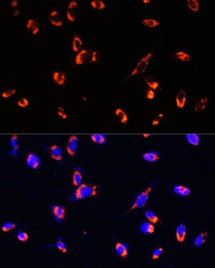
⇄⧉testing_protocols => string (5239) "FCM (Flow Cytometry)||Figure 6. Flow Cytometry analysis of U2OS cells using ...
$value['testing_protocols']
FCM (Flow Cytometry)||Figure 6. Flow Cytometry analysis of U2OS cells using anti-Sorbitol Dehydrogenase/SORD antibody (AAA19716).<br><br>Overlay histogram showing U2OS cells stained with AAA19716 (Blue line). The cells were blocked with 10% normal goat serum. And then incubated with mouse anti-Sorbitol Dehydrogenase/SORD Antibody (AAA19716, 1 ug/1x10^6 cells) for 30 min at 20 degree C. DyLight488 conjugated goat anti-mouse IgG was used as secondary antibody for 30 minutes at 20 degree C. Isotype control antibody (Green line) was mouse IgG (1 ug/1x10^6) used under the same conditions. Unlabelled sample (Red line) was also used as a control.||AAA19716_FCM6.png!!IHC (Immunohistochemistry)||Figure 5. IHC analysis of Sorbitol Dehydrogenase/SORD using anti-Sorbitol Dehydrogenase/SORD antibody (AAA19716).<br><br>Sorbitol Dehydrogenase/SORD was detected in a paraffin-embedded section of rat kidney tissue. Heat mediated antigen retrieval was performed in EDTA buffer (pH 8.0, epitope retrieval solution). The tissue section was blocked with 10% goat serum. The tissue section was then incubated with 2 ug/ml mouse anti-Sorbitol Dehydrogenase/SORD Antibody (AAA19716) overnight at 4 degree C. Peroxidase Conjugated Goat Anti-mouse IgG was used as secondary antibody and incubated for 30 minutes at 37 degree C. The tissue section was developed using HRP Conjugated Mouse IgG Super Vision Assay Kit with DAB as the chromogen.||AAA19716_IHC5.jpg!!IHC (Immunohistochemistry)||Figure 4. IHC analysis of Sorbitol Dehydrogenase/SORD using anti-Sorbitol Dehydrogenase/SORD antibody (AAA19716).<br><br>Sorbitol Dehydrogenase/SORD was detected in a paraffin-embedded section of human SM fatty carcinoma of the left kidney tissue. Heat mediated antigen retrieval was performed in EDTA buffer (pH 8.0, epitope retrieval solution). The tissue section was blocked with 10% goat serum. The tissue section was then incubated with 2 ug/ml mouse anti-Sorbitol Dehydrogenase/SORD Antibody (AAA19716) overnight at 4 degree C. Peroxidase Conjugated Goat Anti-mouse IgG was used as secondary antibody and incubated for 30 minutes at 37 degree C. The tissue section was developed using HRP Conjugated Mouse IgG Super Vision Assay Kit with DAB as the chromogen.||AAA19716_IHC4.jpg!!IHC (Immunohistochemistry)||Figure 3. IHC analysis of Sorbitol Dehydrogenase/SORD using anti-Sorbitol Dehydrogenase/SORD antibody (AAA19716).<br><br>Sorbitol Dehydrogenase/SORD was detected in a paraffin-embedded section of human renal clear cell carcinoma tissue. Heat mediated antigen retrieval was performed in EDTA buffer (pH 8.0, epitope retrieval solution). The tissue section was blocked with 10% goat serum. The tissue section was then incubated with 2 ug/ml mouse anti-Sorbitol Dehydrogenase/SORD Antibody (AAA19716) overnight at 4 degree C. Peroxidase Conjugated Goat Anti-mouse IgG was used as secondary antibody and incubated for 30 minutes at 37 degree C. The tissue section was developed using HRP Conjugated Mouse IgG Super Vision Assay Kit with DAB as the chromogen.||AAA19716_IHC3.jpg!!IHC (Immunohistochemistry)||Figure 2. IHC analysis of Sorbitol Dehydrogenase/SORD using anti-Sorbitol Dehydrogenase/SORD antibody (AAA19716).<br><br>Sorbitol Dehydrogenase/SORD was detected in a paraffin-embedded section of human liver cancer tissue. Heat mediated antigen retrieval was performed in EDTA buffer (pH 8.0, epitope retrieval solution). The tissue section was blocked with 10% goat serum. The tissue section was then incubated with 2 ug/ml mouse anti-Sorbitol Dehydrogenase/SORD Antibody (AAA19716) overnight at 4 degree C. Peroxidase Conjugated Goat Anti-mouse IgG was used as secondary antibody and incubated for 30 minutes at 37 degree C. The tissue section was developed using HRP Conjugated Mouse IgG Super Vision Assay Kit with DAB as the chromogen.||AAA19716_IHC2.jpg!!WB (Western Blot)||Figure 1. Western blot analysis of Sorbitol Dehydrogenase/SORD using anti-Sorbitol Dehydrogenase/SORD antibody (AAA19716).<br><br>Electrophoresis was performed on a 5-20% SDS-PAGE gel at 70V (Stacking gel)/90V (Resolving gel) for 2-3 hours. The sample well of each lane was loaded with 30 ug of sample under reducing conditions.<br><br>Lane 1: human Hela whole cell lysates,<br>Lane 2: human HepG2 whole cell lysates,<br>Lane 3: rat liver tissue tissue lysates,<br>Lane 4: rat kidney tissue lysates,<br>Lane 5: mouse liver tissue lysates,<br>Lane 6: mouse kidney tissue lysates.<br><br>After electrophoresis, proteins were transferred to a nitrocellulose membrane at 150 mA for 50-90 minutes. Blocked the membrane with 5% non-fat milk/TBS for 1.5 hour at RT. The membrane was incubated with mouse anti-Sorbitol Dehydrogenase/SORD antigen affinity purified monoclonal antibody (#AAA19716) at 0.5 ug/mL overnight at 4 degree C, then washed with TBS-0.1%Tween 3 times with 5 minutes each and probed with a goat anti-mouse IgG-HRP secondary antibody at a dilution of 1:10000 for 1.5 hour at RT. The signal is developed using an Enhanced Chemiluminescent detection (ECL) kit with Tanon 5200 system. A specific band was detected for Sorbitol Dehydrogenase/SORD at approximately 40 kDa. The expected band size for Sorbitol Dehydrogenase/SORD is at 40 kDa.||AAA19716_WB.jpg
⇄⧉etc_term1 => string (89) "Reconstitution||Adding 0.2 ml of distilled water will yield a concentration ...
$value['etc_term1']
Reconstitution||Adding 0.2 ml of distilled water will yield a concentration of 500 ug/ml.
⇄⧉products_description => string (549) "Sorbitol dehydrogenase is an enzyme that in humans is encoded by the SORD ge...
$value['products_description']
Sorbitol dehydrogenase is an enzyme that in humans is encoded by the SORD gene. Sorbitol dehydrogenase (SORD) catalyzes the interconversion of polyols and their corresponding ketoses, and together with aldose reductase, makes up the sorbitol pathway that is believed to play an important role in the development of diabetic complications. The first reaction of the pathway (also called the polyol pathway) is the reduction of glucose to sorbitol by ALDR1 with NADPH as the cofactor. SORD then oxidizes the sorbitol to fructose using NAD(+) cofactor.
⇄concentration => string (73) "Adding 0.2 ml of distilled water will yield a concentration of 500 ug/ml."
$value->a['concentration']
⇄⧉storage_stability => string (220) "Store at -20 degree C for one year from date of receipt. After reconstitutio...
$value->a['storage_stability']
Store at -20 degree C for one year from date of receipt. After reconstitution, at 4 degree C for one month. It can also be aliquotted and stored frozen at -20 degree C for six months. Avoid repeated freezing and thawing.
WB: 0.25-0.5 ug/ml, Human, Mouse, Rat<br>IHC-P: 2-5 ug/ml, Human, Rat<br>FC/FACS: 1-3 ug/1x10^6 cells, Human<br>Tested Species: In-house tested species with positive results.<br>Enhanced Chemiluminescent Kit with anti-Mouse IgG for Western blot, and HRP Conjugated anti-Mouse IgG Super Vision Assay Kit for IHC(P).
⇄⧉testing_protocols => string (5239) "FCM (Flow Cytometry)||Figure 6. Flow Cytometry analysis of U2OS cells using ...
$value->a['testing_protocols']
FCM (Flow Cytometry)||Figure 6. Flow Cytometry analysis of U2OS cells using anti-Sorbitol Dehydrogenase/SORD antibody (AAA19716).<br><br>Overlay histogram showing U2OS cells stained with AAA19716 (Blue line). The cells were blocked with 10% normal goat serum. And then incubated with mouse anti-Sorbitol Dehydrogenase/SORD Antibody (AAA19716, 1 ug/1x10^6 cells) for 30 min at 20 degree C. DyLight488 conjugated goat anti-mouse IgG was used as secondary antibody for 30 minutes at 20 degree C. Isotype control antibody (Green line) was mouse IgG (1 ug/1x10^6) used under the same conditions. Unlabelled sample (Red line) was also used as a control.||AAA19716_FCM6.png!!IHC (Immunohistochemistry)||Figure 5. IHC analysis of Sorbitol Dehydrogenase/SORD using anti-Sorbitol Dehydrogenase/SORD antibody (AAA19716).<br><br>Sorbitol Dehydrogenase/SORD was detected in a paraffin-embedded section of rat kidney tissue. Heat mediated antigen retrieval was performed in EDTA buffer (pH 8.0, epitope retrieval solution). The tissue section was blocked with 10% goat serum. The tissue section was then incubated with 2 ug/ml mouse anti-Sorbitol Dehydrogenase/SORD Antibody (AAA19716) overnight at 4 degree C. Peroxidase Conjugated Goat Anti-mouse IgG was used as secondary antibody and incubated for 30 minutes at 37 degree C. The tissue section was developed using HRP Conjugated Mouse IgG Super Vision Assay Kit with DAB as the chromogen.||AAA19716_IHC5.jpg!!IHC (Immunohistochemistry)||Figure 4. IHC analysis of Sorbitol Dehydrogenase/SORD using anti-Sorbitol Dehydrogenase/SORD antibody (AAA19716).<br><br>Sorbitol Dehydrogenase/SORD was detected in a paraffin-embedded section of human SM fatty carcinoma of the left kidney tissue. Heat mediated antigen retrieval was performed in EDTA buffer (pH 8.0, epitope retrieval solution). The tissue section was blocked with 10% goat serum. The tissue section was then incubated with 2 ug/ml mouse anti-Sorbitol Dehydrogenase/SORD Antibody (AAA19716) overnight at 4 degree C. Peroxidase Conjugated Goat Anti-mouse IgG was used as secondary antibody and incubated for 30 minutes at 37 degree C. The tissue section was developed using HRP Conjugated Mouse IgG Super Vision Assay Kit with DAB as the chromogen.||AAA19716_IHC4.jpg!!IHC (Immunohistochemistry)||Figure 3. IHC analysis of Sorbitol Dehydrogenase/SORD using anti-Sorbitol Dehydrogenase/SORD antibody (AAA19716).<br><br>Sorbitol Dehydrogenase/SORD was detected in a paraffin-embedded section of human renal clear cell carcinoma tissue. Heat mediated antigen retrieval was performed in EDTA buffer (pH 8.0, epitope retrieval solution). The tissue section was blocked with 10% goat serum. The tissue section was then incubated with 2 ug/ml mouse anti-Sorbitol Dehydrogenase/SORD Antibody (AAA19716) overnight at 4 degree C. Peroxidase Conjugated Goat Anti-mouse IgG was used as secondary antibody and incubated for 30 minutes at 37 degree C. The tissue section was developed using HRP Conjugated Mouse IgG Super Vision Assay Kit with DAB as the chromogen.||AAA19716_IHC3.jpg!!IHC (Immunohistochemistry)||Figure 2. IHC analysis of Sorbitol Dehydrogenase/SORD using anti-Sorbitol Dehydrogenase/SORD antibody (AAA19716).<br><br>Sorbitol Dehydrogenase/SORD was detected in a paraffin-embedded section of human liver cancer tissue. Heat mediated antigen retrieval was performed in EDTA buffer (pH 8.0, epitope retrieval solution). The tissue section was blocked with 10% goat serum. The tissue section was then incubated with 2 ug/ml mouse anti-Sorbitol Dehydrogenase/SORD Antibody (AAA19716) overnight at 4 degree C. Peroxidase Conjugated Goat Anti-mouse IgG was used as secondary antibody and incubated for 30 minutes at 37 degree C. The tissue section was developed using HRP Conjugated Mouse IgG Super Vision Assay Kit with DAB as the chromogen.||AAA19716_IHC2.jpg!!WB (Western Blot)||Figure 1. Western blot analysis of Sorbitol Dehydrogenase/SORD using anti-Sorbitol Dehydrogenase/SORD antibody (AAA19716).<br><br>Electrophoresis was performed on a 5-20% SDS-PAGE gel at 70V (Stacking gel)/90V (Resolving gel) for 2-3 hours. The sample well of each lane was loaded with 30 ug of sample under reducing conditions.<br><br>Lane 1: human Hela whole cell lysates,<br>Lane 2: human HepG2 whole cell lysates,<br>Lane 3: rat liver tissue tissue lysates,<br>Lane 4: rat kidney tissue lysates,<br>Lane 5: mouse liver tissue lysates,<br>Lane 6: mouse kidney tissue lysates.<br><br>After electrophoresis, proteins were transferred to a nitrocellulose membrane at 150 mA for 50-90 minutes. Blocked the membrane with 5% non-fat milk/TBS for 1.5 hour at RT. The membrane was incubated with mouse anti-Sorbitol Dehydrogenase/SORD antigen affinity purified monoclonal antibody (#AAA19716) at 0.5 ug/mL overnight at 4 degree C, then washed with TBS-0.1%Tween 3 times with 5 minutes each and probed with a goat anti-mouse IgG-HRP secondary antibody at a dilution of 1:10000 for 1.5 hour at RT. The signal is developed using an Enhanced Chemiluminescent detection (ECL) kit with Tanon 5200 system. A specific band was detected for Sorbitol Dehydrogenase/SORD at approximately 40 kDa. The expected band size for Sorbitol Dehydrogenase/SORD is at 40 kDa.||AAA19716_WB.jpg
⇄⧉etc_term1 => string (89) "Reconstitution||Adding 0.2 ml of distilled water will yield a concentration ...
$value->a['etc_term1']
Reconstitution||Adding 0.2 ml of distilled water will yield a concentration of 500 ug/ml.
⇄⧉products_description => string (549) "Sorbitol dehydrogenase is an enzyme that in humans is encoded by the SORD ge...
$value->a['products_description']
Sorbitol dehydrogenase is an enzyme that in humans is encoded by the SORD gene. Sorbitol dehydrogenase (SORD) catalyzes the interconversion of polyols and their corresponding ketoses, and together with aldose reductase, makes up the sorbitol pathway that is believed to play an important role in the development of diabetic complications. The first reaction of the pathway (also called the polyol pathway) is the reduction of glucose to sorbitol by ALDR1 with NADPH as the cofactor. SORD then oxidizes the sorbitol to fructose using NAD(+) cofactor.
⇄concentration => string (73) "Adding 0.2 ml of distilled water will yield a concentration of 500 ug/ml."
$value->d['concentration']
⇄⧉storage_stability => string (220) "Store at -20 degree C for one year from date of receipt. After reconstitutio...
$value->d['storage_stability']
Store at -20 degree C for one year from date of receipt. After reconstitution, at 4 degree C for one month. It can also be aliquotted and stored frozen at -20 degree C for six months. Avoid repeated freezing and thawing.
WB: 0.25-0.5 ug/ml, Human, Mouse, Rat<br>IHC-P: 2-5 ug/ml, Human, Rat<br>FC/FACS: 1-3 ug/1x10^6 cells, Human<br>Tested Species: In-house tested species with positive results.<br>Enhanced Chemiluminescent Kit with anti-Mouse IgG for Western blot, and HRP Conjugated anti-Mouse IgG Super Vision Assay Kit for IHC(P).
⇄⧉testing_protocols => string (5239) "FCM (Flow Cytometry)||Figure 6. Flow Cytometry analysis of U2OS cells using ...
$value->d['testing_protocols']
FCM (Flow Cytometry)||Figure 6. Flow Cytometry analysis of U2OS cells using anti-Sorbitol Dehydrogenase/SORD antibody (AAA19716).<br><br>Overlay histogram showing U2OS cells stained with AAA19716 (Blue line). The cells were blocked with 10% normal goat serum. And then incubated with mouse anti-Sorbitol Dehydrogenase/SORD Antibody (AAA19716, 1 ug/1x10^6 cells) for 30 min at 20 degree C. DyLight488 conjugated goat anti-mouse IgG was used as secondary antibody for 30 minutes at 20 degree C. Isotype control antibody (Green line) was mouse IgG (1 ug/1x10^6) used under the same conditions. Unlabelled sample (Red line) was also used as a control.||AAA19716_FCM6.png!!IHC (Immunohistochemistry)||Figure 5. IHC analysis of Sorbitol Dehydrogenase/SORD using anti-Sorbitol Dehydrogenase/SORD antibody (AAA19716).<br><br>Sorbitol Dehydrogenase/SORD was detected in a paraffin-embedded section of rat kidney tissue. Heat mediated antigen retrieval was performed in EDTA buffer (pH 8.0, epitope retrieval solution). The tissue section was blocked with 10% goat serum. The tissue section was then incubated with 2 ug/ml mouse anti-Sorbitol Dehydrogenase/SORD Antibody (AAA19716) overnight at 4 degree C. Peroxidase Conjugated Goat Anti-mouse IgG was used as secondary antibody and incubated for 30 minutes at 37 degree C. The tissue section was developed using HRP Conjugated Mouse IgG Super Vision Assay Kit with DAB as the chromogen.||AAA19716_IHC5.jpg!!IHC (Immunohistochemistry)||Figure 4. IHC analysis of Sorbitol Dehydrogenase/SORD using anti-Sorbitol Dehydrogenase/SORD antibody (AAA19716).<br><br>Sorbitol Dehydrogenase/SORD was detected in a paraffin-embedded section of human SM fatty carcinoma of the left kidney tissue. Heat mediated antigen retrieval was performed in EDTA buffer (pH 8.0, epitope retrieval solution). The tissue section was blocked with 10% goat serum. The tissue section was then incubated with 2 ug/ml mouse anti-Sorbitol Dehydrogenase/SORD Antibody (AAA19716) overnight at 4 degree C. Peroxidase Conjugated Goat Anti-mouse IgG was used as secondary antibody and incubated for 30 minutes at 37 degree C. The tissue section was developed using HRP Conjugated Mouse IgG Super Vision Assay Kit with DAB as the chromogen.||AAA19716_IHC4.jpg!!IHC (Immunohistochemistry)||Figure 3. IHC analysis of Sorbitol Dehydrogenase/SORD using anti-Sorbitol Dehydrogenase/SORD antibody (AAA19716).<br><br>Sorbitol Dehydrogenase/SORD was detected in a paraffin-embedded section of human renal clear cell carcinoma tissue. Heat mediated antigen retrieval was performed in EDTA buffer (pH 8.0, epitope retrieval solution). The tissue section was blocked with 10% goat serum. The tissue section was then incubated with 2 ug/ml mouse anti-Sorbitol Dehydrogenase/SORD Antibody (AAA19716) overnight at 4 degree C. Peroxidase Conjugated Goat Anti-mouse IgG was used as secondary antibody and incubated for 30 minutes at 37 degree C. The tissue section was developed using HRP Conjugated Mouse IgG Super Vision Assay Kit with DAB as the chromogen.||AAA19716_IHC3.jpg!!IHC (Immunohistochemistry)||Figure 2. IHC analysis of Sorbitol Dehydrogenase/SORD using anti-Sorbitol Dehydrogenase/SORD antibody (AAA19716).<br><br>Sorbitol Dehydrogenase/SORD was detected in a paraffin-embedded section of human liver cancer tissue. Heat mediated antigen retrieval was performed in EDTA buffer (pH 8.0, epitope retrieval solution). The tissue section was blocked with 10% goat serum. The tissue section was then incubated with 2 ug/ml mouse anti-Sorbitol Dehydrogenase/SORD Antibody (AAA19716) overnight at 4 degree C. Peroxidase Conjugated Goat Anti-mouse IgG was used as secondary antibody and incubated for 30 minutes at 37 degree C. The tissue section was developed using HRP Conjugated Mouse IgG Super Vision Assay Kit with DAB as the chromogen.||AAA19716_IHC2.jpg!!WB (Western Blot)||Figure 1. Western blot analysis of Sorbitol Dehydrogenase/SORD using anti-Sorbitol Dehydrogenase/SORD antibody (AAA19716).<br><br>Electrophoresis was performed on a 5-20% SDS-PAGE gel at 70V (Stacking gel)/90V (Resolving gel) for 2-3 hours. The sample well of each lane was loaded with 30 ug of sample under reducing conditions.<br><br>Lane 1: human Hela whole cell lysates,<br>Lane 2: human HepG2 whole cell lysates,<br>Lane 3: rat liver tissue tissue lysates,<br>Lane 4: rat kidney tissue lysates,<br>Lane 5: mouse liver tissue lysates,<br>Lane 6: mouse kidney tissue lysates.<br><br>After electrophoresis, proteins were transferred to a nitrocellulose membrane at 150 mA for 50-90 minutes. Blocked the membrane with 5% non-fat milk/TBS for 1.5 hour at RT. The membrane was incubated with mouse anti-Sorbitol Dehydrogenase/SORD antigen affinity purified monoclonal antibody (#AAA19716) at 0.5 ug/mL overnight at 4 degree C, then washed with TBS-0.1%Tween 3 times with 5 minutes each and probed with a goat anti-mouse IgG-HRP secondary antibody at a dilution of 1:10000 for 1.5 hour at RT. The signal is developed using an Enhanced Chemiluminescent detection (ECL) kit with Tanon 5200 system. A specific band was detected for Sorbitol Dehydrogenase/SORD at approximately 40 kDa. The expected band size for Sorbitol Dehydrogenase/SORD is at 40 kDa.||AAA19716_WB.jpg
⇄⧉etc_term1 => string (89) "Reconstitution||Adding 0.2 ml of distilled water will yield a concentration ...
$value->d['etc_term1']
Reconstitution||Adding 0.2 ml of distilled water will yield a concentration of 500 ug/ml.
⇄⧉products_description => string (549) "Sorbitol dehydrogenase is an enzyme that in humans is encoded by the SORD ge...
$value->d['products_description']
Sorbitol dehydrogenase is an enzyme that in humans is encoded by the SORD gene. Sorbitol dehydrogenase (SORD) catalyzes the interconversion of polyols and their corresponding ketoses, and together with aldose reductase, makes up the sorbitol pathway that is believed to play an important role in the development of diabetic complications. The first reaction of the pathway (also called the polyol pathway) is the reduction of glucose to sorbitol by ALDR1 with NADPH as the cofactor. SORD then oxidizes the sorbitol to fructose using NAD(+) cofactor.
⇄⧉products_description => string (1327) "Intended Uses: This KLH ELISA kit is a 1.5 hour solid-phase ELISA designed f...
$value[0]['_source']['products_description']
Intended Uses: This KLH ELISA kit is a 1.5 hour solid-phase ELISA designed for the quantitative determination of HumanKLH. This ELISA kit for research use only!<br><br>Principle of the Assay: KLH ELISA kit applies the competitive enzyme immunoassay technique utilizing a polyclonal anti-KLH antibody and an KLH-HRP conjugate. The assay sample and buffer are incubated together with KLH-HRP conjugate in pre-coated plate for one hour. After the incubation period, the wells are decanted and washed five times. The wells are then incubated with a substrate for HRP enzyme. The product of the enzyme-substrate reaction forms a blue colored complex. Finally, a stop solution is added to stop the reaction, which will then turn the solution yellow. The intensity of color is measured spectrophotometrically at 450nm in a microplate reader. The intensity of the color is inversely proportional to the KLH concentration since KLH from samples and KLH-HRP conjugate compete for the anti-KLH antibody binding site. Since the number of sites is limited, as more sites are occupied by KLH from the sample, fewer sites are left to bind KLH-HRP conjugate. A standard curve is plotted relating the intensity of the color (O.D.) to the concentration of standards. The KLH concentration in each sample is interpolated from this standard curve.
kallikrein-10; protease serine-like 1; kallikrein 10 protein 1; kallikrein 10 protein 2; kallikrein 10 protein 3; kallikrein 10 protein 4; kallikrein 10 protein 7; kallikrein 10 protein 8; kallikrein 10 protein 9; kallikrein 10 protein 12; protease, serine-like, 1; normal epithelial cell-specific 1; breast normal epithelial cell associated serine protease
⇄⧉search_terms => string (551) "aaa16119 mouse typical testing data standard curve for reference only aaa161...
$value[0]['_source']['search_terms']
aaa16119 mouse typical testing data standard curve for reference only aaa16119_sc elisa kit kallikrein 10 klk10 preproprotein related peptidase nes1 prssl1 protease serine like 1 protein 2 3 4 7 8 9 12 normal epithelial cell specific breast associated 30,170 da klk10_human 117422433 np_001070968.1 o43240 nm_001077500.1 q53yl3 q99920 q9gzw9 a6nc12 602673 signal transduction samples serum plasma culture supernatants body fluid and tissue homogenate assay type quantitative competitive sensitivity 0.1 ng ml kallikrein10 like1 protein2 sensitivity0.1
⇄⧉specificity => string (181) "This assay has high sensitivity and excellent specificity for detection of h...
$value[1]['_source']['specificity']
This assay has high sensitivity and excellent specificity for detection of human KLK7. No significant cross-reactivity or interference between human KLK7 and analogues was observed.
⇄purity => string (3) "N/A"
$value[1]['_source']['purity']
⇄form => string (3) "N/A"
$value[1]['_source']['form']
⇄concentration => string (3) "N/A"
$value[1]['_source']['concentration']
⇄⧉storage_stability => string (129) "Unopened test kits should be stored at 2 to 8 degree C upon receipt. Please ...
$value[1]['_source']['storage_stability']
Unopened test kits should be stored at 2 to 8 degree C upon receipt. Please refer to pdf manual for further storage instructions.
Samples||Serum, plasma, tissue homogenates!!Assay Type||Sandwich!!Detection Range||0.625 ng/ml -40 ng/ml.!!Sensitivity||The minimum detectable dose of human KLK7 is typically less than 0.156 ng/ml. The sensitivity of this assay, or Lower Limit of Detection (LLD) was defined as the lowest protein concentration that could be differentiated from zero. It was determined the mean O.D value of 20 replicates of the zero standard added by their three standard deviations.
⇄⧉etc_term2 => string (327) "Intra-assay Precision||Intra-assay Precision (Precision within an assay): CV...
$value[1]['_source']['etc_term2']
Intra-assay Precision||Intra-assay Precision (Precision within an assay): CV%<8%. Three samples of known concentration were tested twenty times on one plate to assess.!!Inter-assay Precision||Inter-assay Precision (Precision between assays): CV%<10%. Three samples of known concentration were tested in twenty assays to assess.
⇄⧉products_description => string (742) "<b>Principle of the assay: </b>This assay employs the quantitative sandwich ...
$value[1]['_source']['products_description']
<b>Principle of the assay: </b>This assay employs the quantitative sandwich enzyme immunoassay technique. Antibody specific for KLK 7 has been pre-coated onto a microplate. Standards and samples are pipetted into the wells and any KLK 7 present is bound by the immobilized antibody. After removing any unbound substances, a biotin-conjugated antibody specific for KLK 7 is added to the wells. After washing, avidin conjugated Horseradish Peroxidase (HRP) is added to the wells. Following a wash to remove any unbound avidin-enzyme reagent, a substrate solution is added to the wells and color develops in proportion to the amount of KLK 7 bound in the initial step. The color development is stopped and the intensity of the color is measured.
⇄⧉search_terms => string (841) "aaa15318 human this assay has high sensitivity and excellent specificity for...
$value[1]['_source']['search_terms']
aaa15318 human this assay has high sensitivity and excellent specificity for detection of klk7 no significant cross reactivity or interference between analogues was observed typical testing data standard curve reference only aaa15318_td elisa kit kallikrein related peptidase 7 klk prss6 scce chymotryptic stratum corneum protease serine 6 signal protein enzyme isoform 2 hk7 27,525 da hscce klk7_human 333609286 np_001193982.1 p49862 nm_001207053.1 q8n5n9 q8nfv7 a8k0u5 604438 samples serum plasma tissue homogenates type sandwich range 0.625 ng ml 40 the minimum detectable dose is typically less than 0.156 lower limit lld defined as lowest concentration that could be differentiated from zero it determined mean o.d value 20 replicates added by their three deviations intra precision within an cv peptidase7 serine6 isoform2 ml40 value20
⇄⧉specificity => string (198) "The Human Kallikrein 5 ELISA Kit allows for the detection and quantification...
$value[2]['_source']['specificity']
The Human Kallikrein 5 ELISA Kit allows for the detection and quantification of endogenous levels of natural and/or recombinant Human Kallikrein 5 proteins within the range of 78 pg/ml - 5000 pg/ml.
⇄purity => string (3) "N/A"
$value[2]['_source']['purity']
⇄form => string (3) "N/A"
$value[2]['_source']['form']
⇄concentration => string (3) "N/A"
$value[2]['_source']['concentration']
⇄⧉storage_stability => string (107) "Shipped and store at 4 degree C for 6 months, store at -20 degree C for one ...
$value[2]['_source']['storage_stability']
Shipped and store at 4 degree C for 6 months, store at -20 degree C for one year. Avoid freeze/thaw cycles.
SCTE; Kallikrein-5; Kallikrein-like protein 2; KLK-L2; Stratum corneum tryptic enzyme
⇄products_gene_name => string (4) "KLK5"
$value[2]['_source']['products_gene_name']
⇄products_gene_name_syn => string (3) "N/A"
$value[2]['_source']['products_gene_name_syn']
⇄⧉products_description => string (1565) "Background: Kallikrein gene 5 (KLK5, also known as KLK-L2), located on chrom...
$value[2]['_source']['products_description']
Background: Kallikrein gene 5 (KLK5, also known as KLK-L2), located on chromosome 19q13.4, is one of the newly identified members of the kallikrein gene family, which is a subgroup of the serine protease enzyme family. In normal human tissues, KLK5 is highly expressed in skin, mammary gland and testis. KLK5 has been suggested to regulate cell shedding (desquamation) in conjunction with KLK7 and KLK14, given its ability to degrade proteins which form the extracellular component of cell junctions in the stratum corneum. It is proposed that KLK5 regulates this process since it is able to self-activate in addition to activating KLK7 and KLK14.<br><br>Principle of the Assay: The Human Kallikrein 5 ELISA (Enzyme-Linked Immunosorbent Assay) kit is an in vitro enzyme-linked immunosorbent assay for the quantitative measurement of Human Kallikrein 5 in Cell Culture Supernatants, Serum, Plasma. This assay employs an antibody specific for Human Kallikrein 5 coated on a 96-well plate. Standards and samples are pipetted into the wells and Kallikrein 5 present in a sample is bound to the wells by the immobilized antibody. The wells are washed and biotinylated anti-Human Kallikrein 5 antibody is added. After washing away unbound biotinylated antibody, HRP-conjugated streptavidin is pipetted to the wells. The wells are again washed, a TMB substrate solution is added to the wells and color develops in proportion to the amount of Kallikrein 5 bound. The Stop Solution changes the color from blue to yellow, and the intensity of the color is measured at 450 nm.
⇄⧉search_terms => string (531) "aaa17862 human the kallikrein 5 elisa kit allows for detection and quantific...
$value[2]['_source']['search_terms']
aaa17862 human the kallikrein 5 elisa kit allows for detection and quantification of endogenous levels natural or recombinant proteins within range 78 pg ml 5000 sandwich se typical testing data standard curve reference only aaa17862_td scte like protein 2 klk l2 stratum corneum tryptic enzyme klk5 preproprotein related peptidase klkl2 32,019 da klk5_human 117306172 np_001070959.1 q9y337 nm_001077491.1 q53zr3 q9hbg8 605643 samples cell culture supernatants serum plasma sensitivity 10 kallikrein5 range78 protein2 sensitivity10
⇄⧉specificity => string (169) "This assay has high sensitivity and excellent specificity for detection of K...
$value[3]['_source']['specificity']
This assay has high sensitivity and excellent specificity for detection of KLK2. No significant cross-reactivity or interference between KLK2 and analogues was observed.
⇄purity => string (3) "N/A"
$value[3]['_source']['purity']
⇄form => string (3) "N/A"
$value[3]['_source']['form']
⇄concentration => string (3) "N/A"
$value[3]['_source']['concentration']
⇄⧉storage_stability => string (156) "Store entire kit at 2-8C for short-term. For longer-term, please store the m...
$value[3]['_source']['storage_stability']
Store entire kit at 2-8C for short-term. For longer-term, please store the microplate & standard at -20C, while the remaining reagents can be stored at 2-8C
⇄⧉products_description => string (825) "Principle of the Assay: This kit was based on sandwich enzyme-linked immune-...
$value[3]['_source']['products_description']
Principle of the Assay: This kit was based on sandwich enzyme-linked immune-sorbent assay technology. Capture antibody was pre-coated onto 96-well plates. And the biotin conjugated antibody was used as detection antibodies. The standards, test samples and biotin conjugated detection antibody were added to the wells subsequently, and washed with wash buffer. HRP-Streptavidin was added and unbound conjugates were washed away with wash buffer. TMB substrates were used to visualize HRP enzymatic reaction. TMB was catalyzed by HRP to produce a blue color product that changed into yellow after adding acidic stop solution. The density of yellow is proportional to the target amount of sample captured in plate. Read the O.D. absorbance at 450nm in a microplate reader, and then the concentration of target can be calculated.
⇄⧉ncbi_pathways => string (603) "Activated PKN1 Stimulates Transcription Of AR (androgen Receptor) Regulated ...
$value[3]['_source']['ncbi_pathways']
Activated PKN1 Stimulates Transcription Of AR (androgen Receptor) Regulated Genes KLK2 And KLK3 Pathway||1269513!!Activation Of Matrix Metalloproteinases Pathway||1270258!!Coregulation Of Androgen Receptor Activity Pathway||138085!!Degradation Of The Extracellular Matrix Pathway||1270257!!Endocrine And Other Factor-regulated Calcium Reabsorption Pathway||213307!!Endocrine And Other Factor-regulated Calcium Reabsorption Pathway||213276!!Extracellular Matrix Organization Pathway||1270244!!Metabolism Of Proteins Pathway||1268677!!Prostate Cancer Pathway||755440!!RHO GTPase Effectors Pathway||1269509
⇄⧉search_terms => string (589) "aaa17597 human this assay has high sensitivity and excellent specificity for...
$value[3]['_source']['search_terms']
aaa17597 human this assay has high sensitivity and excellent specificity for detection of klk2 no significant cross reactivity or interference between analogues was observed typical testing data standard curve reference only aaa17597_sc elisa kit kallikrein 2 glandular 1 hgk tissue isoform preproprotein related peptidase hk2 klk2a2 17,301 da klk2_human 5031829 np_005542.1 p20151 nm_005551.4 q15946 q9ujz9 b4du93 b4dub0 f5h8l3 147960 samples serum plasma homogenates other biological fluids type quantitative sandwich range 0.156 10ng ml 0.094ng intra precision cv kallikrein2 glandular1
⇄⧉specificity => string (379) "This assay has high sensitivity and excellent specificity for detection of F...
$value[4]['_source']['specificity']
This assay has high sensitivity and excellent specificity for detection of FGF-7. No significant cross-reactivity or interference between FGF-7 and analogues was observed. NOTE: Limited by current skills and knowledge, it is impossible for us to complete the cross-reactivity detection between FGF-7 and all the analogues, therefore, cross reaction may still exist in some cases.
⇄purity => string (3) "N/A"
$value[4]['_source']['purity']
⇄form => string (3) "N/A"
$value[4]['_source']['form']
⇄concentration => string (3) "N/A"
$value[4]['_source']['concentration']
⇄storage_stability => string (35) "Store all reagents at 2-8 degree C."
⇄⧉products_description => string (1397) "Intended Uses: This FGF-7 ELISA kit is a 1.5 hour solid-phase ELISA designed...
$value[4]['_source']['products_description']
Intended Uses: This FGF-7 ELISA kit is a 1.5 hour solid-phase ELISA designed for the quantitative determination of PorcineFGF-7. This ELISA kit for research use only, not for therapeutic or test applications!<br><br>Principle of the Assay: FGF-7 ELISA kit applies the competitive enzyme immunoassay technique utilizing a polyclonal anti-FGF-7 antibody and an FGF-7-HRP conjugate. The assay sample and buffer are incubated together with FGF-7-HRP conjugate in pre-coated plate for one hour. After the incubation period, the wells are decanted and washed five times. The wells are then incubated with a substrate for HRP enzyme. The product of the enzyme-substrate reaction forms a blue colored complex. Finally, a stop solution is added to stop the reaction, which will then turn the solution yellow. The intensity of color is measured spectrophotometrically at 450nm in a microplate reader. The intensity of the color is inversely proportional to the FGF-7 concentration since FGF-7 from samples and FGF-7-HRP conjugate compete for the anti-FGF-7 antibody binding site. Since the number of sites is limited, as more sites are occupied by FGF-7 from the sample, fewer sites are left to bind FGF-7-HRP conjugate. A standard curve is plotted relating the intensity of the color (O.D.) to the concentration of standards. The FGF-7 concentration in each sample is interpolated from this standard curve.
⇄⧉ncbi_pathways => string (467) "Activated Point Mutants Of FGFR2 Pathway||645281!!Adaptive Immune System Pat...
$value[4]['_source']['ncbi_pathways']
Activated Point Mutants Of FGFR2 Pathway||645281!!Adaptive Immune System Pathway||366160!!Constitutive PI3K/AKT Signaling In Cancer Pathway||685535!!DAP12 Interactions Pathway||685549!!DAP12 Signaling Pathway||685550!!Disease Pathway||530764!!Downstream Signal Transduction Pathway||106385!!Downstream Signaling Events Of B Cell Receptor (BCR) Pathway||576250!!Downstream Signaling Of Activated FGFR Pathway||160957!!FGFR Ligand Binding And Activation Pathway||106344
⇄⧉search_terms => string (682) "aaa16754 porcine this assay has high sensitivity and excellent specificity f...
$value[4]['_source']['search_terms']
aaa16754 porcine this assay has high sensitivity and excellent specificity for detection of fgf 7 no significant cross reactivity or interference between analogues was observed note limited by current skills knowledge it is impossible us to complete the all therefore reaction may still exist in some cases typical testing data standard curve reference only aaa16754_sc elisa kit fibroblast growth factor fgf7 partial kgf hbgf keratinocyte heparin binding 11,520 da fgf7_human 49456957 cag46799.1 p21781 q6fgv5 q96fg5 h0yny5 148180 signal transduction samples serum plasma cell culture supernatants body fluid tissue homogenate type quantitative competitive 0.1 ng ml competitive0.1
⇄⧉specificity => string (379) "This assay has high sensitivity and excellent specificity for detection of K...
$value[5]['_source']['specificity']
This assay has high sensitivity and excellent specificity for detection of KLKB1. No significant cross-reactivity or interference between KLKB1 and analogues was observed. NOTE: Limited by current skills and knowledge, it is impossible for us to complete the cross-reactivity detection between KLKB1 and all the analogues, therefore, cross reaction may still exist in some cases.
⇄purity => string (3) "N/A"
$value[5]['_source']['purity']
⇄form => string (3) "N/A"
$value[5]['_source']['form']
⇄concentration => string (3) "N/A"
$value[5]['_source']['concentration']
⇄storage_stability => string (35) "Store all reagents at 2-8 degree C."
⇄⧉products_description => string (1400) "Principle of the Assay: KLKB1 ELISA kit applies the competitive enzyme immun...
$value[5]['_source']['products_description']
Principle of the Assay: KLKB1 ELISA kit applies the competitive enzyme immunoassay technique utilizing a polyclonal anti-KLKB1 antibody and an KLKB1-HRP conjugate. The assay sample and buffer are incubated together with KLKB1-HRP conjugate in pre-coated plate for one hour. After the incubation period, the wells are decanted and washed five times. The wells are then incubated with a substrate for HRP enzyme. The product of the enzyme-substrate reaction forms a blue colored complex. Finally, a stop solution is added to stop the reaction, which will then turn the solution yellow. The intensity of color is measured spectrophotometrically at 450nm in a microplate reader. The intensity of the color is inversely proportional to the KLKB1 concentration since KLKB1 from samples and KLKB1-HRP conjugate compete for the anti-KLKB1 antibody binding site. Since the number of sites is limited, as more sites are occupied by KLKB1 from the sample, fewer sites are left to bind KLKB1-HRP conjugate. A standard curve is plotted relating the intensity of the color (O.D.) to the concentration of standards. The KLKB1 concentration in each sample is interpolated from this standard curve.<br><br>Intended Uses: This KLKB1 ELISA kit is a 1.5 hour solid-phase ELISA designed for the quantitative determination of Rat KLKB1. This ELISA kit for research use only, not for therapeutic or diagnostic applications!
⇄⧉ncbi_pathways => string (440) "Activation Of Matrix Metalloproteinases Pathway||1001029!!Complement And Coa...
$value[5]['_source']['ncbi_pathways']
Activation Of Matrix Metalloproteinases Pathway||1001029!!Complement And Coagulation Cascades Pathway||198335!!Complement And Coagulation Cascades Pathway||83270!!Complement And Coagulation Cascades Pathway||484!!Degradation Of The Extracellular Matrix Pathway||1001028!!Extracellular Matrix Organization Pathway||1001015!!Formation Of Fibrin Clot (Clotting Cascade) Pathway||1001318!!Hemostasis Pathway||1001295!!Intrinsic Pathway||1001320
⇄⧉search_terms => string (531) "aaa27127 rat typical testing data standard curve for reference only aaa27127...
$value[5]['_source']['search_terms']
aaa27127 rat typical testing data standard curve for reference only aaa27127_sc elisa kit plasma kallikrein klkb1 b 1 aps psa kal3 klk3 kal 3 kininogenin fletcher factor prekallikrein antigen prostate specific 71,383 da prekallikreincleaved into the following 2 chains:plasma heavy chain light pk klkb1_mouse 236465805 np_032481.2 p26262 nm_008455.2 q8r0p5 cardiovascular samples serum cell culture supernatants body fluid and tissue homogenate assay type quantitative competitive sensitivity 0.1 ng ml b1 following2 sensitivity0.1
⇄⧉specificity => string (183) "This assay has high sensitivity and excellent specificity for detection of m...
$value[6]['_source']['specificity']
This assay has high sensitivity and excellent specificity for detection of mouse KLKB1. No significant cross-reactivity or interference between mouse KLKB1 and analogues was observed.
⇄purity => string (3) "N/A"
$value[6]['_source']['purity']
⇄form => string (3) "N/A"
$value[6]['_source']['form']
⇄concentration => string (3) "N/A"
$value[6]['_source']['concentration']
⇄⧉storage_stability => string (129) "Unopened test kits should be stored at 2 to 8 degree C upon receipt. Please ...
$value[6]['_source']['storage_stability']
Unopened test kits should be stored at 2 to 8 degree C upon receipt. Please refer to pdf manual for further storage instructions.
⇄⧉etc_term2 => string (327) "Intra-assay Precision||Intra-assay Precision (Precision within an assay): CV...
$value[6]['_source']['etc_term2']
Intra-assay Precision||Intra-assay Precision (Precision within an assay): CV%<8%. Three samples of known concentration were tested twenty times on one plate to assess.!!Inter-assay Precision||Inter-assay Precision (Precision between assays): CV%<10%. Three samples of known concentration were tested in twenty assays to assess.
⇄⧉products_description => string (735) "Principle of the Assay: This assay employs the quantitative sandwich enzyme ...
$value[6]['_source']['products_description']
Principle of the Assay: This assay employs the quantitative sandwich enzyme immunoassay technique. Antibody specific for KLKB1 has been pre-coated onto a microplate. Standards and samples are pipetted into the wells and any KLKB1 present is bound by the immobilized antibody. After removing any unbound substances, a biotin-conjugated antibody specific for KLKB1 is added to the wells. After washing, avidin conjugated Horseradish Peroxidase (HRP) is added to the wells. Following a wash to remove any unbound avidin-enzyme reagent, a substrate solution is added to the wells and color develops in proportion to the amount of KLKB1 bound in the initial step. The color development is stopped and the intensity of the color is measured.
⇄⧉ncbi_pathways => string (440) "Activation Of Matrix Metalloproteinases Pathway||1001029!!Complement And Coa...
$value[6]['_source']['ncbi_pathways']
Activation Of Matrix Metalloproteinases Pathway||1001029!!Complement And Coagulation Cascades Pathway||198335!!Complement And Coagulation Cascades Pathway||83270!!Complement And Coagulation Cascades Pathway||484!!Degradation Of The Extracellular Matrix Pathway||1001028!!Extracellular Matrix Organization Pathway||1001015!!Formation Of Fibrin Clot (Clotting Cascade) Pathway||1001318!!Hemostasis Pathway||1001295!!Intrinsic Pathway||1001320
⇄⧉search_terms => string (745) "aaa15782 mouse this assay has high sensitivity and excellent specificity for...
$value[6]['_source']['search_terms']
aaa15782 mouse this assay has high sensitivity and excellent specificity for detection of klkb1 no significant cross reactivity or interference between analogues was observed typical testing data standard curve reference only aaa15782_td elisa kit plasma kallikrein klk3 ppk fletcher factor kininogenin b1 heavy chain light b 1 aps psa kal3 kal 3 prekallikrein antigen prostate specific 71,383 da prekallikreincleaved into the following 2 chains:plasma pk klkb1_mouse 236465805 np_032481.2 p26262 nm_008455.2 q8r0p5 samples serum tissue homogenates type quantitative sandwich range 3.12 ng ml 200 <0.78 intra precision within an cv <8 three known concentration were tested twenty times on one plate to assess inter assays <10 in following2 ml200
⇄⧉specificity => string (379) "This assay has high sensitivity and excellent specificity for detection of K...
$value[7]['_source']['specificity']
This assay has high sensitivity and excellent specificity for detection of KLKB1. No significant cross-reactivity or interference between KLKB1 and analogues was observed. NOTE: Limited by current skills and knowledge, it is impossible for us to complete the cross-reactivity detection between KLKB1 and all the analogues, therefore, cross reaction may still exist in some cases.
⇄purity => string (3) "N/A"
$value[7]['_source']['purity']
⇄form => string (3) "N/A"
$value[7]['_source']['form']
⇄concentration => string (3) "N/A"
$value[7]['_source']['concentration']
⇄storage_stability => string (35) "Store all reagents at 2-8 degree C."
⇄⧉products_description => string (1396) "Intended Uses: This KLKB1 ELISA kit is a 1.5 hour solid-phase ELISA designed...
$value[7]['_source']['products_description']
Intended Uses: This KLKB1 ELISA kit is a 1.5 hour solid-phase ELISA designed for the quantitative determination of Human KLKB1. This ELISA kit for research use only, not for therapeutic or test applications!<br><br>Principle of the Assay: KLKB1 ELISA kit applies the competitive enzyme immunoassay technique utilizing a polyclonal anti-KLKB1 antibody and an KLKB1-HRP conjugate. The assay sample and buffer are incubated together with KLKB1-HRP conjugate in pre-coated plate for one hour. After the incubation period, the wells are decanted and washed five times. The wells are then incubated with a substrate for HRP enzyme. The product of the enzyme-substrate reaction forms a blue colored complex. Finally, a stop solution is added to stop the reaction, which will then turn the solution yellow. The intensity of color is measured spectrophotometrically at 450nm in a microplate reader. The intensity of the color is inversely proportional to the KLKB1 concentration since KLKB1 from samples and KLKB1-HRP conjugate compete for the anti-KLKB1 antibody binding site. Since the number of sites is limited, as more sites are occupied by KLKB1 from the sample, fewer sites are left to bind KLKB1-HRP conjugate. A standard curve is plotted relating the intensity of the color (O.D.) to the concentration of standards. The KLKB1 concentration in each sample is interpolated from this standard curve.
⇄⧉ncbi_pathways => string (440) "Activation Of Matrix Metalloproteinases Pathway||1001029!!Complement And Coa...
$value[7]['_source']['ncbi_pathways']
Activation Of Matrix Metalloproteinases Pathway||1001029!!Complement And Coagulation Cascades Pathway||198335!!Complement And Coagulation Cascades Pathway||83270!!Complement And Coagulation Cascades Pathway||484!!Degradation Of The Extracellular Matrix Pathway||1001028!!Extracellular Matrix Organization Pathway||1001015!!Formation Of Fibrin Clot (Clotting Cascade) Pathway||1001318!!Hemostasis Pathway||1001295!!Intrinsic Pathway||1001320
⇄⧉search_terms => string (787) "aaa27203 human this assay has high sensitivity and excellent specificity for...
$value[7]['_source']['search_terms']
aaa27203 human this assay has high sensitivity and excellent specificity for detection of klkb1 no significant cross reactivity or interference between analogues was observed note limited by current skills knowledge it is impossible us to complete the all therefore reaction may still exist in some cases typical testing data standard curve reference only aaa27203_sc elisa kit plasma kallikrein b 1 aps psa kal3 klk3 kal 3 kininogenin fletcher factor prekallikrein antigen prostate specific 71,383 da prekallikreincleaved into following 2 chains:plasma heavy chain light pk klkb1_mouse 236465805 np_032481.2 p26262 nm_008455.2 q8r0p5 cardiovascular samples serum cell culture supernatants body fluid tissue homogenate type quantitative competitive 1.0 ng ml b1 following2 competitive1.0
⇄⧉specificity => string (379) "This assay has high sensitivity and excellent specificity for detection of K...
$value[8]['_source']['specificity']
This assay has high sensitivity and excellent specificity for detection of KLKB1. No significant cross-reactivity or interference between KLKB1 and analogues was observed. NOTE: Limited by current skills and knowledge, it is impossible for us to complete the cross-reactivity detection between KLKB1 and all the analogues, therefore, cross reaction may still exist in some cases.
⇄purity => string (3) "N/A"
$value[8]['_source']['purity']
⇄form => string (3) "N/A"
$value[8]['_source']['form']
⇄concentration => string (3) "N/A"
$value[8]['_source']['concentration']
⇄storage_stability => string (35) "Store all reagents at 2-8 degree C."
⇄⧉products_description => string (1403) "Principle of the Assay: KLKB1 ELISA kit applies the competitive enzyme immun...
$value[8]['_source']['products_description']
Principle of the Assay: KLKB1 ELISA kit applies the competitive enzyme immunoassay technique utilizing a polyclonal anti-KLKB1 antibody and an KLKB1-HRP conjugate. The assay sample and buffer are incubated together with KLKB1-HRP conjugate in pre-coated plate for one hour. After the incubation period, the wells are decanted and washed five times. The wells are then incubated with a substrate for HRP enzyme. The product of the enzyme-substrate reaction forms a blue colored complex. Finally, a stop solution is added to stop the reaction, which will then turn the solution yellow. The intensity of color is measured spectrophotometrically at 450nm in a microplate reader. The intensity of the color is inversely proportional to the KLKB1 concentration since KLKB1 from samples and KLKB1-HRP conjugate compete for the anti-KLKB1 antibody binding site. Since the number of sites is limited, as more sites are occupied by KLKB1 from the sample, fewer sites are left to bind KLKB1-HRP conjugate. A standard curve is plotted relating the intensity of the color (O.D.) to the concentration of standards. The KLKB1 concentration in each sample is interpolated from this standard curve.<br><br>Intended Uses: This KLKB1 ELISA kit is a 1.5 hour solid-phase ELISA designed for the quantitative determination of Rabbit KLKB1. This ELISA kit for research use only, not for therapeutic or diagnostic applications!
⇄⧉ncbi_pathways => string (440) "Activation Of Matrix Metalloproteinases Pathway||1001029!!Complement And Coa...
$value[8]['_source']['ncbi_pathways']
Activation Of Matrix Metalloproteinases Pathway||1001029!!Complement And Coagulation Cascades Pathway||198335!!Complement And Coagulation Cascades Pathway||83270!!Complement And Coagulation Cascades Pathway||484!!Degradation Of The Extracellular Matrix Pathway||1001028!!Extracellular Matrix Organization Pathway||1001015!!Formation Of Fibrin Clot (Clotting Cascade) Pathway||1001318!!Hemostasis Pathway||1001295!!Intrinsic Pathway||1001320
⇄⧉search_terms => string (788) "aaa27160 rabbit this assay has high sensitivity and excellent specificity fo...
$value[8]['_source']['search_terms']
aaa27160 rabbit this assay has high sensitivity and excellent specificity for detection of klkb1 no significant cross reactivity or interference between analogues was observed note limited by current skills knowledge it is impossible us to complete the all therefore reaction may still exist in some cases typical testing data standard curve reference only aaa27160_sc elisa kit plasma kallikrein b 1 aps psa kal3 klk3 kal 3 kininogenin fletcher factor prekallikrein antigen prostate specific 71,383 da prekallikreincleaved into following 2 chains:plasma heavy chain light pk klkb1_mouse 236465805 np_032481.2 p26262 nm_008455.2 q8r0p5 cardiovascular samples serum cell culture supernatants body fluid tissue homogenate type quantitative competitive 0.1 ng ml b1 following2 competitive0.1
⇄⧉specificity => string (181) "This assay has high sensitivity and excellent specificity for detection of h...
$value[9]['_source']['specificity']
This assay has high sensitivity and excellent specificity for detection of human hK 2. No significant cross-reactivity or interference between human hK 2 and analogues was observed.
⇄purity => string (3) "N/A"
$value[9]['_source']['purity']
⇄form => string (3) "N/A"
$value[9]['_source']['form']
⇄concentration => string (3) "N/A"
$value[9]['_source']['concentration']
⇄⧉storage_stability => string (129) "Unopened test kits should be stored at 2 to 8 degree C upon receipt. Please ...
$value[9]['_source']['storage_stability']
Unopened test kits should be stored at 2 to 8 degree C upon receipt. Please refer to pdf manual for further storage instructions.
⇄⧉etc_term2 => string (327) "Intra-assay Precision||Intra-assay Precision (Precision within an assay): CV...
$value[9]['_source']['etc_term2']
Intra-assay Precision||Intra-assay Precision (Precision within an assay): CV%<8%. Three samples of known concentration were tested twenty times on one plate to assess.!!Inter-assay Precision||Inter-assay Precision (Precision between assays): CV%<10%. Three samples of known concentration were tested in twenty assays to assess.
⇄⧉products_description => string (731) "Principle of the Assay: This assay employs the quantitative sandwich enzyme ...
$value[9]['_source']['products_description']
Principle of the Assay: This assay employs the quantitative sandwich enzyme immunoassay technique. Antibody specific for hK 2 has been pre-coated onto a microplate. Standards and samples are pipetted into the wells and any hK 2 present is bound by the immobilized antibody. After removing any unbound substances, a biotin-conjugated antibody specific for hK 2 is added to the wells. After washing, avidin conjugated Horseradish Peroxidase (HRP) is added to the wells. Following a wash to remove any unbound avidin-enzyme reagent, a substrate solution is added to the wells and color develops in proportion to the amount of hK 2 bound in the initial step. The color development is stopped and the intensity of the color is measured.
⇄⧉ncbi_pathways => string (645) "Activation Of Matrix Metalloproteinases Pathway||576264!!Coregulation Of And...
$value[9]['_source']['ncbi_pathways']
Activation Of Matrix Metalloproteinases Pathway||576264!!Coregulation Of Androgen Receptor Activity Pathway||138085!!Degradation Of The Extracellular Matrix Pathway||576263!!Endocrine And Other Factor-regulated Calcium Reabsorption Pathway||213307!!Endocrine And Other Factor-regulated Calcium Reabsorption Pathway||213276!!Extracellular Matrix Organization Pathway||576262!!Metabolism Of Proteins Pathway||106230!!Prostate Cancer Pathway||755440!!Regulation Of Androgen Receptor Activity Pathway||138027!!Regulation Of Insulin-like Growth Factor (IGF) Transport And Uptake By Insulin-like Growth Factor Binding Proteins (IGFBPs) Pathway||105908
⇄⧉search_terms => string (617) "aaa15640 human this assay has high sensitivity and excellent specificity for...
$value[9]['_source']['search_terms']
aaa15640 human this assay has high sensitivity and excellent specificity for detection of hist1h3a no significant cross reactivity or interference between analogues was observed typical testing data standard curve reference only aaa15640_td elisa kit kallikrein related peptidase 2 hk klk2a2 mgc12201 hk2 glandular prostatic klk2 isoform preproprotein hgk 1 tissue 28,671 da klk2_human 50363237 np_001002231.1 p20151 nm_001002231.2 q15946 q9ujz9 b4du93 b4dub0 f5h8l3 147960 samples serum plasma homogenates cell lysates type quantitative sandwich range 23.5 pg ml 1500 5.8 intra precision within an cv peptidase2 hgk1
⇄⧉specificity => string (181) "This assay has high sensitivity and excellent specificity for detection of h...
$value[10]['_source']['specificity']
This assay has high sensitivity and excellent specificity for detection of human KLK4. No significant cross-reactivity or interference between human KLK4 and analogues was observed.
⇄purity => string (3) "N/A"
$value[10]['_source']['purity']
⇄form => string (3) "N/A"
$value[10]['_source']['form']
⇄concentration => string (3) "N/A"
$value[10]['_source']['concentration']
⇄⧉storage_stability => string (129) "Unopened test kits should be stored at 2 to 8 degree C upon receipt. Please ...
$value[10]['_source']['storage_stability']
Unopened test kits should be stored at 2 to 8 degree C upon receipt. Please refer to pdf manual for further storage instructions.
⇄⧉etc_term2 => string (327) "Intra-assay Precision||Intra-assay Precision (Precision within an assay): CV...
$value[10]['_source']['etc_term2']
Intra-assay Precision||Intra-assay Precision (Precision within an assay): CV%<8%. Three samples of known concentration were tested twenty times on one plate to assess.!!Inter-assay Precision||Inter-assay Precision (Precision between assays): CV%<10%. Three samples of known concentration were tested in twenty assays to assess.
⇄⧉products_description => string (731) "Principle of the Assay: This assay employs the quantitative sandwich enzyme ...
$value[10]['_source']['products_description']
Principle of the Assay: This assay employs the quantitative sandwich enzyme immunoassay technique. Antibody specific for KLK4 has been pre-coated onto a microplate. Standards and samples are pipetted into the wells and any KLK4 present is bound by the immobilized antibody. After removing any unbound substances, a biotin-conjugated antibody specific for KLK4 is added to the wells. After washing, avidin conjugated Horseradish Peroxidase (HRP) is added to the wells. Following a wash to remove any unbound avidin-enzyme reagent, a substrate solution is added to the wells and color develops in proportion to the amount of KLK4 bound in the initial step. The color development is stopped and the intensity of the color is measured.
⇄products_references => string (3) "N/A"
$value[10]['_source']['products_references']
⇄⧉products_related_diseases => string (231) "Neoplasms, Glandular and Epithelial||8!!Inflammation||3!!Hyperplasia||3!!Liv...
⇄⧉search_terms => string (657) "aaa18147 human this assay has high sensitivity and excellent specificity for...
$value[10]['_source']['search_terms']
aaa18147 human this assay has high sensitivity and excellent specificity for detection of klk4 no significant cross reactivity or interference between analogues was observed elisa kit kallikrein related peptidase 4 ai2a1 arm1 emsp emsp1 klk l1 mgc116827 mgc116828 prostase prss17 psts androgen regulated message 1 enamel matrix serine protease prostate kallikre preproprotein 17 like protein proteinase 27,032 da klk4_human 89142741 np_004908.3 q9y5k2 nm_004917.3 q4vb16 q9gzl6 q9ubj6 603767 samples serum plasma tissue homogenates type quantitative sandwich range 0.156 ng ml 10 < 0.039 intra precision within an cv peptidase4 message1 preproprotein17 ml10
⇄⧉specificity => string (177) "This assay has high sensitivity and excellent specificity for detection of r...
$value[11]['_source']['specificity']
This assay has high sensitivity and excellent specificity for detection of rat Klk3. No significant cross-reactivity or interference between rat Klk3 and analogues was observed.
⇄purity => string (3) "N/A"
$value[11]['_source']['purity']
⇄form => string (3) "N/A"
$value[11]['_source']['form']
⇄concentration => string (3) "N/A"
$value[11]['_source']['concentration']
⇄⧉storage_stability => string (129) "Unopened test kits should be stored at 2 to 8 degree C upon receipt. Please ...
$value[11]['_source']['storage_stability']
Unopened test kits should be stored at 2 to 8 degree C upon receipt. Please refer to pdf manual for further storage instructions.
⇄⧉etc_term2 => string (327) "Intra-assay Precision||Intra-assay Precision (Precision within an assay): CV...
$value[11]['_source']['etc_term2']
Intra-assay Precision||Intra-assay Precision (Precision within an assay): CV%<8%. Three samples of known concentration were tested twenty times on one plate to assess.!!Inter-assay Precision||Inter-assay Precision (Precision between assays): CV%<10%. Three samples of known concentration were tested in twenty assays to assess.
⇄⧉products_name_syn => string (103) "Rat prostate specific antigen; PSA ELISA Kit; prostate specific antigen; PSA...
$value[11]['_source']['products_name_syn']
Rat prostate specific antigen; PSA ELISA Kit; prostate specific antigen; PSA; prostate specific antigen
⇄products_gene_name => string (4) "Klk3"
$value[11]['_source']['products_gene_name']
⇄products_gene_name_syn => string (3) "PSA"
$value[11]['_source']['products_gene_name_syn']
⇄⧉products_description => string (731) "Principle of the Assay: This assay employs the quantitative sandwich enzyme ...
$value[11]['_source']['products_description']
Principle of the Assay: This assay employs the quantitative sandwich enzyme immunoassay technique. Antibody specific for Klk3 has been pre-coated onto a microplate. Standards and samples are pipetted into the wells and any Klk3 present is bound by the immobilized antibody. After removing any unbound substances, a biotin-conjugated antibody specific for Klk3 is added to the wells. After washing, avidin conjugated Horseradish Peroxidase (HRP) is added to the wells. Following a wash to remove any unbound avidin-enzyme reagent, a substrate solution is added to the wells and color develops in proportion to the amount of Klk3 bound in the initial step. The color development is stopped and the intensity of the color is measured.
Androgen Receptor Signaling Pathway||198806!!Coregulation Of Androgen Receptor Activity Pathway||138085!!FOXA1 Transcription Factor Network Pathway||137979!!Metabolism Of Proteins Pathway||106230!!Pathways In Cancer||83105!!Prostate Cancer Pathway||755440!!Prostate Cancer Pathway||83111!!Prostate Cancer Pathway||523!!Regulation Of Androgen Receptor Activity Pathway||138027!!Regulation Of Insulin-like Growth Factor (IGF) Transport And Uptake By Insulin-like Growth Factor Binding Proteins (IGFBPs) Pathway||105908
⇄⧉search_terms => string (659) "aaa15635 rat this assay has high sensitivity and excellent specificity for d...
$value[11]['_source']['search_terms']
aaa15635 rat this assay has high sensitivity and excellent specificity for detection of psa no significant cross reactivity or interference between analogues was observed typical testing data standard curve reference only aaa15635_td elisa kit prostate specific antigen isoform 1 preproprotein kallikrein related peptidase 3 klk3 aps hk3 klk2a1 seminin p 30 semenogelase gamma seminoprotein 25,116 da klk3_human 4502173 np_001639.1 p07288 nm_001648.2 q15096 q16272 q86tg8 q8ixi4 c9jxh3 g3v0h4 g3xae3 176820 samples serum plasma tissue homogenates type quantitative sandwich range 0.78 pg ml 50 < 0.195 intra precision within an cv isoform1 peptidase3 p30 ml50
<b>Storage:</b><br>Avoid repeated freeze/thaw cycles.<br>Store at 2-8 degree C for one month. <br>Aliquot and store at -80 degree C for 12 months.<br><br><b>Stability Test:</b><br>The thermal stability is described by the loss rate. The loss rate was determined by accelerated thermal degradation test, that is, incubate the protein at 37 degree C for 48h, and no obvious degradation and precipitation were observed. The loss rate is less than 5% within the expiration date under appropriate storage condition.
⇄app_tested => string (57) "Positive Control; Immunogen; SDS-PAGE; Western Blot (WB)."
$value[12]['_source']['app_tested']
⇄app_notes => string (75) "(May be suitable for use in other assays to be determined by the end user.)"
Traits||Freeze-dried powder!!Predicted isoelectric point||6.0!!Usage||Reconstitute in 20mM Tris, 150mM NaCl (pH8.0) to a concentration of 0.1-1.0 mg/mL. Do not vortex.
⇄products_price => string (6) "0.0000"
$value[12]['_source']['products_price']
⇄products_weight => string (4) "5.00"
$value[12]['_source']['products_weight']
⇄products_status => boolean true
$value[12]['_source']['products_status']
⇄products_tax_class_id => string (1) "1"
$value[12]['_source']['products_tax_class_id']
⇄manufacturers_id => string (4) "2000"
$value[12]['_source']['manufacturers_id']
⇄products_ordered => string (1) "0"
$value[12]['_source']['products_ordered']
⇄language_id => string (1) "1"
$value[12]['_source']['language_id']
⇄products_name => string (19) "Gasdermin D (GSDMD)"
$value[12]['_source']['products_name']
⇄products_name_oem => string (31) "Recombinant Gasdermin D (GSDMD)"
aaa21147 e.coli mus musculus mouse > 95 pbs ph7.4 containing 0.01 skl 5 trehalose positive control immunogen sds page western blot wb may be suitable for use in other assays to determined by the end user sequence aaa21147_td4 gene sequencing extract aaa21147_td3 aaa21147_sds recombinant protein gasdermin d gsdmd df5l dfna5l fksg10 gsdmdc1 predicted molecular mass 39.5kda accurate 40kda as reducing conditions domain 1 gsdmd_human 83320070 p57764 nm_024736.7 signal transduction source prokaryotic expression residues met1~gln325 tags n terminal his tag subcellular location membrane cytoplasm extracellular matrix traits freeze dried powder isoelectric point 6.0 usage reconstitute 10mm a concentration of 0.1 1.0 mg ml do not vortex >95 skl5 domain1 point6.0 of0.1
⇄⧉products_references => string (5557) "Morquio disease isolation, characterization and expression of full-length cD...
$value[13]['_source']['products_references']
Morquio disease

isolation, characterization and expression of full-length cDNA for human N-acetylgalactosamine-6-sulfate sulfatase.Tomatsu S., Fukuda S., Masue M., Sukegawa K., Fukao T., Yamagishi A., Hori T., Iwata H., Ogawa T., Nakashima Y., Hanyu Y., Hashimoto T., Titani K., Oyama R., Suzuki M., Yagi K., Hayashi Y., Orii T.Biochem. Biophys. Res. Commun. 181:677-683(1991)

 Morquio A syndrome

cloning, sequence, and structure of the human N-acetylgalactosamine 6-sulfatase (GALNS)

 gene.Morris C.P., Guo X.H., Apostolou S., Hopwood J.J., Scott H.S.Genomics 22:652-654(1994)


 Glycoproteomics analysis of human liver tissue by combination of multiple enzyme digestion and hydrazide chemistry.Chen R., Jiang X., Sun D., Han G., Wang F., Ye M., Wang L., Zou H.J. Proteome Res. 8:651-661(2009)

 The structure of human GALNS reveals the molecular basis for mucopolysaccharidosis IV A.Rivera-Colon Y., Schutsky E.K., Kita A.Z., Garman S.C.J. Mol. Biol. 423:736-751(2012)

 Mucopolysaccharidosis type IVA. N-acetylgalactosamine-6-sulfate sulfatase exonic point mutations in classical Morquio and mild cases.Fukuda S., Tomatsu S., Masue M., Sukegawa K., Iwata H., Ogawa T., Nakashima Y., Hori T., Yamagishi A., Hanyu Y., Morooka K., Kiman T., Hashimoto T., Orii T.J. Clin. Invest. 90:1049-1053(1992)

 Mucopolysaccharidosis IVA

identification of a common missense mutation I113F in the N-Acetylgalactosamine-6-sulfate sulfatase gene.Tomatsu S., Fukuda S., Cooper A., Wraith J.E., Maruf Rezvi G., Yamagishi A., Yamada N., Kato Z., Isogai K., Sukegawa K., Kondo N., Suzuki Y., Shimozawa N., Orii T.Am. J. Hum. Genet. 57:556-563(1995)

 Mucopolysaccharidosis IVA

screening and identification of mutations of the N-acetylgalactosamine-6-sulfate sulfatase gene.Ogawa T., Tomatsu S., Fukuda S., Yamagishi A., Maruf Rezvi G., Sukegawa K., Kondo N., Suzuki Y., Shimozawa N., Orii T.Hum. Mol. Genet. 4:341-349(1995)

 Mucopolysaccharidosis type IVA

identification of six novel mutations among non-Japanese patients.Tomatsu S., Fukuda S., Cooper A., Wraith J.E., Maruf Rezvi G., Yamagishi A., Yamada N., Kato Z., Isogai K., Sukegawa K., Kondo N., Suzuki Y., Shimozawa N., Orii T.Hum. Mol. Genet. 4:741-743(1995)

 Two new mutations, Q473X and N487S, in a Caucasian patient with mucopolysaccharidosis IVA (Morquio disease)

.Tomatsu S., Fukuda S., Cooper A., Wraith J.E., Yamada N., Isogai K., Kato Z., Sukegawa K., Kondo N., Suzuki Y., Shimozawa N., Orii T.Hum. Mutat. 6:195-196(1995)

 Mucopolysaccharidosis IVA

four new exonic mutations in patients with N-acetylgalactosamine-6-sulfate sulfatase deficiency.Tomatsu S., Fukuda S., Yamagishi A., Cooper A., Wraith J.E., Hori T., Kato Z., Yamada N., Isogai K., Sukegawa K., Kondo N., Suzuki Y., Shimozawa N., Orii T.Am. J. Hum. Genet. 58:950-962(1996)

 Heteroallelic missense mutations of the galactosamine-6-sulfate sulfatase (GALNS)

 gene in a mild form of Morquio disease (MPS IVA)

.Cole D.E.C., Fukuda S., Gordon B.A., Rip J.W., Lecouteur A.N., Rupar C.A., Tomatsu S., Ogawa T., Sukegawa K., Orii T.3.3.CO;2-V>Am. J. Med. Genet. 63:558-565(1996)

 Identification of 31 novel mutations in the N-acetylgalactosamine-6-sulfatase gene reveals excessive allelic heterogeneity among patients with Morquio A syndrome.Bunge S., Kleijer W.J., Tylki-Szymanska A., Steglich C., Beck M., Tomatsu S., Fukuda S., Poorthuis B.J.H.M., Czartoryska B., Orii T., Gal A.3.3.CO;2-M>Hum. Mutat. 10:223-232(1997)

 Fourteen novel mucopolysaccharidosis IVA producing mutations in GALNS gene.Tomatsu S., Fukuda S., Cooper A., Wraith J.E., Ferreira P., di Natale P., Tortora P., Fujimoto A., Kato Z., Yamada N., Isogai K., Yamagishi A., Sukegawa K., Suzuki Y., Shimozawa N., Kondo N., Sly W.S., Orii T.3.0.CO;2-B>Hum. Mutat. 10:368-375(1997)

 Molecular heterogeneity in mucopolysaccharidosis IVA in Australia and Northern Ireland

nine novel mutations including T312S, a common allele that confers a mild phenotype.Yamada N., Fukuda S., Tomatsu S., Muller V., Hopwood J.J., Nelson J., Kato Z., Yamagishi A., Sukegawa K., Kondo N., Orii T.3.0.CO;2-J>Hum. Mutat. 11:202-208(1998)

 Fifteen polymorphisms in the N-acetylgalactosamine-6-sulfate sulfatase (GALNS)

 gene

diagnostic implications in Morquio disease.Tomatsu S., Fukuda S., Cooper A., Wraith J.E., Yamagishi A., Kato Z., Yamada N., Isogai K., Sukegawa K., Suzuki Y., Shimozawa N., Kondo N., Orii T.Hum. Mutat. Suppl. 1:S42-S46(1998)

 Mutation and polymorphism spectrum of the GALNS gene in mucopolysaccharidosis IVA (Morquio A)

.Tomatsu S., Montano A.M., Nishioka T., Gutierrez M.A., Pena O.M., Tranda Firescu G.G., Lopez P., Yamaguchi S., Noguchi A., Orii T.Hum. Mutat. 26:500-512(2005)

 The consensus coding sequences of human breast and colorectal cancers.Sjoeblom T., Jones S., Wood L.D., Parsons D.W., Lin J., Barber T.D., Mandelker D., Leary R.J., Ptak J., Silliman N., Szabo S., Buckhaults P., Farrell C., Meeh P., Markowitz S.D., Willis J., Dawson D., Willson J.K.V., Gazdar A.F., Hartigan J., Wu L., Liu C., Parmigiani G., Park B.H., Bachman K.E., Papadopoulos N., Vogelstein B., Kinzler K.W., Velculescu V.E.Science 314:268-274(2006)

 Molecular testing of 163 patients with Morquio A (Mucopolysaccharidosis IVA)

 identifies 39 novel GALNS mutations.Morrone A., Tylee K.L., Al-Sayed M., Brusius-Facchin A.C., Caciotti A., Church H.J., Coll M.J., Davidson K., Fietz M.J., Gort L., Hegde M., Kubaski F., Lacerda L., Laranjeira F., Leistner-Segal S., Mooney S., Pajares S., Pollard L., Ribeiro I., Wang R.Y., Miller N.Mol. Genet. Metab. 112:160-170(2014)
⇄⧉search_terms => string (965) "aaa18470 e coli or yeast baculovirus mammalian cell greater equal to 85 puri...
$value[13]['_source']['search_terms']
aaa18470 e coli or yeast baculovirus mammalian cell greater equal to 85 purity as determined by sds page lyophilized liquid format be during the manufacturing process aaa18470_sds apqppnillllmddmgwgdlgvygepsretpnldrmaaegllfpnfysanplcspsraalltgrlpirngfyttnaharnaytpqeivggipdseqllpellkkagyvskivgkwhlghrpqfhplkhgfdewfgspnchfgpydnkarpnipvyrdwemvgryyeefpinlktgeanltqiylqealdfikrqarhhpfflywavdathapvyaskpflgtsqrgrygdavreiddsigkilellqdlhvadntfvfftsdngaalisapeqggsngpflcgkqttfeggmrepalawwpghvtagqvshqlgsimdlfttslalagltppsdraidglnllptllqgrlmdrpifyyrgdtlmaatlgqhkahfwtwtnswenfrqgidfcpgqnvsgvtthnledhtklplifhlgrdpgerfplsfasaeyqealsritsvvqqhqealvpaqpqlnvcnwavmnwappgceklgkcltppesipkkclwsh
 recombinant protein n acetylgalactosamine 6 sulfatase human chondroitinsulfatase chondroitinase galactose sulfate galn6s galnac6s galns galactosamine acetyl gas mps4a 57.4 kda galns_human 4503899 np_000503.1 p34059 nm_000512.4 q86vk3 253000 signal transduction to85 acetylgalactosamine6
⇄⧉specificity => string (391) "This assay has high sensitivity and excellent specificity for detection of N...
$value[14]['_source']['specificity']
This assay has high sensitivity and excellent specificity for detection of NT-proANP. No significant cross-reactivity or interference between NT-proANP and analogues was observed. NOTE: Limited by current skills and knowledge, it is impossible for us to complete the cross-reactivity detection between NT-proANP and all the analogues, therefore, cross reaction may still exist in some cases.
⇄purity => string (3) "N/A"
$value[14]['_source']['purity']
⇄form => string (3) "N/A"
$value[14]['_source']['form']
⇄concentration => string (3) "N/A"
$value[14]['_source']['concentration']
⇄storage_stability => string (35) "Store all reagents at 2-8 degree C."
Assay Type||Quantitative Competitive or Sandwich!!Samples||Serum, plasma, cell culture supernatants, body fluid and tissue homogenate!!Sensitivity||1.0 pg/mL
⇄⧉etc_term2 => string (206) "Intended Uses||This NTproANP ELISA kit is a 1.5 hour solid-phase ELISA desig...
$value[14]['_source']['etc_term2']
Intended Uses||This NTproANP ELISA kit is a 1.5 hour solid-phase ELISA designed for the quantitative determination of Canine NTproANP. This ELISA kit for research use only, not for therapeutic applications!
⇄products_price => string (6) "0.0000"
$value[14]['_source']['products_price']
⇄products_weight => string (4) "5.00"
$value[14]['_source']['products_weight']
⇄products_status => boolean true
$value[14]['_source']['products_status']
⇄products_tax_class_id => string (1) "1"
$value[14]['_source']['products_tax_class_id']
⇄manufacturers_id => string (3) "720"
$value[14]['_source']['manufacturers_id']
⇄products_ordered => string (1) "0"
$value[14]['_source']['products_ordered']
⇄language_id => string (1) "1"
$value[14]['_source']['language_id']
⇄products_name => string (41) "N-Terminal Pro Atrial Natriuretic Peptide"
⇄⧉products_description => string (1455) "Principle of the Assay: NT-proANP ELISA kit applies the competitive enzyme i...
$value[14]['_source']['products_description']
Principle of the Assay: NT-proANP ELISA kit applies the competitive enzyme immunoassay technique utilizing a polyclonal anti-NT-proANP antibody and an NT-proANP-HRP conjugate. The assay sample and buffer are incubated together with NT-proANP-HRP conjugate in pre-coated plate for one hour. After the incubation period, the wells are decanted and washed five times. The wells are then incubated with a substrate for HRP enzyme. The product of the enzyme-substrate reaction forms a blue colored complex. Finally, a stop solution is added to stop the reaction, which will then turn the solution yellow. The intensity of color is measured spectrophotometrically at 450nm in a microplate reader. The intensity of the color is inversely proportional to the NT-proANP concentration since NT-proANP from samples and NT-proANP-HRP conjugate compete for the anti-NT-proANP antibody binding site. Since the number of sites is limited, as more sites are occupied by NT-proANP from the sample, fewer sites are left to bind NT-proANP-HRP conjugate. A standard curve is plotted relating the intensity of the color (O.D.) to the concentration of standards. The NT-proANP concentration in each sample is interpolated from this standard curve.<br><br>Intended Uses: This NT-proANP ELISA kit is a 1.5 hour solid-phase ELISA designed for the quantitative determination of Canine NT-proANP. This ELISA kit for research use only, not for therapeutic or diagnostic applications!
⇄⧉search_terms => string (424) "aaa17102 canine typical testing data standard curve for reference only aaa17...
$value[14]['_source']['search_terms']
aaa17102 canine typical testing data standard curve for reference only aaa17102_td elisa kit n terminal pro atrial natriuretic peptide nt proanp signal transduction samples serum plasma cell culture supernatants body fluid and tissue homogenate assay type competitive or sandwich intended uses this ntproanp is a 1.5 hour solid phase designed the quantitative determination of research use not therapeutic applications! a1.5
⇄⧉specificity => string (183) "This assay has high sensitivity and excellent specificity for detection of h...
$value[15]['_source']['specificity']
This assay has high sensitivity and excellent specificity for detection of human KLKB1. No significant cross-reactivity or interference between human KLKB1 and analogues was observed.
⇄purity => string (3) "N/A"
$value[15]['_source']['purity']
⇄form => string (3) "N/A"
$value[15]['_source']['form']
⇄concentration => string (3) "N/A"
$value[15]['_source']['concentration']
⇄⧉storage_stability => string (129) "Unopened test kits should be stored at 2 to 8 degree C upon receipt. Please ...
$value[15]['_source']['storage_stability']
Unopened test kits should be stored at 2 to 8 degree C upon receipt. Please refer to pdf manual for further storage instructions.
⇄⧉etc_term2 => string (327) "Intra-assay Precision||Intra-assay Precision (Precision within an assay): CV...
$value[15]['_source']['etc_term2']
Intra-assay Precision||Intra-assay Precision (Precision within an assay): CV%<8%. Three samples of known concentration were tested twenty times on one plate to assess.!!Inter-assay Precision||Inter-assay Precision (Precision between assays): CV%<10%. Three samples of known concentration were tested in twenty assays to assess.
⇄⧉products_description => string (735) "Principle of the Assay: This assay employs the quantitative sandwich enzyme ...
$value[15]['_source']['products_description']
Principle of the Assay: This assay employs the quantitative sandwich enzyme immunoassay technique. Antibody specific for KLKB1 has been pre-coated onto a microplate. Standards and samples are pipetted into the wells and any KLKB1 present is bound by the immobilized antibody. After removing any unbound substances, a biotin-conjugated antibody specific for KLKB1 is added to the wells. After washing, avidin conjugated Horseradish Peroxidase (HRP) is added to the wells. Following a wash to remove any unbound avidin-enzyme reagent, a substrate solution is added to the wells and color develops in proportion to the amount of KLKB1 bound in the initial step. The color development is stopped and the intensity of the color is measured.
⇄⧉search_terms => string (580) "aaa15163 human this assay has high sensitivity and excellent specificity for...
$value[15]['_source']['search_terms']
aaa15163 human this assay has high sensitivity and excellent specificity for detection of klkb1 no significant cross reactivity or interference between analogues was observed typical testing data standard curve reference only aaa15163_td elisa kit kallikrein b plasma fletcher factor 1 klk3 ppk kininogenin b1 heavy chain light preproprotein prekallikrein 71,370 da prekallikreincleaved into the following 2 chains:plasma klkb1_human 78191798 np_000883.2 p03952 nm_000892.3 q17re8 q17re9 q4w5c3 a6nh96 b2r8h9 gene 612423 samples serum range 9.38 mu ml 600 factor1 following2 ml600
⇄⧉specificity => string (179) "This assay has high sensitivity and excellent specificity for detection of h...
$value[16]['_source']['specificity']
This assay has high sensitivity and excellent specificity for detection of human PSA. No significant cross-reactivity or interference between human PSA and analogues was observed.
⇄purity => string (3) "N/A"
$value[16]['_source']['purity']
⇄form => string (3) "N/A"
$value[16]['_source']['form']
⇄concentration => string (3) "N/A"
$value[16]['_source']['concentration']
⇄⧉storage_stability => string (129) "Unopened test kits should be stored at 2 to 8 degree C upon receipt. Please ...
$value[16]['_source']['storage_stability']
Unopened test kits should be stored at 2 to 8 degree C upon receipt. Please refer to pdf manual for further storage instructions.
⇄⧉etc_term2 => string (327) "Intra-assay Precision||Intra-assay Precision (Precision within an assay): CV...
$value[16]['_source']['etc_term2']
Intra-assay Precision||Intra-assay Precision (Precision within an assay): CV%<8%. Three samples of known concentration were tested twenty times on one plate to assess.!!Inter-assay Precision||Inter-assay Precision (Precision between assays): CV%<10%. Three samples of known concentration were tested in twenty assays to assess.
Human prostate specific antigen; PSA ELISA Kit; APS; KLK2A1; PSA; hK3; P-30 antigen; gamma-seminoprotein; prostate specific antigen; semenogelase; seminin; Human prostate specific antigen; PSA
⇄products_gene_name => string (3) "PSA"
$value[16]['_source']['products_gene_name']
⇄products_gene_name_syn => string (3) "N/A"
$value[16]['_source']['products_gene_name_syn']
⇄⧉products_description => string (727) "Principle of the Assay: This assay employs the quantitative sandwich enzyme ...
$value[16]['_source']['products_description']
Principle of the Assay: This assay employs the quantitative sandwich enzyme immunoassay technique. Antibody specific for PSA has been pre-coated onto a microplate. Standards and samples are pipetted into the wells and any PSA present is bound by the immobilized antibody. After removing any unbound substances, a biotin-conjugated antibody specific for PSA is added to the wells. After washing, avidin conjugated Horseradish Peroxidase (HRP) is added to the wells. Following a wash to remove any unbound avidin-enzyme reagent, a substrate solution is added to the wells and color develops in proportion to the amount of PSA bound in the initial step. The color development is stopped and the intensity of the color is measured.
Androgen Receptor Signaling Pathway||198806!!Coregulation Of Androgen Receptor Activity Pathway||138085!!FOXA1 Transcription Factor Network Pathway||137979!!Metabolism Of Proteins Pathway||106230!!Pathways In Cancer||83105!!Prostate Cancer Pathway||755440!!Prostate Cancer Pathway||83111!!Prostate Cancer Pathway||523!!Regulation Of Androgen Receptor Activity Pathway||138027!!Regulation Of Insulin-like Growth Factor (IGF) Transport And Uptake By Insulin-like Growth Factor Binding Proteins (IGFBPs) Pathway||105908
⇄⧉search_terms => string (881) "aaa15354 human this assay has high sensitivity and excellent specificity for...
$value[16]['_source']['search_terms']
aaa15354 human this assay has high sensitivity and excellent specificity for detection of klk3 no significant cross reactivity or interference between analogues was observed typical testing data standard curve reference only aaa15354_td elisa kit kallikrein related peptidase 3 prostate specific antigen psa aps klk2a1 hk3 p 30 gamma seminoprotein semenogelase seminin isoform preproprotein 28,741 da klk3_human 71834853 np_001025218.1 p07288 nm_001030047.1 q15096 q16272 q86tg8 q8ixi4 c9jxh3 g3v0h4 g3xae3 176820 samples serum plasma cell culture supernates tissue homogenates range 1 ng ml 32 0.5 intra precision within an cv is less than 8 three known concentration were tested twenty times on one plate to assess inter assays 10 in wavelength 450 nm sample volume 50 100ul protein biological process tumor marker peptidase3 p30 range1 ml32 than8 assays10 wavelength450 volume50
⇄⧉specificity => string (587) "It recognizes an intermediate filament protein (IFP) of 55kDa, which is iden...
$value[17]['_source']['specificity']
It recognizes an intermediate filament protein (IFP) of 55kDa, which is identified as cytokeratin 7. This MAb is highly specific to cytokeratin 7 and shows no cross-reaction with other IFPs. Cytokeratin 7 is a basic cytokeratin, which is found in most glandular and transitional epithelia but not in the stratified squamous epithelia. Keratin 7 is expressed in the epithelial cells of ovary, lung, and breast but not of colon, prostate, or gastrointestinal tract. MAb to cytokeratin 7 is highly useful in distinguishing ovarian carcinomas (keratin 7+) from colon carcinomas (keratin 7-).
⇄⧉purity => string (91) "Purified Ab with BSA and Azide at 200ug/ml OR Purified Ab WITHOUT BSA and Az...
$value[17]['_source']['purity']
Purified Ab with BSA and Azide at 200ug/ml OR Purified Ab WITHOUT BSA and Azide at 1.0mg/ml
⇄⧉form => string (166) "200ug/ml of Ab Purified from Bioreactor Concentrate by Protein A/G. Prepared...
$value[17]['_source']['form']
200ug/ml of Ab Purified from Bioreactor Concentrate by Protein A/G. Prepared in 10mM PBS with 0.05% BSA & 0.05% azide. Also available WITHOUT BSA & azide at 1.0mg/ml.
⇄concentration => string (3) "N/A"
$value[17]['_source']['concentration']
⇄⧉storage_stability => string (173) "Antibody with azide: store at 2 to 8 degree C.<br>Antibody without azide: st...
$value[17]['_source']['storage_stability']
Antibody with azide: store at 2 to 8 degree C.<br>Antibody without azide: store at -20 to -80 degree C.<br>Antibody is stable for 24 months. Non-hazardous. No MSDS required.
⇄app_tested => string (54) "Western Blot (WB), Immunohistochemistry (IHC) Paraffin"
WB: 1-2ug/ml<br>Immunohistochemistry (Formalin-fixed) (1-2ug/ml for 30 minutes at RT) (Staining of formalin-fixed tissues requires boiling tissue sections in 10mM Citrate Buffer, pH 6.0, for 10-20 min followed by cooling at RT for 20 minutes)
SDS-PAGE||SDS-PAGE Analysis Purified Cytokeratin-7 Mouse Monoclonal Antibody (KRT7/2200). Confirmation of Integrity and Purity of Antibody.||AAA23922_SDS_PAGE6.jpg!!WB (Western Blot)||Western Blot Analysis of human HeLa cell lysate using Cytokeratin-7 Mouse Monoclonal Antibody (KRT7/2200).||AAA23922_WB5.jpg!!IHC (Immunohistochemistry)||Formalin-fixed, paraffin-embedded human Placenta stained with Cytokeratin-7 Mouse Monoclonal Antibody (KRT7/2200).||AAA23922_IHC4.jpg!!IHC (Immunohistochemistry)||Formalin-fixed, paraffin-embedded human Lung Carcinoma stained with Cytokeratin-7 Mouse Monoclonal Antibody (KRT7/2200).||AAA23922_IHC3.jpg!!IHC (Immunohistochemistry)||Formalin-fixed, paraffin-embedded human Breast Carcinoma stained with Cytokeratin-7 Mouse Monoclonal Antibody (KRT7/2200).||AAA23922_IHC2.jpg!!IHC (Immunohistochemistry)||Formalin-fixed, paraffin-embedded human Ovarian Carcinoma stained with Cytokeratin-7 Mouse Monoclonal Antibody (KRT7/2200).||AAA23922_IHC.jpg
⇄⧉etc_term1 => string (134) "Positive Control||HeLa cells. Carcinoma of ovary, lung, cervix or breast (IH...
$value[17]['_source']['etc_term1']
Positive Control||HeLa cells. Carcinoma of ovary, lung, cervix or breast (IHC).!!Immunogen||Recombinant full-length human KRT7 protein
⇄⧉products_name_syn => string (198) "CK-7; K2C7; Keratin 55K Type II Cytoskeletal; Keratin Simple Epithelial Type...
$value[17]['_source']['products_name_syn']
CK-7; K2C7; Keratin 55K Type II Cytoskeletal; Keratin Simple Epithelial Type 1 K7; Keratin Type II Cytoskeletal 7; Krt2-7; KRT7; Sarcolectin; SCL; Type II Mesothelial Keratin K7; Type-II Keratin Kb7
⇄products_gene_name => string (4) "KRT7"
$value[17]['_source']['products_gene_name']
⇄products_gene_name_syn => string (3) "N/A"
$value[17]['_source']['products_gene_name_syn']
⇄products_description => string (3) "N/A"
$value[17]['_source']['products_description']
⇄⧉products_references => string (199) "Ramaekers F, van Niekerk C, Poels L, Schaafsma E, Huijsmans A, Robben H, et ...
$value[17]['_source']['products_references']
Ramaekers F, van Niekerk C, Poels L, Schaafsma E, Huijsmans A, Robben H, et al. Use of monoclonal antibodies to keratin 7 in the differential diagnosis of adenocarcinomas. Am J Pathol 1990;136:641-55
⇄⧉search_terms => string (1247) "aaa23922 mouse human monoclonal igg1 kappa krt7 2200 purified ab with bsa an...
$value[17]['_source']['search_terms']
aaa23922 mouse human monoclonal igg1 kappa krt7 2200 purified ab with bsa and azide at 200ug ml or without 1.0mg of from bioreactor concentrate by protein a g prepared in 10mm pbs 0.05 also available it recognizes an intermediate filament ifp 55kda which is identified as cytokeratin 7 this mab highly specific to shows no cross reaction other ifps basic found most glandular transitional epithelia but not the stratified squamous keratin expressed epithelial cells ovary lung breast colon prostate gastrointestinal tract useful distinguishing ovarian carcinomas 7+ western blot wb immunohistochemistry ihc paraffin 1 2ug formalin fixed for 30 minutes rt staining tissues requires boiling tissue sections citrate buffer ph 6.0 10 20 min followed cooling embedded carcinoma stained antibody aaa23922_ihc aaa23922_ihc2 aaa23922_ihc3 placenta aaa23922_ihc4 analysis hela cell lysate using aaa23922_wb5 sds page confirmation integrity purity aaa23922_sds6 marker ck k2c7 55k type ii cytoskeletal simple k7 krt2 sarcolectin scl mesothelial kb7 ck7 k2c7_human 67782365 np_005547.3 p08729 148059 positive control cervix immunogen recombinant full length cellular localization cytoplasmic hu chromosome location 12q13.13 cytokeratin7 paraffin1 for30 ph6.0
⇄⧉specificity => string (181) "This assay has high sensitivity and excellent specificity for detection of m...
$value[18]['_source']['specificity']
This assay has high sensitivity and excellent specificity for detection of mouse Klk3. No significant cross-reactivity or interference between mouse Klk3 and analogues was observed.
⇄purity => string (3) "N/A"
$value[18]['_source']['purity']
⇄form => string (3) "N/A"
$value[18]['_source']['form']
⇄concentration => string (3) "N/A"
$value[18]['_source']['concentration']
⇄⧉storage_stability => string (129) "Unopened test kits should be stored at 2 to 8 degree C upon receipt. Please ...
$value[18]['_source']['storage_stability']
Unopened test kits should be stored at 2 to 8 degree C upon receipt. Please refer to pdf manual for further storage instructions.
⇄⧉etc_term2 => string (327) "Intra-assay Precision||Intra-assay Precision (Precision within an assay): CV...
$value[18]['_source']['etc_term2']
Intra-assay Precision||Intra-assay Precision (Precision within an assay): CV%<8%. Three samples of known concentration were tested twenty times on one plate to assess.!!Inter-assay Precision||Inter-assay Precision (Precision between assays): CV%<10%. Three samples of known concentration were tested in twenty assays to assess.
⇄products_name_syn => string (78) "Mouse prostate specific antigen; PSA ELISA Kit; prostate specific antigen; PSA"
$value[18]['_source']['products_name_syn']
⇄products_gene_name => string (4) "Klk3"
$value[18]['_source']['products_gene_name']
⇄products_gene_name_syn => string (3) "N/A"
$value[18]['_source']['products_gene_name_syn']
⇄⧉products_description => string (731) "Principle of the Assay: This assay employs the quantitative sandwich enzyme ...
$value[18]['_source']['products_description']
Principle of the Assay: This assay employs the quantitative sandwich enzyme immunoassay technique. Antibody specific for Klk3 has been pre-coated onto a microplate. Standards and samples are pipetted into the wells and any Klk3 present is bound by the immobilized antibody. After removing any unbound substances, a biotin-conjugated antibody specific for Klk3 is added to the wells. After washing, avidin conjugated Horseradish Peroxidase (HRP) is added to the wells. Following a wash to remove any unbound avidin-enzyme reagent, a substrate solution is added to the wells and color develops in proportion to the amount of Klk3 bound in the initial step. The color development is stopped and the intensity of the color is measured.
Androgen Receptor Signaling Pathway||198806!!Coregulation Of Androgen Receptor Activity Pathway||138085!!FOXA1 Transcription Factor Network Pathway||137979!!Metabolism Of Proteins Pathway||106230!!Pathways In Cancer||83105!!Prostate Cancer Pathway||755440!!Prostate Cancer Pathway||83111!!Prostate Cancer Pathway||523!!Regulation Of Androgen Receptor Activity Pathway||138027!!Regulation Of Insulin-like Growth Factor (IGF) Transport And Uptake By Insulin-like Growth Factor Binding Proteins (IGFBPs) Pathway||105908
⇄⧉search_terms => string (672) "aaa15085 mouse this assay has high sensitivity and excellent specificity for...
$value[18]['_source']['search_terms']
aaa15085 mouse this assay has high sensitivity and excellent specificity for detection of klk3 no significant cross reactivity or interference between analogues was observed typical testing data standard curve reference only aaa15085_td elisa kit kallikrein 1 related peptidase b3 prostate specific antigen psa isoform preproprotein 3 aps hk3 klk2a1 seminin p 30 semenogelase gamma seminoprotein 25,116 da klk3_human 4502173 np_001639.1 p07288 nm_001648.2 q15096 q16272 q86tg8 q8ixi4 c9jxh3 g3v0h4 g3xae3 176820 samples serum plasma tissue homogenates type quantitative sandwich range 0.312 ng ml 20 < 0.078 intra precision within an cv kallikrein1 preproprotein3 p30 ml20
⇄⧉specificity => string (160) "This SRC antibody is generated from rabbits immunized with a KLH conjugated ...
$value[19]['_source']['specificity']
This SRC antibody is generated from rabbits immunized with a KLH conjugated synthetic peptide between 24-52 amino acids from the N-terminal region of human SRC.
⇄⧉form => string (172) "Purified polyclonal antibody supplied in PBS with 0.09% (W/V) sodium azide. ...
$value[19]['_source']['form']
Purified polyclonal antibody supplied in PBS with 0.09% (W/V) sodium azide. This antibody is purified through a protein A column, followed by peptide affinity purification.
IF (Immunofluorescence)||Confocal immunofluorescent analysis of SRC Antibody (N-term) with A431 cell followed by Alexa Fluor 488-conjugated goat anti-rabbit lgG (green). Actin filaments have been labeled with Alexa Fluor 555 phalloidin (red). DAPI was used to stain the cell nuclear (blue).||AAA28721_IF6.jpg!!IF (Immunofluorescence)||Confocal immunofluorescent analysis of SRC Antibody (N-term) with MCF-7 cell followed by Alexa Fluor 488-conjugated goat anti-rabbit lgG (green). Actin filaments have been labeled with Alexa Fluor 555 phalloidin (red). DAPI was used to stain the cell nuclear (blue).||AAA28721_IF5.jpg!!FCM (Flow Cytometry)||SRC Antibody (N-term) flow cytometric analysis of MCF-7 cells (right histogram) compared to a negative control cell (left histogram).Alexa Fluor 488-conjugated donkey anti-rabbit lgG secondary antibodies were used for the analysis.||AAA28721_FCM4.jpg!!IHC (Immunohistochemistry)||SRC Antibody (N-term) immunohistochemistry analysis in formalin fixed and paraffin embedded human brain tissue followed by peroxidase conjugation of the secondary antibody and DAB staining.This data demonstrates the use of SRC Antibody (N-term) for immunohistochemistry. Clinical relevance has not been evaluated.||AAA28721_IHC3.jpg!!IHC (Immunohistochemistry)||SRC Antibody (N-term) immunohistochemistry analysis in formalin fixed and paraffin embedded human small intestine tissue followed by peroxidase conjugation of the secondary antibody and DAB staining.This data demonstrates the use of SRC Antibody (N-term) for immunohistochemistry. Clinical relevance has not been evaluated.||AAA28721_IHC2.jpg!!WB (Western Blot)||SRC Antibody (N-term) western blot analysis in MCF-7,A431 cell line lysates (35ug/lane).This demonstrates the SRC antibody detected the SRC protein (arrow).||AAA28721_WB.jpg
⇄⧉products_description => string (462) "This gene is highly similar to the v-src gene of Rous sarcoma virus. This pr...
$value[19]['_source']['products_description']
This gene is highly similar to the v-src gene of Rous sarcoma virus. This proto-oncogene may play a role in the regulation of embryonic development and cell growth. The protein encoded by this gene is a tyrosine-protein kinase whose activity can be inhibited by phosphorylation by c-SRC kinase. Mutations in this gene could be involved in the malignant progression of colon cancer. Two transcript variants encoding the same protein have been found for this gene.
⇄⧉products_references => string (332) "Pradhan, S., et al. J. Biol. Chem. 285(38):29059-29068(2010) Dergai, M., et ...
$value[19]['_source']['products_references']
Pradhan, S., et al. J. Biol. Chem. 285(38):29059-29068(2010) Dergai, M., et al. Biochem. Biophys. Res. Commun. 399(2):307-312(2010) Liu, H., et al. J. Biol. Chem. 285(29):21934-21942(2010) Pene-Dumitrescu, T., et al. J. Biol. Chem. 285(28):21446-21457(2010) Liu, X., et al. Acta Biochim. Biophys. Sin. (Shanghai) 42(7):496-501(2010)
aaa28721 rabbit human predicted reactivity mouse polyclonal ig peptide affinity purified antibody pab supplied in pbs with 0.09 w v sodium azide this is through a protein column followed by purification src generated from rabbits immunized klh conjugated synthetic between 24 52 amino acids the n terminal region of western blot wb elisa eia immunohistochemistry ihc flow cytometry fc facs immunofluorescence if wb~~1:2000 ihc~~1:50 fc~~1:50 if~~1:50 term analysis mcf 7,a431 cell line lysates 35ug lane demonstrates detected arrow aaa28721_wb formalin fixed and paraffin embedded small intestine tissue peroxidase conjugation secondary dab staining.this data use for clinical relevance has not been evaluated aaa28721_ihc2 brain aaa28721_ihc3 cytometric 7 cells right histogram compared to negative control left alexa fluor 488 donkey anti lgg antibodies were used aaa28721_fc4 confocal immunofluorescent goat green actin filaments have labeled 555 phalloidin red dapi was stain nuclear blue aaa28721_if5 a431 aaa28721_if6 proto oncogene tyrosine kinase c pp60c p60 src1 non receptor asv 59835 src_human 4885609 np_005408.1 np_938033.1 p12931 nm_005417.4 q76p87 q86vb9 q9h5a8 e1p5v4 190090 cancer signal transduction antigen type source between24 cytometric7 fluor488 labeled555