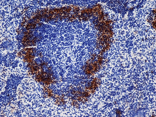
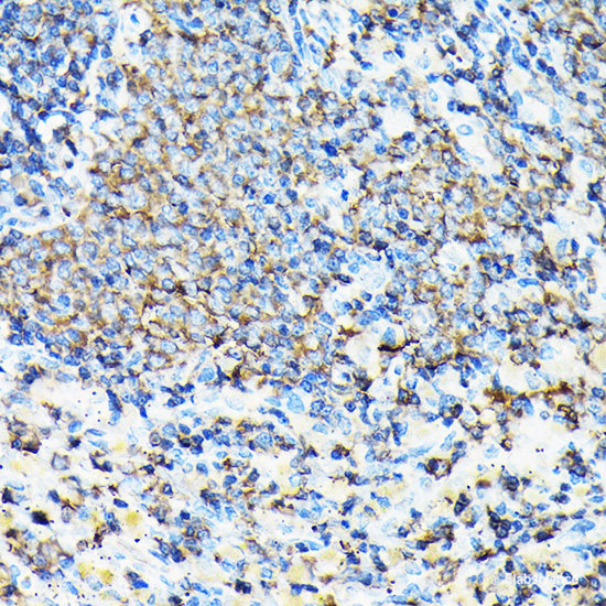
Rabbit anti-Mouse Pdgfra Polyclonal Antibody | anti-Pdgfra antibody
Pdgfra, ID (Pdgfra, Platelet-derived growth factor receptor alpha, Alpha platelet-derived growth factor receptor, Alpha-type platelet-derived growth factor receptor, CD140 antigen-like family member A, Platelet-derived growth factor alpha receptor, CD140a
NCBI and Uniprot Product Information
Uniprot Description
Function: Tyrosine-protein kinase that acts as a cell-surface receptor for PDGFA, PDGFB and PDGFC and plays an essential role in the regulation of embryonic development, cell proliferation, survival and chemotaxis. Depending on the context, promotes or inhibits cell proliferation and cell migration. Plays an important role in the differentiation of bone marrow-derived mesenchymal stem cells. Required for normal skeleton development and cephalic closure during embryonic development. Required for normal development of the mucosa lining the gastrointestinal tract, and for recruitment of mesenchymal cells and normal development of intestinal villi. Plays a role in cell migration and chemotaxis in wound healing. Plays a role in platelet activation, secretion of agonists from platelet granules, and in thrombin-induced platelet aggregation. Binding of its cognate ligands - homodimeric PDGFA, homodimeric PDGFB, heterodimers formed by PDGFA and PDGFB or homodimeric PDGFC -leads to the activation of several signaling cascades; the response depends on the nature of the bound ligand and is modulated by the formation of heterodimers between PDGFRA and PDGFRB. Phosphorylates PIK3R1, PLCG1, and PTPN11. Activation of PLCG1 leads to the production of the cellular signaling molecules diacylglycerol and inositol 1,4,5-trisphosphate, mobilization of cytosolic Ca2+ and the activation of protein kinase C. Phosphorylates PIK3R1, the regulatory subunit of phosphatidylinositol 3-kinase, and thereby mediates activation of the AKT1 signaling pathway. Mediates activation of HRAS and of the MAP kinases MAPK1/ERK2 and/or MAPK3/ERK1. Promotes activation of STAT family members STAT1, STAT3 and STAT5A and/or STAT5B. Receptor signaling is down-regulated by protein phosphatases that dephosphorylate the receptor and its down-stream effectors, and by rapid internalization of the activated receptor. Ref.5 Ref.6 Ref.10 Ref.11
Catalytic activity: ATP + a [protein]-L-tyrosine = ADP + a [protein]-L-tyrosine phosphate.
Enzyme regulation: Present in an inactive conformation in the absence of bound ligand. Binding of PDGFA and/or PDGFB leads to dimerization and activation by autophosphorylation on tyrosine residues. Inhibited by imatinib, nilotinib and sorafenib
By similarity.
Subunit structure: Interacts with homodimeric PDGFA, PDGFB and PDGFC, and with heterodimers formed by PDGFA and PDGFB. Monomer in the absence of bound ligand. Interaction with dimeric PDGFA, PDGFB and/or PDGFC leads to receptor dimerization, where both PDGFRA homodimers and heterodimers with PDGFRB are observed. Interacts (tyrosine phosphorylated) with SHB (via SH2 domain). Interacts (tyrosine phosphorylated) with SHF (via SH2 domain). Interacts (tyrosine phosphorylated) with SRC (via SH2 domain). Interacts (tyrosine phosphorylated) with PIK3R1. Interacts (tyrosine phosphorylated) with PLCG1 (via SH2 domain). Interacts (tyrosine phosphorylated) with CRK, GRB2 and GRB7
By similarity. Ref.7
Subcellular location: Cell membrane; Single-pass type I membrane protein. Note: The activated receptor is rapidly internalized and degraded
By similarity.
Tissue specificity: Focally expressed in cortical interstitial cells and highly expressed in the interstitium of the papillary region. Also expressed by adventitial cells in arterial vessels. Up-regulated in areas of renal fibrosis. In mice with unilateral ureteral obstruction, expression in cortical interstitial cells becomes prominent at day 4 which increases progressively until day 14. Ref.8
Post-translational modification: Ubiquitinated, leading to its degradation
By similarity.Autophosphorylated on tyrosine residues upon ligand binding. Autophosphorylation occurs in trans, i.e. one subunit of the dimeric receptor phosphorylates tyrosine residues on the other subunit. Phosphorylation at Tyr-731 and Tyr-742 is important for interaction with PIK3R1. Phosphorylation at Tyr-720 and Tyr-754 is important for interaction with PTPN11. Phosphorylation at Tyr-762 is important for interaction with CRK. Phosphorylation at Tyr-572 and Tyr-574 is important for interaction with SRC and SRC family members. Phosphorylation at Tyr-988 and Tyr-1018 is important for interaction with PLCG1
By similarity.
Disruption phenotype: Embryonically lethal. Most embryos survive up to 13 dpc, but display important defects in skeleton development, including spina bifida, fusions of cervical vertebrae and ribs, and incomplete fusion of the skull parietal bone. Embryos display also abnormal mucosa lining the gastrointestinal tract, including fewer and misshapen villi and loss of pericryptal mesenchyme. At about 16 dpc, embryos display extensive hemorrhaging. Ref.5 Ref.6 Ref.9
Sequence similarities: Belongs to the protein kinase superfamily. Tyr protein kinase family. CSF-1/PDGF receptor subfamily.Contains 5 Ig-like C2-type (immunoglobulin-like) domains.Contains 1 protein kinase domain.
Similar Products
Product Notes
The Pdgfra pdgfra (Catalog #AAA6332277) is an Antibody produced from Rabbit and is intended for research purposes only. The product is available for immediate purchase. The Pdgfra, ID (Pdgfra, Platelet-derived growth factor receptor alpha, Alpha platelet-derived growth factor receptor, Alpha-type platelet-derived growth factor receptor, CD140 antigen-like family member A, Platelet-derived growth factor alpha receptor, CD140a reacts with Mouse and may cross-react with other species as described in the data sheet. AAA Biotech's Pdgfra can be used in a range of immunoassay formats including, but not limited to, Western Blot (WB), FLISA. Applications are based on unconjugated antibody. Researchers should empirically determine the suitability of the Pdgfra pdgfra for an application not listed in the data sheet. Researchers commonly develop new applications and it is an integral, important part of the investigative research process. It is sometimes possible for the material contained within the vial of "Pdgfra, Polyclonal Antibody" to become dispersed throughout the inside of the vial, particularly around the seal of said vial, during shipment and storage. We always suggest centrifuging these vials to consolidate all of the liquid away from the lid and to the bottom of the vial prior to opening. Please be advised that certain products may require dry ice for shipping and that, if this is the case, an additional dry ice fee may also be required.Precautions
All products in the AAA Biotech catalog are strictly for research-use only, and are absolutely not suitable for use in any sort of medical, therapeutic, prophylactic, in-vivo, or diagnostic capacity. By purchasing a product from AAA Biotech, you are explicitly certifying that said products will be properly tested and used in line with industry standard. AAA Biotech and its authorized distribution partners reserve the right to refuse to fulfill any order if we have any indication that a purchaser may be intending to use a product outside of our accepted criteria.Disclaimer
Though we do strive to guarantee the information represented in this datasheet, AAA Biotech cannot be held responsible for any oversights or imprecisions. AAA Biotech reserves the right to adjust any aspect of this datasheet at any time and without notice. It is the responsibility of the customer to inform AAA Biotech of any product performance issues observed or experienced within 30 days of receipt of said product. To see additional details on this or any of our other policies, please see our Terms & Conditions page.Item has been added to Shopping Cart
If you are ready to order, navigate to Shopping Cart and get ready to checkout.