


TUBB recombinant antibody
TUBB Recombinant Monoclonal Antibody
IHC: 1:50-1:200
IF: 1:50-1:200
FC/FACS: 1:50-1:200
FCM (Flow Cytometry)
(Overlay Peak curve showing K562 cells stained with AAA28078 (red line) at 1:100.The cells were fixed in 4% formaldehyde and permeated by 0.2% TritonX-100. Then 10% normal goat serum to block non-specific protein-protein interactions followed by the antibody (1ug/1*106cells) for 45min at 4 degree C. The secondary antibody used was FITC-conjugated Goat Anti-human IgG(H+L) at 1:200 dilution for 35min at 4 degree C.Control antibody (green line) was human IgG (1ug/1*106cells) used under the same conditions. Acquisition of >10,000 events was performed.)
IF (Immunofluorescence)
(Immunofluorescence staining of Hela cell with AAA28078 at 1:100, counter-stained with DAPI. The cells were fixed in 4% formaldehyde and blocked in 10% normal Goat Serum. The cells were then incubated with the antibody overnight at 4C. The secondary antibody was FITC-conjugated AffiniPure Goat Anti-Human IgG(H+L).)
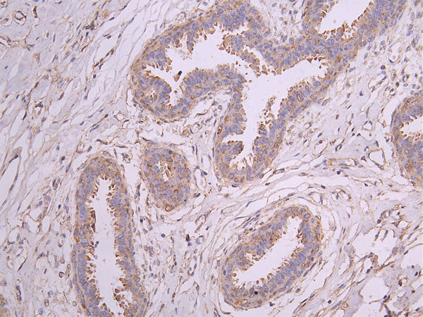
IHC (Immunohistochemistry)
(IHC image diluted at 1?200 and staining in paraffin-embedded human breast cancer performed on a Leica BondTM system. After dewaxing and hydration, antigen retrieval was mediated by high pressure in a citrate buffer (pH 6.0). Section was blocked with 10% normal goat serum 30min at RT. Then primary antibody (1% BSA) was incubated at 4 degree C overnight. The primary is detected by a Goat anti-human polymer IgG labeled by HRP and visualized using 0.05% DAB.)
IHC (Immunohistochemistry)
(IHC image diluted at 1?200 and staining in paraffin-embedded human brain tissue performed on a Leica BondTM system. After dewaxing and hydration, antigen retrieval was mediated by high pressure in a citrate buffer (pH 6.0). Section was blocked with 10% normal goat serum 30min at RT. Then primary antibody (1% BSA) was incubated at 4 degree C overnight. The primary is detected by a Goat anti-human polymer IgG labeled by HRP and visualized using 0.05% DAB.)
IHC (Immunohistochemistry)
(IHC image diluted at 1?200 and staining in paraffin-embedded human tonsil tissue performed on a Leica BondTM system. After dewaxing and hydration, antigen retrieval was mediated by high pressure in a citrate buffer (pH 6.0). Section was blocked with 10% normal goat serum 30min at RT. Then primary antibody (1% BSA) was incubated at 4 degree C overnight. The primary is detected by a Goat anti-human polymer IgG labeled by HRP and visualized using 0.05% DAB.)
WB (Western Blot)
(Western BlotPositive WB detected in:A431 whole cell lysate (20ug), SH-SY5Y whole cell lysate (20ug), U251 whole cell lysate (20ug), 293T whole cell lysate (20ug), HepG2 whole cell lysate (20ug), K562 whole cell lysate (20ug), MCF7 whole cell lysate (20ug), NIH/3T3 whole cell lysate (20ug)All lanes: TUBB antibody at 1:1000SecondaryGoat polyclonal to human IgG at 1/40000 dilutionPredicted band size:49 kDaObserved band size:55 kDaExposure time:5s)


